https://doi.org/10.5049/EBP.2019.17.2.45
Incidence of Acute Kidney Injury after Adrenalectomy in Patients with Primary Aldosteronism
Jee Young Lee1, Hyoungnae Kim2, Hyung Woo Kim1, Geun Woo Ryu1, Yooju Nam1, Seonyeong Lee1, Young Su Joo1, Sangmi Lee1, Jung Tak Park1, Seung Hyeok Han1, Shin-Wook Kang1,3, Tae-Hyun Yoo1 and Hae-Ryong Yun1
1Department of Internal Medicine, Institute of Kidney Disease Research, Yonsei University College of Medicine, Seoul; 2Division of Nephrology, Soonchunhyang University Hospital, Soonchunhyang University College of Medicine, Seoul, 3Department of Internal Medicine, Severance Biomedical Science Institute, Brain Korea 21 PLUS, Yonsei University College of Medicine, Seoul, Korea
Received: October 1, 2019 Revised: October 29, 2019 Accepted: October 30, 2019
Corresponding Author: Hae-Ryong Yun, MD Department of Internal Medicine, Institute of Kidney Disease Research, Yonsei University College of Medicine, 50-1 Yonsei-ro, Seodaemun-Gu, Seoul 03722, Korea
Tel: +82-2-2228-5345 Fax: +82-2-393-6884 E-mail: [email protected]
Background: Aldosterone-induced glomerular hyperfiltration can lead to mas- ked preoperative renal dysfunction in primary aldosteronism (PA) patients. We evaluated whether PA patients had a higher prevalence of acute kidney injury (AKI) after unilateral adrenalectomy. In addition, we identified risk factors for AKI in these subjects.
Methods: This retrospective study included 107 PA patients, and 186 pheo- chromocytoma patients as a control group, all of whom underwent adrena- lectomy between January 2006 and November 2017 at Yonsei University Severance Hospital. The primary outcome was AKI within 48 hours after ad- renalectomy. Univariate and multivariate logistic regression analyses were performed to identify predictors of AKI after adrenalectomy.
Results: Overall incidence of AKI was 49/293 (16.7%). In PA patients, the in- cidence of AKI was 29/107 (27.1%). In contrast, incidence of AKI was 20/
186 (10.7%) in pheochromocytoma patients. Univariate and multivariate lo- gistic regression analysis both showed a higher risk of postoperative AKI in PA patients compared to pheochromocytoma patients. In addition, old age, diabetes, longer duration of hypertension, lower preoperative estimated glo- merular filtration rate, high aldosterone-cortisol ratio (ACR) and lateraliza- tion index (LI) were identified as independent risk factors for postoperative AKI in PA patients after unilateral adrenalectomy.
Conclusion: Incidence and risk of postoperative AKI were significantly high- er in PA patients after surgical treatment. High ACR on the tumor side and high LI were associated with higher risk of AKI in PA patients compared to pheochromocytoma patients.
Key Words: Primary aldosteronism, Adrenalectomy, Renal insufficiency, Pheochromocytoma
This is an Open Access article distributed under the terms of the Creative Commons Attribution Non-Commercial License (https://creativecommons.org/licenses/by-nc/4.0/) which permits unrestricted non-commercial use, distribution, and reproduction in any medium, provided the original work is properly cited.
Introduction
Primary aldosteronism (PA) is one of the most frequent causes of secondary hypertension and is characterized by high blood pressure, hypokalemia, low plasma renin ac- tivity, and excessive aldosterone production by breaking away from the renin-angiotensin system1,2). PA has long been considered to be a relatively benign form of hyper-
tension due to the suppression of the renin-angiotensin axis which plays an important role in cardiovascular re- modeling and damage3). However, other studies have shown that long-term exposure to high levels of aldoster- one is associated with tissue fibrosis, vascular remodeling, and endothelial dysfunction4-7). In addition, TAPAI Study Group et al. reported that even mild impairment of renal function may predict residual hypertension after unilat-
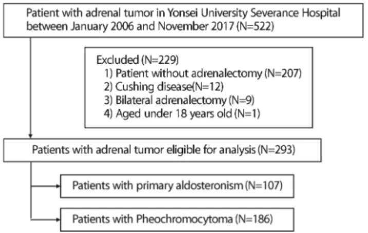
Fig. 1. Flow diagram of study design.
eral adrenalectomy in patients with aldosterone-produc- ing adenoma8).
Aldosterone-induced renal impairments in PA patients present clinically with increased urinary albumin excre- tion and estimated glomerular filtration rate (eGFR)9). Urinary albumin excretion and eGFR in PA patients de- crease after adrenalectomy or starting treatment with mi- neralocorticoid receptor antagonists. This can be explai- ned by aldosterone’s effects on glomerular hyperfiltra- tion, inflammation, fibrosis, mesangial cell proliferation, podocyte injury, and endothelial inflammation10).
Since aldosterone-induced glomerular hyperfiltration can lead to masked preoperative renal dysfunction in PA patients, it is important to be able to predict which pati- ents are at risk of adverse renal outcomes. Thus, we evalu- ated whether PA patients had a higher prevalence of renal impairment after unilateral adrenalectomy compared to pheochromocytoma patients, who represented secondary hypertension not caused by PA. In addition, we evaluated risk factors for renal impairment after adrenalectomy in PA patients.
Methods
1. Study participantsBetween January 2006 and November 2017, 522 pati- ents with adrenal tumors were admitted to Yonsei Univer- sity Severance Hospital. We excluded patients diagnosed with Cushing syndrome (n=12), patients who did not un- dergo adrenalectomy (n=207), patients who underwent bilateral adrenalectomy (n=9), and patients who were aged under 18 years old (n=1). Finally, a total of 293 patients were selected (Fig. 1). Of these patients, 107 were diagnosed with PA by a saline loading test, which was then localized by adrenal or abdominopelvic computed tomography (CT)11). The remaining 186 patients were di- agnosed with pheochromocytoma and set as a control group. Pheochromocytoma was confirmed by serum or urine catecholamine, metanephrine, and vanillylmandelic acid testing and localized by adrenal or abdominopelvic CT12). Patients with PA or pheochromocytoma under- went unilateral adrenalectomy between January 2006 and
November 2017 at Yonsei University Severance Hospital.
This study was conducted in accordance with the princi- ples of the Declaration of Helsinki, and the study pro- tocol was approved by the Institutional Review Board (IRB) of Yonsei University Health System’s Clinical Trial Center (approval number: 4-2019-0922). Because this was a retrospective observational study, the informed consent requirement was waived.
2. Data collection
Demographic details including sex, age, body mass in- dex (BMI), smoking status, history of hypertension, and history of diabetes mellitus (DM) were collected by retro- spective review of electrical medical records from the day of adrenalectomy before the operation. BMI was calcu- lated by dividing body weight (kg) by height squared (m2). After 5 minutes of seated rest, blood pressure was measured using an electronic sphygmomanometer.
After overnight fasting, venous samples were collected to determine blood urea nitrogen, creatinine, sodium, potassium, chloride, total CO2, uric acid, and total cho- lesterol levels, as well as eGFR. Albuminuria was meas- ured by dipstick. We used a creatinine measurement method which was calibrated to be traceable to isotope dilution mass spectrometry. The eGFRs were calcula- ted using the Chronic Kidney Disease-Epidemiology Col- laboration equation (CKD-EPI)13). In all PA patients, adre- nal vein sampling (AVS) was performed to evaluate aldos- terone and cortisol levels in each adrenal gland. We as-
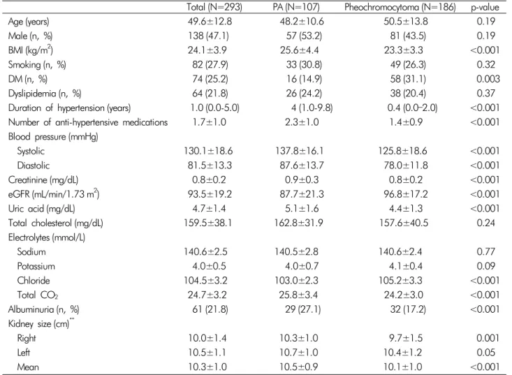
Table 1. Baseline characteristics of patients
Total (N=293) PA (N=107) Pheochromocytoma (N=186) p-value
Age (years) 49.6±12.8 48.2±10.6 50.5±13.8 0.19
Male (n, %) 138 (47.1) 57 (53.2) 81 (43.5) 0.19
BMI (kg/m2) 24.1±3.9 25.6±4.4 23.3±3.3 <0.001
Smoking (n, %) 82 (27.9) 33 (30.8) 49 (26.3) 0.32
DM (n, %) 74 (25.2) 16 (14.9) 58 (31.1) 0.003
Dyslipidemia (n, %) 64 (21.8) 26 (24.2) 38 (20.4) 0.37
Duration of hypertension (years) 1.0 (0.0-5.0) 4 (1.0-9.8) 0.4 (0.0–2.0) <0.001 Number of anti-hypertensive medications 1.7±1.0 2.3±1.0 1.4±0.9 <0.001 Blood pressure (mmHg)
Systolic 130.1±18.6 137.8±16.1 125.8±18.6 <0.001
Diastolic 81.5±13.3 87.6±13.7 78.0±11.8 <0.001
Creatinine (mg/dL) 0.8±0.2 0.9±0.3 0.8±0.2 <0.001
eGFR (mL/min/1.73 m2) 93.5±19.2 87.7±21.3 96.8±17.2 <0.001
Uric acid (mg/dL) 4.7±1.4 5.1±1.6 4.4±1.3 <0.001
Total cholesterol (mg/dL) 159.5±38.1 162.8±31.9 157.6±40.5 0.24
Electrolytes (mmol/L)
Sodium 140.6±2.5 140.5±2.8 140.6±2.4 0.77
Potassium 4.0±0.5 4.0±0.7 4.1±0.4 0.09
Chloride 104.5±3.2 103.0±2.3 105.2±3.3 <0.001
Total CO2 24.7±3.2 25.8±3.4 24.2±3.0 <0.001
Albuminuria (n, %) 61 (21.8) 29 (27.1) 32 (17.2) <0.001
Kidney size (cm)**
Right 10.0±1.4 10.3±1.0 9.7±1.5 0.001
Left 10.5±1.1 10.7±1.0 10.4±1.2 0.05
Mean 10.3±1.0 10.5±0.9 10.1±1.0 <0.001
Note: Values for categorical variables are given as number (percentage); values for continuous variables, as mean±standard devia- tion or median (interquartile range). eGFR was calculated using the CKD-EPI equation.
**Kidney size was measured by computed tomography.
PA, primary aldosteronism; BMI, body mass index; DM, diabetes mellitus; eGFR, estimated glomerular filtration rate; CO2, carbon dioxide; CKD, chronic kidney disease; CKD-EPI, chronic kidney disease-epidemiology collaboration.
sessed peripheral cortisol, adrenal vein cortisol, inferior vena cava cortisol, peripheral aldosterone, adrenal vein aldosterone, and inferior vena cava aldosterone. The late- ralization index (LI) was defined as the aldosterone-to- cortisol ratio of the adrenal vein on the tumor side divi- ded by the aldosterone-to-cortisol ratio of the contralate- ral adrenal vein14).
3. Statistical analysis
Continuous variables were expressed as means and standard deviations (SDs) or as medians with interquar- tile ranges (IQRs). Categorical variables were expressed as
numbers and percentages. Comparisons between groups were made by way of analysis of variance or Student’s t-test for continuous variables and by the chi-squared test or Fisher’s exact test for categorical variables. The Kolmogorov-Smirnov test was performed to determine the normality of distribution of the parameters. If the re- sulting data did not show a normal distribution, the geo- metric mean±standard deviation was reported; the Mann- Whitney U test or Kruskal-Wallis test was used for multi- ple comparisons. Multivariate logistic regression analysis was performed to identify independent predictors of AKI after adrenalectomy. The results of the logistic regression analysis were presented as odds ratios (ORs) and 95%
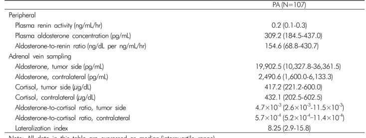
Table 2. Hormonal indexes in patients with PA
PA (N=107) Peripheral
Plasma renin activity (ng/mL/hr) 0.2 (0.1-0.3)
Plasma aldosterone concentration (pg/mL) 309.2 (184.5-437.0)
Aldosterone-to-renin ratio (ng/dL per ng/mL/hr) 154.6 (68.8-430.7) Adrenal vein sampling
Aldosterone, tumor side (pg/mL) 19,902.5 (10,327.8-36,361.5)
Aldosterone, contralateral (pg/mL) 2,490.6 (1,600.0-6,133.3)
Cortisol, tumor side (μg/dL) 417.2 (221.2-600.0)
Cortisol, contralateral (μg/dL) 432.1 (202.5-602.5)
Aldosterone-to-cortisol ratio, tumor side 4.7×10⁻³(2.6×10⁻³-11.5×10⁻³) Aldosterone-to-cortisol ratio, contralateral 5.7×10⁻⁴(5.2×10⁻⁴–11.4×10⁻⁴)
Lateralization index 8.25 (2.9-15.8)
Note: All data in this table are expressed as median (interquartile range).
PA, primary aldosteronism.
confidence intervals (CIs). Statistical significance was de- fined as p<0.05. Data were analyzed using IBM SPSS sta- tistical analysis software for Windows version 23.0 (IBM Corporation, Armonk, NY, USA) and SAS software ver- sion 9.4 (SAS Institute, Cary, NC, USA).
4. Primary outcomes
The primary outcome of interest in this study was AKI within 48 hours after adrenalectomy. Postoperative AKI was defined as an increase in serum creatinine level ≥0.3 mg/dL or a decrease in eGFR >25% from preoperative baseline values, in accordance with KDIGO (Kidney Dis- ease Improving Global Outcomes) guidelines15) and a pre- vious study16).
Results
1. Baseline characteristicsBaseline characteristics are shown in Table 1. The mean age of study participants was 49.6±12.8 years and 138 (47.1%) were men. Baseline BMI was 24.1±3.9 kg/m2 and mean eGFR was 93.5±19.2 mL/min per 1.73 m2. Of the 293 patients, 107 (35.9%) were diagnosed with PA and 186 (63.5%) were diagnosed with pheochromocytoma.
Baseline BMI was significantly higher in PA patients (25.6
kg/m2 vs. 23.3 kg/m2, p<0.001). The duration of hyper- tension was significantly longer in PA patients (4 years vs. 0.4 years, p<0.001) and systolic blood pressure was significantly higher (137.8±16.1 mmHg vs. 125.8±18.6
mmHg, p<0.001). Baseline eGFR was significantly lower in PA patients (87.7±21.3 mL/min per 1.73 m2 vs.
96.8±17.2 mL/min per 1.73 m2, p<0.001) and albumi- nuria was more frequent (27.1% vs. 17.2%, p<0.001).
In addition, mean kidney size was slightly larger in PA patients (10.5±0.9 cm vs. 10.1±1.0 cm, p<0.001).
2. Hormonal indexes in patients with PA In PA patients, median (IQRs) plasma renin activity was 0.2 (0.1-0.3) ng/mL/hr and median plasma aldosterone concentration was 309.2 (184.5-437.0) pg/mL. The aldos- terone-to-renin ratio was 154.6 (68.8-430.7) ng/dL per ng/ml/hr. The median LI was 8.25 (2.9-15.8) (Table 2).
3. Incidence of postoperative AKI
In both PA and pheochromocytoma patients, postope- rative eGFR was significantly lower than preoperative eGFR (postoperative eGFR: 73.0±1.97 mL/min per 1.73 m2 in PA patients and 85.1±2.07 mL/min per 1.73 m2 in pheochromocytoma patients) (Fig. 2). However, the in- cidence of postoperative AKI was 29/107 (27.1%) in PA
Fig. 2. Comparison of baseline and postoperative eGFR in PA and pheochromocytoma patients. Note: Baseline and postopera- tive eGFR were compared using the Student’s t-test. (A) Primary aldosteronism, (B) Pheochromocytoma. *p-value=0.001. PA, pri- mary aldosteronism; eGFR, estimated glomerular filtration rate.
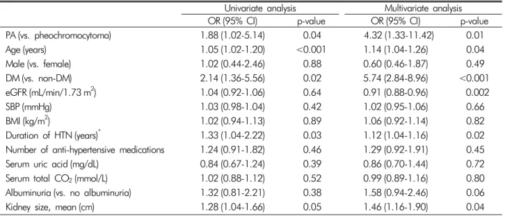
Table 3. Logistic regression analyses for risk of postoperative AKI
Univariate analysis Multivariate analysis
OR (95% CI) p-value OR (95% CI) p-value
PA (vs. pheochromocytoma) 1.88 (1.02-5.14) 0.04 4.32 (1.33-11.42) 0.01
Age (years) 1.05 (1.02-1.20) <0.001 1.14 (1.04-1.26) 0.04
Male (vs. female) 1.02 (0.44-2.46) 0.88 0.60 (0.46-1.87) 0.49
DM (vs. non-DM) 2.14 (1.36-5.56) 0.02 5.74 (2.84-8.96) <0.001
eGFR (mL/min/1.73 m2) 1.04 (0.92-1.06) 0.64 0.91 (0.88-0.96) 0.002
SBP (mmHg) 1.03 (0.98-1.04) 0.42 1.02 (0.95-1.06) 0.66
BMI (kg/m2) 1.02 (0.94-1.13) 0.89 1.06 (0.92-1.14) 0.82
Duration of HTN (years)* 1.33 (1.04-2.22) 0.03 1.12 (1.04-1.16) 0.02
Number of anti-hypertensive medications 1.24 (0.91-1.82) 0.46 1.29 (0.92-1.91) 0.45
Serum uric acid (mg/dL) 0.84 (0.67-1.24) 0.39 0.86 (0.70-1.44) 0.72
Serum total CO2(mmol/L) 1.02 (0.88-1.12) 0.52 0.99 (0.89-1.16) 0.80
Albuminuria (vs. no albuminuria) 1.32 (0.81-2.21) 0.38 1.58 (0.94-2.46) 0.06
Kidney size, mean (cm) 1.28 (1.04-1.66) 0.05 1.46 (1.16-1.90) 0.04
Note: Multivariate logistic regression analysis was performed after adjustment for confounding factors including age, sex, DM, eGFR, SBP, BMI, duration of HTN, albuminuria, and mean kidney size.
*Variable was log transformed.
AKI, acute kidney injury; OR, odds ratio; CI, confidence interval; PA, primary aldosteronism; DM, diabetes mellitus; eGFR, estimated glomerular filtration rate; SBP, systolic blood pressure; BMI, body mass index; HTN, hypertension; CO2, carbon dioxide.
patients but 20/186 (10.7%) in pheochromocytoma pati- ents. The incidence of postoperative AKI was significantly higher in PA patients than in pheochromocytoma patients (p<0.001).
4. Predictors of postoperative AKI
First, risk factors for AKI after adrenalectomy were de- termined by univariate logistic regression analysis. Accor-
ding to univariate logistic regression analysis, PA patients had a significantly higher risk of AKI compared to pheo- chromocytoma patients (OR 1.88, 95% CI 1.02-5.14, p=
0.04). Older age (OR 1.05, 95% CI 1.02-1.20, p<0.001), previous history of diabetes (OR 2.14, 95% CI 1.36-5.56, p=0.02), and longer duration of hypertension (OR 1.33, 95% CI 1.04-2.22, p=0.03) were also significantly asso- ciated with postoperative AKI. In addition, larger kidney size increased the risk of AKI (OR 1.28, 95% CI 1.04- 1.66, p=0.05) (Table 3). Multivariate logistic regression analysis revealed that patients with PA were still at incre- ased risk of postoperative AKI even after adjustment for confounding factors such as age, sex, DM, eGFR, systolic blood pressure, BMI, duration of hypertension, albu- minuria, and mean kidney size (OR 4.32, 95% CI 1.33- 11.42, p=0.01). In addition, lower eGFR was associated with an increased risk of postoperative AKI (OR 0.91, 95% CI 0.88-0.96, p=0.002). We further evaluated the risk factors in PA patients according to AVS values.
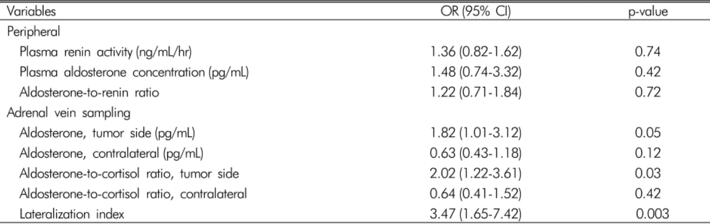
Among the AVS values, high aldosterone-cortisol ratio (ACR) (OR 2.02, 95% CI 1.22-3.61, p=0.03) and high LI (OR 3.47, 95% CI 1.65-7.42, p=0.003) may be inde- pendently associated with AKI after adrenalectomy in PA
Table 4. Relationships between hormonal indexes and postoperative AKI in patients with PA
Variables OR (95% CI) p-value
Peripheral
Plasma renin activity (ng/mL/hr) 1.36 (0.82-1.62) 0.74
Plasma aldosterone concentration (pg/mL) 1.48 (0.74-3.32) 0.42
Aldosterone-to-renin ratio 1.22 (0.71-1.84) 0.72
Adrenal vein sampling
Aldosterone, tumor side (pg/mL) 1.82 (1.01-3.12) 0.05
Aldosterone, contralateral (pg/mL) 0.63 (0.43-1.18) 0.12
Aldosterone-to-cortisol ratio, tumor side 2.02 (1.22-3.61) 0.03
Aldosterone-to-cortisol ratio, contralateral 0.64 (0.41-1.52) 0.42
Lateralization index 3.47 (1.65-7.42) 0.003
Note: The relationship between each hormonal activity index and AKI development was analyzed by multivariate logistic regression analysis with the following covariates: age, sex, DM, eGFR, SBP, BMI, duration of HTN, albuminuria, and mean kidney size.
*All hormonal activity indexes were log transformed.
AKI, acute kidney injury; PA, primary aldosteronism; OR, odds ratio; CI, confidence interval; DM, diabetes mellitus; eGFR, estimated glomerular filtration rate; SBP, systolic blood pressure; BMI, body mass index; HTN, hypertension.
patients (Table 4).
Discussion
In this study, we demonstrated that AKI occurred in PA patients after unilateral adrenalectomy. The risk of AKI in PA patients was significantly higher than in pheo- chromocytoma patients. In addition, higher tumor-side ACR and LI were independent risk factors for AKI after unilateral adrenalectomy in PA patients. Furthermore, we identified old age, previous history of diabetes, longer duration of hypertension, and lower preoperative eGFR as risk factors for postoperative AKI.
Renal function impairment is often observed in PA pa- tients after adrenalectomy. A previous study reported that nearly 40% of PA patients showed postoperative eGFR decline after unilateral adrenalectomy. On the other hand, renal function impairment was not seen in the control group who underwent the same surgical treatment16). This suggests that unilateral adrenalectomy was not the cause of postoperative renal function decline. In our study, the incidence of postoperative AKI in PA patients was 27.1%.
This was relatively lower than in the previous report. This difference in the incidence of AKI between studies could have been due to differences in sample size, follow-up duration, and baseline characteristics of the study groups.
In the previous study, PA patients and pheochromocytoma
patients were followed up for 6 months after unilateral adrenalectomy. The eGFR decline after unilateral adrena- lectomy in PA patients continued for up to 1 month, stabi- lizing afterwards17). The primary outcome in our study was creatinine decline within 48 hours post-adrenalec- tomy. With longer follow-up duration, further decline in eGFR would be observed, leading to more recorded AKI events. Although lower than in the previous study, the incidence of AKI in our study was significantly higher in PA patients than in pheochromocytoma patients with the same surgical treatment. Thus, these findings imply that a decrease in renal function after unilateral adrenalecto- my occurs mainly in PA patients rather than in pheochro- mocytoma patients, possibly due to physiologic changes before and after surgery.
Physiologic changes in PA patients after adrenalectomy are well documented. Normally, aldosterone is a key hor- mone in electrolyte and water homeostasis1). Patients with PA are exposed to uncontrolled high aldosterone levels due to pathologic adrenocortical lesions2,18). Excess aldos- terone stimulates sodium and water reabsorption, result- ing in extracellular fluid volume expansion, causing sec- ondary hypertension19,20). This condition increases renal perfusion, which leads to glomerular hyperfiltration21). Thus, it is hard to evaluate actual renal function in PA patients before treatment because renal dysfunction in PA patients is usually masked by increased eGFR, due to the
hyperfiltration effect of aldosterone22,23). When PA pa- tients are treated surgically or medically, their actual renal function is revealed8,18,24). This is consistent with the changes that were observed in our study.
We also identified risk factors for AKI in PA patients after unilateral adrenalectomy. Old age, previous history of diabetes, longer duration of hypertension, and lower preoperative eGFR were identified as risk factors for post- operative AKI. Old age, history of diabetes, and longer duration of hypertension are well-known risk factors for renal impairment25-29). However, previous studies re- ported that higher preoperative eGFR increased the risk of postoperative AKI, while lower preoperative eGFR was associated with the development of CKD in PA pati- ents after adrenalectomy16). Our results differ somewhat from these previous reports, possibly due to the following reason. PA patients in our study had a significantly longer duration of hypertension compared to pheochromocyto- ma patients. Chronic hypertension and long-term aldos- terone exposure can cause structural kidney damage such as tubulointerstitial fibrosis30-32). Thus, PA patients in this study may have had subclinical renal impairments that were concealed by the effect of aldosterone, leading to lower eGFR compared to pheochromocytoma patients who had a shorter duration of hypertension. As a result, lower eGFR caused by long-term hypertension was asso- ciated with postoperative AKI which was revealed after adrenalectomy when the aldosterone level dropped.
Long-term aldosterone exposure promotes endothelial dysfunction, as well as cardiovascular and renal inflam- mation and fibrosis4,33-36). According to multivariate logis- tic regression analysis for the evaluation of risk factors for renal impairment after adrenalectomy, higher ACR on the tumor side and higher LI were associated with an increased risk of renal impairment in PA patients com- pared to in pheochromocytoma patients. ACR and LI are both calculated using cortisol levels collected by AVS to negate the dilution of aldosterone caused by tributa- ries and malposition of the catheter when carrying out AVS37,38). Thereby, ACR and LI reflect more accurate as- sessments of aldosterone levels than the actual aldoster- one levels. This is why aldosterone level was not exam- ined as a risk factor for renal impairment, whereas ACR
and LI were examined. Therefore, it can be said that high- er aldosterone levels, when corrected according to corti- sol levels, resulted in increased risk of AKI. This result is compatible with the results of previous studies.
The strengths of our study are as follows. First, previ- ous studies about renal dysfunction in PA patients mostly compared PA patients to essential hypertension patients to evaluate the effect of aldosterone, since both patient groups had hypertension39-41). In contrast, our study com- pared PA patients with pheochromocytoma patients in which both groups underwent unilateral adrenalectomy.
By comparing PA and pheochromocytoma patients, we compared the effect of aldosterone on renal function since PA and pheochromocytoma induce hypertension by di- fferent mechanisms. Second, we used AVS to assess hor- mone levels. Most previous studies used samples from peripheral blood vessels, which do not reflect actual aldo- sterone levels. By using AVS, we more accurately meas- ured aldosterone levels in PA patients. Thus, we identified that higher ACR and LI were independent risk factors for AKI after unilateral adrenalectomy in these patients.
There are some limitations to this study. First, we only included PA patients who underwent unilateral adrena- lectomy with AVS. Thus, the study population was rela- tively small. Second, aldosterone-induced renal impair- ment presents clinically with urinary albumin excretion.
In this study, urine albumin measurement was performed by urine dipstick and quantitative measurement was not performed. Third, postoperative AKI was defined as an increase in serum creatinine ≥0.3 mg/dL or a decrease in eGFR >25% from baseline. However, urine output was not available as a definition for AKI. In addition, sur- gery involves bleeding, hypotension, and use of inter- mittent nephrotoxic agents which may affect renal func- tion postoperatively. We did not consider perioperative complications. Finally, we did not evaluate long-term re- nal outcomes in PA patients who underwent adrenalec- tomy. A further study should be performed to evaluate long-term renal damage using quantitative tests for uri- nary albumin or protein excretion and following the AKI or CKD criteria.
In conclusion, the incidence and risk of AKI were sig- nificantly higher in PA patients after unilateral adrenalec-
tomy. Old age, previous history of diabetes, longer dura- tion of hypertension, and lower preoperative eGFR were risk factors for postoperative AKI. In addition, higher tu- mor-side ACR and LI were risk factor for AKI in PA patients. Considering that AKI is a potent risk factor for CKD, proper management and regular follow-up should be performed on PA patients with unilateral adrenalec- tomy. Further studies are needed to evaluate the associa- tion between postoperative AKI and development of CKD.
Financial Support
This research did not receive any specific grants from funding agencies in the public, commercial, or not-for- profit sectors.
Conflict of interest
The authors declare that they have no conflicts of inte- rest. This material has not been published previously and will not be submitted for publication elsewhere.
Acknowledgements
We express our sincere gratitude to the staff of the Department of Internal Medicine, College of Medicine, Institute of Kidney Disease Research, Yonsei University, Seoul, Korea.
References
1. Conn JW: Part I. Painting background. Part II. Primary aldosteronism, a new clinical syndrome, 1954. J Lab Clin Med 116(2):253-267, 1990
2. Funder JW, Carey RM, Fardella C, et al.: Case detection, diagnosis, and treatment of patients with primary aldos- teronism: an endocrine society clinical practice guideline.
J Clin Endocrinol Metab 93(9):3266-3281, 2008 3. Monticone S, Sconfienza E, D'Ascenzo F, et al.: Renal
damage in primary aldosteronism: a systematic review and meta-analysis. J Hypertens, 2019
4. Hung CS, Sung SH, Liao CW, et al.: Aldosterone induces vascular damage. Hypertension 74(3):623-629, 2019 5. Catena C, Colussi G, Sechi LA: Treatment of primary al-
dosteronism and organ protection. Int J Endocrinol 2015:
597247, 2015
6. Ma TK, Szeto CC: Mineralocorticoid receptor antagonist for renal protection. Ren Fail 34(6):810-817, 2012 7. Rossi GP, Sacchetto A, Pavan E, et al.: Remodeling of
the left ventricle in primary aldosteronism due to Conn’s adenoma. Circulation 95(6):1471-1478, 1997
8. Wu VC, Chueh SC, Chang HW, et al.: Association of kidney function with residual hypertension after treat- ment of aldosterone-producing adenoma. Am J Kidney Dis 54(4):665-673, 2009
9. Tuck ML, Corry DB: Renal damage in primary aldoste- ronism: results of the PAPY study. Curr Hypertens Rep 9(2):87-89, 2007, doi: 0.1007/s11906-007-0016-4 10. Kramers BJ, Kramers C, Lenders JW, Deinum J: Effects of
treating primary aldosteronism on renal function. J Clin Hypertens (Greenwich) 19(3):290-295, 2017
11. Stowasser M, Gordon RD, Rutherford JC, Nikwan NZ, Daunt N, Slater GJ: Diagnosis and management of pri- mary aldosteronism. Journal of the Renin-Angiotensin- Aldosterone System 2(3):156-169, 2001
12. Davison AS, Jones DM, Ruthven S, Helliwell T, Shore SL:
Clinical evaluation and treatment of phaeochromocy- toma. Annals of Clinical Biochemistry 55(1):34-48, 2018 13. Schwandt A, Denkinger M, Fasching P, et al.: Comparison
of MDRD, CKD-EPI, and Cockcroft-Gault equation in relation to measured glomerular filtration rate among a large cohort with diabetes. Journal of Diabetes and its Complications 31(9):1376-1383, 2017
14. Tanemoto M: Diagnosis of unilateral aldosterone hyper- secretion in adrenal venous sampling. J Hypertens 37(10):
2110, 2019
15. Khwaja A: KDIGO clinical practice guidelines for acute kidney injury. Nephron Clin Pract 120(4):c179-184, 2012 16. Kim DH, Kwon HJ, Ji SA, et al.: Risk factors for renal
impairment revealed after unilateral adrenalectomy in patients with primary aldosteronism. Medicine (Baltimore) 95(27):e3930, 2016
17. Kim IY, Park IS, Kim MJ, et al.: Change in kidney func- tion after unilateral adrenalectomy in patients with pri- mary aldosteronism: identification of risk factors for de- creased kidney function. Int Urol Nephrol 50(10):1887- 1895, 2018
18. Sechi LA, Novello M, Lapenna R, et al.: Long-term renal outcomes in patients with primary aldosteronism. JAMA 295(22):2638-2645, 2006
19. Fogari R, Preti P, Zoppi A, Rinaldi A, Fogari E, Mugellini
A: Prevalence of primary aldosteronism among unselec- ted hypertensive patients: a prospective study based on the use of an aldosterone/renin ratio above 25 as a scree- ning test. Hypertens Res 30(2):111-117, 2007
20. Rossi GP, Bernini G, Caliumi C, et al.: A prospective study of the prevalence of primary aldosteronism in 1,125 hypertensive patients. J Am Coll Cardiol 48(11):
2293-2300, 2006
21. Ribstein J, Du Cailar G, Fesler P, Mimran A: Relative glomerular hyperfiltration in primary aldosteronism. J Am Soc Nephrol 16(5):1320-1325, 2005
22. Utsumi T, Kamiya N, Kaga M, et al.: Development of novel nomograms to predict renal functional outcomes after laparoscopic adrenalectomy in patients with prima- ry aldosteronism. World J Urol 35(10):1577-1583, 2017 23. Utsumi T, Kawamura K, Imamoto T, et al.: Preoperative
masked renal damage in Japanese patients with primary aldosteronism: identification of predictors for chronic kidney disease manifested after adrenalectomy. Int J Urol 20(7):685-691, 2013
24. Onohara T, Takagi T, Yoshida K, et al.: Assessment of postoperative renal function after adrenalectomy in pa- tients with primary aldosteronism. Int J Urol 26(2):229- 233, 2019
25. Maertens S, Van Den Noortgate N: Kidney in old age.
Acta Clinica Belgica 63(1):8-15, 2008
26. Gargiulo R, Suhail F, Lerma EV: Hypertension and ch- ronic kidney disease. Disease-a-month: DM 61(9):387, 2015
27. Hamrahian SM: Management of hypertension in patients with chronic kidney disease. Current hypertension re- ports 19(5):43, 2017
28. Edeghere S, English P: Management of type 2 diabetes:
now and the future. Clin Med (Lond) 19(5):403-405, 2019 29. Zafari N, Churilov L, MacIsaac RJ, et al.: Diagnostic per- formance of the Chronic Kidney Disease Epidemiology Collaboration (CKD-EPI) equation at estimating glome- rular filtration rate in adults with diabetes mellitus: a systematic review and meta-analysis protocol. BMJ Open 9(8):e031558, 2019
30. Hollenberg NK: Aldosterone in the development and progression of renal injury. Kidney Int 66(1):1-9, 2004 31. Remuzzi G, Cattaneo D, Perico N: The aggravating me-
chanisms of aldosterone on kidney fibrosis. J Am Soc Nephrol 19(8):1459-1462, 2008
32. Danforth DN, Orlando MM, Bartter FC, Javadpour N:
Renal changes in primary aldosteronism. The Journal of Urology 117(2):140-144, 1977
33. Brown NJ: Contribution of aldosterone to cardiovascular and renal inflammation and fibrosis. Nat Rev Nephrol 9(8):459-469, 2013
34. Tomaschitz A, Pilz S, Ritz E, Meinitzer A, Boehm BO, Marz W: Plasma aldosterone levels are associated with increased cardiovascular mortality: the Ludwigshafen Risk and Cardiovascular Health (LURIC) study. Eur Heart J 31(10):1237-1247, 2010
35. Rocha R, Stier CT, Jr.: Pathophysiological effects of al- dosterone in cardiovascular tissues. Trends Endocrinol Metab 12(7):308-314, 2001
36. Rossi G, Boscaro M, Ronconi V, Funder JW: Aldoste- rone as a cardiovascular risk factor. Trends in Endocri- nology & Metabolism 16(3):104-107, 2005
37. Steichen O, Amar L: Diagnostic criteria for adrenal ve- nous sampling. Curr Opin Endocrinol Diabetes Obes 23(3):218-224, 2016
38. Kline G, Holmes DT: Adrenal venous sampling for pri- mary aldosteronism: laboratory medicine best practice.
J Clin Pathol 70(11):911-916, 2017
39. Chen YY, Lin YH, Huang WC, et al.: Adrenalectomy improves the long-term risk of end-stage renal disease and mortality of primary aldosteronism. J Endocr Soc 3(6):
1110-1126, 2019
40. Hundemer GL, Curhan GC, Yozamp N, Wang M, Vaidya A: Renal outcomes in medically and surgically treated primary aldosteronism. Hypertension 72(3):658-666, 2018 41. Li H, Liu J, Liu J, et al.: The association between a
24-hour blood pressure pattern and circadian change in plasma aldosterone concentration for patients with al- dosterone-producing adenoma. Int J Endocrinol 2019:
4828402, 2019