서 론
인공 고관절 삽입물 주위 골절은 평균 수명의 연장으로 고령 인 구가 증가하고 골절이나 관절염 등으로 인한 인공관절이나 내고 정물 등의 삽입 빈도가 증가함에 따라 발생률이 급격히 증가하 고 있다.1,2) 용어상으로 인공관절 주위에 발생하는 골절을 인공관 절 주위 골절(periprosthetic fracture)이라 하고 금속판, 나사못, 금속정 등의 인공 삽입물 주위에 생기는 골절을 삽입물 주위 골 절(peri-implant fracture)이라 한다.3) 인공관절 삽입물 주위 골 절은 삽입물 주위 대퇴골의 골강도가 저하되어 있는 고령의 환
자에게서 일상생활중 낙상과 같은 저에너지 손상에 의해서도 흔 히 발생한다.4) 골절에 관계된 요인으로는 환자의 나이, 골용해 또 는 무균성 해리의 여부, 재치환 수술의 횟수, 치환술 당시 골절 진 단의 여부, 인공관절 삽입물의 종류와 수술자의 경험에 따라서 도 차이가 난다.4) 일반적으로 수술 중 또는 수술 후 발생하는 인 공관절 주위 골절로 구분하며 골질(bone quality)이 약하고 인 공 삽입물이 골수강 내에 존재하므로 치료하기 어렵고 예후도 좋 지 않아 골절이 발생한 후 치료 받는 것보다 골절이 발생하는 것 을 미리 예방하는 것이 중요하다.5) 골절이 발생하면 정확한 골절 분류를 통하여 적절한 치료 방법을 선택하여야 하고, 특히 삽입 물의 안정성을 정확히 판단하여 골절의 정복과 고정을 시도할 것 인지, 아니면 삽입물의 교체가 필요한지를 판단하는 것이 중요하 다.6) 이에 저자들은 술 후 발생하는 고관절 인공관절 삽입물 주위 골절 중 대퇴골에 발생하는 골절의 치료 원칙과 대퇴부 삽입물의 안정성 판단에 대해 문헌고찰과 함께 저자들의 경험을 서술하고 자 한다.
Copyright © 2021 by The Korean Orthopaedic Association
“This is an Open Access article distributed under the terms of the Creative Commons Attribution Non-Commercial License (http://creativecommons.org/licenses/by-nc/4.0/) which permits unrestricted non-commercial use, distribution, and reproduction in any medium, provided the original work is properly cited.”
The Journal of the Korean Orthopaedic Association Volume 56 Number 3 2021 Received August 31, 2020 Revised October 13, 2020
Accepted October 19, 2020
Correspondence to: Byung-Woo Min, M.D., Ph.D.
Department of Orthopedic Surgery, Keimyung University Dongsan Hospital, Keimyung University School of Medicine, 1035 Dalgubeol-daero, Dalseo-gu, Daegu 42601, Korea TEL: +82-53-258-4772 FAX: +82-53-258-4773 E-mail: [email protected] ORCID: https://orcid.org/0000-0002-1727-8872
고관절 치환술 후 삽입물의 안정성 판단과 대퇴 삽입물 주위 골절의 치료 원칙
김범수 • 이경재 • 민병우
계명대학교 의과대학 정형외과학교실
DecisionMaking and Principle of Management in Periprosthetic Femoral Fracture after Total Hip Arthroplasty
Beom-Soo Kim, M.D., Kyung-Jae Lee, M.D., and Byung-Woo Min, M.D., Ph.D.
Department of Orthopedic Surgery, Keimyung University Dongsan Hospital, Keimyung University School of Medicine, Daegu, Korea
Periprosthetic femoral fractures remain as one of the most challenging complications following total hip arthroplasty. A thorough clinical and radiographic evaluation, precise classification, and understanding of modern management principles are essential to obtain optimal results for these fractures. The Vancouver classification system is a simple, effective, and reproducible method for the planning treatments of these injuries. The fractures associated with a stable femoral stem can be treated effectively with osteosynthesis, but periprosthetic femoral fractures associated with a loose stem require revision arthroplasty. This paper describes the principle of the treatment of patients with periprosthetic femoral fractures and how to assess the stability of the femoral stem.
Key words: hip joint, arthroplasty, femur, periprosthetic fractures, decision making, treatment
발생 빈도와 역학
수술 후 발생하는 인공관절 주위 골절은 고관절 전치환술 후 시 행하는 재수술 중 세 번째로 많은 원인(9.5%)이며, 고관절 전치환 술 후 골절이 발생하는 빈도는 0.4%–1.1%이고, 고관절 재치환술 후 발생 빈도는 2.1%–4%이다.7) 수술 도중 발생하는 골절은 일차 고관절 전치환술 시 시멘트형 치환술에서는 약 1%, 무시멘트형 치환술에서는 약 3%–20%의 빈도로 일어난다.8) 고관절 재치환 술 중에 골절이 발생하는 빈도는 조금 더 높아서 시멘트형에서는 6.3%이며, 무시멘트형에서는 17.6% 정도로 발생한다.4)
인공 고관절 주위 골절은 주로 골다공증이 있는 노년기에 잘 발생하며 인공 삽입물 자체로 인한 응력 방패(stress shielding) 현상으로 인해 골질이 약화되어 있어 골절의 고정력이 약화되고, 인공 삽입물이 나사못이나 골수정 등의 내고정물을 삽입하는 데 방해물로 작용할 수 있으며, 나사못이나 인공 삽입물이 들어간 자리가 골절을 일으키는 응력 집중 요인(stress-riser)으로도 작 용할 수 있다.9) 또한 인공물의 삽입 과정이나 인공관절을 고정하 기 위한 시멘트 등을 삽입하는 과정에서 골수강 내 혈류 순환을 차단하여 골절 부위의 불유합을 초래할 수도 있어 인공관절 주위 골절의 치료 시 세심한 주의를 요한다.10)
인공관절 주위 골절의 원인은 전신적 요인으로 골다공증, 골감 소증, 골이형성증 등의 전신질환으로 인한 골 변형, 평형감각이 상으로 인한 낙상 등이 있으며, 국소적인 요인으로는 인공관절 재치환술, 최소 침습법(minimally invasive plate osteosynthe- sis technique)을 이용한 수술, 국소적인 응력 집중 요인(나사못 구멍, 금속판의 끝부분, 불안정한 내고정물의 고정 상태), 그리고 국소적인 골결손 등이 있다.11,12) 이러한 요인들 외에 인공관절 주 위 골절은 많은 경우에서 인공물의 삽입 도중 시술상의 기술적인 잘못에 기인하는 경우가 많다.13) 인공관절 삽입 시 대퇴 스템이 피질골에 손상을 주어 응력 집중을 유발하여 골절이 오는 경우와 골절 등의 치료를 위해 삽입하였던 내고정물 제거 후에 나사못이 제거된 구멍을 통해 골절이 발생하는 경우 등이 대표적이다.6) 따 라서 삽입물 주위 골절을 예방하려면 수술 중 갈라진 틈과 골결 손 등이 생기지 않도록 하고, 시멘트가 갈라진 틈으로 새어나가 는 것을 방지하며, 수술 도중 미세 골절이 발생한 경우 골절의 전 위가 없어도 환형 강선으로 고정한다. 골손실이 있는 부위는 골 이식으로 보정하며 수술 후 골용해, 통증, 또는 기능에 변화가 있 는지 관찰하여야 한다.
분 류
인공 고관절 주위 대퇴골 골절은 크게 수술 중 골절(intraopera- tive fracture) 및 수술 후 골절(postoperative fracture)로 나누 게 되며, 본 종설에서는 수술 후 발생하는 삽입물 주위 골절에 대
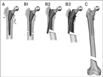
해서만 기술하고자 한다. 대퇴 삽입물 주위 골절의 치료 방침을 결정하기 위해서는 정확한 진단이 우선되어야 하며, 대퇴 스템에 대한 골절의 위치, 골절 양상(분쇄 또는 단순 골절), 대퇴 스템의 안정성, 골질, 골절을 유발할 수 있는 골용해 및 응력 차단 현상 등의 유무, 기존 대퇴 스템의 고정 방법(시멘트형 또는 무시멘트 형) 등 여러 요소들을 동시에 고려하여야 한다. 현재 수술 후 발생 하는 대퇴 삽입물 주위 골절에서 가장 널리 사용되고 있는 Van- couver 분류법은 비교적 단순하고, 신뢰할 수 있으며 재현성이 높은 것으로 알려져 있고, 1) 골절의 위치, 2) 대퇴 스템의 안정성, 3) 대퇴골 상태(bone stock)에 따라 A, B, C의 세 가지 형으로 구 분한다.14) A형은 삽입물 근위부의 골절로 대전자의 골절은 AG형 으로, 소전자의 골절은 AL형으로 다시 세부 분류하였다. B형 골 절은 대퇴 삽입물 또는 삽입물 말단의 바로 아래에 일어난 골절 이며, C형은 대퇴 삽입물 말단의 원위부에 발생한 골절이다. B형 골절은 삽입물의 안정성과 주위 골의 골질에 따라 다시 세부 분 류하였는데, B1 골절은 삽입물의 고정이 안정된 골절, B2 골절은 삽입물이 불안정한 골절이며, B3 골절은 전신적 골감소증, 골용 해, 또는 심한 분쇄 골절 등으로 심한 골결손이 있는 경우를 말한 다(Fig. 1). Duncan과 Masri15)는 수술 후 발생하는 대퇴 삽입물 주위 골절 중 A형이 4%, B형이 86.7%, C형이 9.3%의 빈도로 발 생한다고 보고하였으며, Lindahl 등6)은 A형이 2%, B1형이 28%, B2형이 49%, B3형이 11%, C형이 10%의 빈도로 발생하였다고 보고하여 전체 골절 중 B형 골절이 가장 많이 발생함을 알 수 있다.
수술 전 고려 사항
대퇴 삽입물 주위 골절은 대부분 B형인데 이 중 B1으로 분류된
A B1 B2 B3 C
Figure 1. The Vancouver classification of periprosthetic hip fractures is the most widely used classification system. It considers the fracture site, status of the femoral implant, and quality of surrounding femoral bone stock.
환자 중 50%에서 실제로는 B2로 판정되고 있으므로 삽입물 주 위 골절의 치료 시 가장 먼저 판단하여야 할 것은 삽입물의 고정 상태, 즉 대퇴 삽입물의 안정성 여부이다.3,6,9,16) 이러한 대퇴 스템 의 안정성 여부를 판단하기 위해 임상증상뿐만 아니라 단순 방사 선 촬영, 컴퓨터 단층촬영, 자기공명영상, 골주사 등도 시행할 수 있으나 금속의 간섭효과 때문에 충분한 정보를 얻을 수 없는 경 우도 많다. 인공관절 주위 골절의 진단 및 수술 전 처치 시 또 한 가지 주의할 점은 인공 삽입물의 존재로 인한 감염 가능성이 잔 존하므로 혈액 검사를 시행하고 경우에 따라 골절 부위의 천자 (aspiration)를 시행하여 감염 가능성을 미리 예측하고, 필요하면 수술 도중 현미경검사를 통하여 감염 가능성을 배제하여야 한다.17) 수술 전 환자에 대한 세심한 진찰과 최근 방사선 사진에 대한 정확한 평가가 필요하며, 전신적인 그리고 국소적인 골절 발생 위험인자를 파악하고 그에 따른 적당한 삽입물과 수술 도달법을 선택해야 한다. 수술 중에는 지나친 견인보다는 적절한 절개를 통해 충분한 시야를 확보하여야 한다. 수술과정에서는 고관절 탈 구, 골수강 확공, 시험 정복, 특히 무시멘트형 스템을 삽입할 경우 골절이 일어나지 않도록 주의하여야 한다. 재치환술 시 기존 삽 입물 또는 시멘트를 제거할 때는 의도하지 않은 골절이 일어나지 않도록 세심한 주의가 필요하다. 대퇴골과 비구 삽입물 주위의 골질과 골결손의 정도가 예후에 큰 영향을 미치므로 관절면의 마 모로 인한 골용해가 있다면 치료시기를 놓치지 않도록 주의를 기 울여야 할 것이다.
대퇴 삽입물의 안정성 판단
대퇴 삽입물의 안정성 판단은 Vancouver 분류 중 B1과 B2를 구 별하는 것인데, 이에 따라 수술의 방법과 원칙이 변하기 때문에 아주 중요하다. 골절로 인한 대퇴 스템의 안정성 판단을 위해서 는 대퇴골 골절 발생 전 촬영한 방사선 사진과 비교해 보는 것이 중요하며, 대퇴 스템의 침강(subsidence) 소견이 있을 경우 삽 입물이 불안정할 수 있으므로 재치환술에 대한 준비가 필요하다 (Fig. 2). 방사선적으로 대퇴 스템의 안정성을 판단하는 데 있어 서 기존에 사용된 대퇴 스템의 종류와 골절선의 위치를 파악하는 것이 중요하며, 이때 컴퓨터 단층촬영을 통한 골절선 파악이 도 움된다(Fig. 3)18,19). 무시멘트성 대퇴 스템의 경우 Khanuja 등20) 의 분류에 따른 대퇴 스템의 종류를 파악하고, 대퇴골 골절의 골 절선이 대퇴 스템의 고정이 일어나는 부위를 침범한 경우나 골 이입성 스템의 피복 주변에 골절이 발생하는 경우에서 불안정할 가능성이 높다. 시멘트형 대퇴 스템의 경우 뼈-시멘트 또는 시멘 트-대퇴 스템 사이에 연속된 방사선 투과선(continuous radio- graphic lucency)이 있거나 시멘트 맨틀(mantle)의 골절이 있는 경우 삽입물의 불안정성을 의심해 볼 수 있다.21) 하지만 방사선적 으로 B1과 B2 골절을 구분하기가 쉽지 않기 때문에, 안정성 여부 가 불명확한 경우 수술 중 대퇴 스템의 안정성 여부를 확인하여 수술 방법을 선택하는 것이 좋다.3,21)
추가적으로 골절 전 대퇴부 통증 유무를 확인하는 것이 삽입물 의 안정성 판단에 도움을 줄 수 있으며, 평소 체중 부하 시 악화되 거나 의자에서 일어설 때 발생하는 통증이 있었다면 대퇴 스템의 해리가 있었을 가능성이 있어 주의하여야 한다.
Figure 2. Anteroposterior radiographs of the femur obtained pre- fracture (A) and post-fracture (B). Note that the distance between the tip of the greater trochanter and stem shoulder was increased after the periprosthetic fracture (stem subsidence). This suggests that the stem is unstable.
A B
Figure 3. (A) Radiograph shows a periprosthetic femoral fracture with a spiral pattern around the stem. (B) Images of the computed tomographyscan show that the fracture line (arrows) crosses from the distal medial side to the proximal lateral side at the posterior side of the stem.
A B
골절의 치료 원칙
골절의 위치, 삽입물과 골절의 안정성, 대퇴 골질, 환자의 전신 상 태와 나이, 수술의사의 경험에 따라 치료 방법을 결정하여야 한 다. Vancouver 분류에 따라 대퇴 삽입물이 안정되어 있는 경우 에는 골절 치료만으로 충분하나 대퇴 삽입물이 불안정한 경우에 는 대퇴 삽입물을 교체하여야 하며 인공 삽입물 주위의 대퇴골 골질이 충분하지 않은 경우에는 골이식술을 고려하여야 한다.3) 대퇴골 삽입물 주위 골절의 정복 시 가장 피해야 할 것은 내반 정 복(varus reduction)이며, 추가 골절을 피하기 위하여 응력 집중 을 만들지 말아야 한다.22) 인공관절 주위 골절의 고정 원칙은 일 반 골절과 마찬가지로 골절의 형태에 따라 절대적 안정성(abso- lute stability)을 얻을 것인지 상대적 안정성(relative stability) 을 얻을 것인지 결정하여야 한다.23) 일반적으로 단순 골절은 해부 학적인 정복과 함께 절대적 안정성을 얻기 위한 고정술을 시도하 고(Fig. 4), 비전위 골절 및 분쇄 골절은 상대적 안정성을 얻기 위 해 간접 정복을 통해 생물학적 고정술(biologic fixation)을 시도 한다(Fig. 5). 금속판과 나사못으로 고정할 경우 골질이 약한 뼈 의 고정력을 높이기 위해 필요한 경우 잠김 나사못 등을 사용하 여 고정하여야 하며, 대퇴 삽입물이 골수강 내에 존재하는 대퇴 근위부에는 고정력을 높이기 위하여 locking attachment plate 나 케이블 등을 사용하여 고정력을 높일 수 있다. 골다공증 등으 로 인한 골질 손상이 심할 경우 충분한 고정력을 얻기 위해 노력 하여야 하며 때로는 대퇴골 전장을 고정하여야 할 경우도 있다.
시멘트형 대퇴 스템을 고정할 경우 시멘트의 손상을 줄이기 위해 blunt screw tip을 사용하는 것이 좋으며, 스템 말단부 주위의 횡 골절이나 심한 분쇄 골절의 경우에는 필요하면 이중 금속판이나 구조적인 골이식을 추가한다.24,25)
Vancouver 분류에 따른 골절의 치료
Vancouver A형 골절은 비수술적 치료가 원칙이나 골절의 원인 이 골용해인 경우 원인을 교정하여야 한다.3) AG형 골절은 대부 분 안정적이어서 약 6–12주간 체중 부하와 능동적 고관절 외전 운동을 제한하면 된다.26) 골편이 2.5 cm 이상 전위되었거나, 대 전자의 불유합으로 인한 통증, 관절의 불안정, 그리고 외전력의 약화가 있으면 골이식과 내고정을 고려해야 한다.3,26,27) 그리고 골 결손이 심한 경우 통증 감소 및 보행 기능 개선 등을 위해 대둔근 피판 이전술(gluteus maximus flap transfer)을 시행해 볼 수도 있다.28) AL형 골절은 매우 드물지만, 이 형태의 단독 골절은 대부 분 전위가 심하지 않고 대퇴 스템의 안정성에 영향을 미치지 않 기 때문에 보존적 치료가 가능하나,21) 골편의 크기가 커서 대퇴 스템의 내측 지지대(medial buttress)가 소실되어 불안정해질 가 능성이 있거나 골절 전 존재하는 골용해 등으로 인해 삽입물이 불안정하였던 경우 등에서는 대퇴 스템 재치환술과 내고정술을 동시에 시행해야 한다.3)
대퇴 삽입물이 안정된 B1형이나 C형인 경우 골절을 보존적으 로 치료할 경우 불유합, 부정유합, 삽입물의 해리 및 장기간 침상 안정 등으로 인한 합병증의 발생 빈도가 높아 골절 정복과 견고
A B
Figure 4. (A) Preoperative radiograph shows periprosthetic femoral fractures with short oblique simple fracture lines around the stem.
(B) Open reduction and internal fixation with a locking plate (absolute stability) were applied, and a well-united fracture site and stable stem can be seen.
Figure 5. (A) Minimally invasive plate osteosynthesis with locking plate (relatively stability) was applied for nondisplaced Vancouver type B1 fractures. (B) Clinical photograph shows two incisions for plate fixation without exposure of the fracture site.
A B
한 내고정으로 치료하는 것을 원칙으로 한다.3,12,21) 그러므로 관 혈적 정복술 및 금속내고정이 원칙이며,29,30) 경우에 따라서는 연 부조직 손상을 최소화할 수 있는 최소 침습법을 이용한 비관혈 적 정복술이 효과적인 경우도 있다.23,31,32) 근위 대퇴부에서 충분 한 고정력을 얻기 위해서는 케이블 단독 고정만으로는 부족하며 잠김 부착 금속판, 잠김 나사(locking screw), 여러 방향으로 나 사 고정을 할 수 있는 금속판(polyaxial locking plate) 등과 병 행하여 부가적인 고정법으로 사용하는 것이 좋다.3,21) 또한 가능 하다면 금속판을 대전자 부위까지 충분히 올려서 고정력을 높일
수 있도록 하는 것이 좋다. 저자들은 B1형과 C형 골절 치료에 있 어서 가급적 연부조직 손상을 줄이기 위해 노력하고 단순 골절 의 경우 해부학적 정복을 통한 절대적 안정성을 얻고, 전위가 없 는 골절이거나 분쇄가 심한 골절일 경우 충분한 길이의 금속판을 이용하여 상대적 안정성을 얻을 수 있도록 고정한다. 하지만 대 퇴 스템 끝부분에서 발생한 횡골절의 경우 단일 금속판만으로 충 분한 고정력을 얻기 어려워 실패율이 높기 때문에 동종지주골이 나 이중 금속판을 이용하여 고정력을 높이고 골절 부위의 골질이 좋지 못한 경우에도 동종지주골 이식을 병행한다.8,33-35) 응력 집 중 부위를 없애기 위해서 골절부 아래위로 충분한 길이의 금속판 을 이용하고, 특히 근위부 고정력을 높이기 위해 다양한 고정법 을 함께 사용하는 것이 좋을 것으로 생각된다(Fig. 6).
대퇴 삽입물의 안정성이 없는 B2형은 대퇴 삽입물의 교체가 불가피하며 새로운 삽입물은 기존의 삽입물이 있던 곳을 완전 히 우회(bypass)하여야 한다. 사용할 대퇴 삽입물의 선택은 골절 의 양상, 환자의 활동성에 대한 예상기대치, 골의 질, 골수강 직경 의 크기, 골수강의 모양에 따라 결정하며, 간혹 시멘트형 대퇴 삽 입물이 필요할 경우도 있다. 대부분의 경우 원위고정 무시멘트형 삽입물을 사용하여 골절 부위를 지나 원위 골간부에서 고정력을 얻는다.2) 대퇴 스템 재치환 시 골절 부위를 충분히 노출하여 기 존 대퇴 스템을 포함하여 골수강 내에 남아있는 시멘트, 플러그 (plug), 육아조직 등 수술에 방해가 될 수 있는 것들을 모두 제거 하여야 하며, 재치환 대퇴 스템을 위한 원위 대퇴골 확공(ream- ing) 전에 예방적 환형 강선이나 케이블 고정을 시행하여 원위 대 퇴골에 골절이 발생되지 않도록 해야 한다(Fig. 7).3) B2 골절 치 료 시 시멘트형 대퇴 스템은 골절 부위로 시멘트 누출이 일어나 장기적으로 대퇴 스템의 생존에 문제를 야기할 수 있으므로 주로
A B C D E F
R
Figure 6. A 72-year-old woman underwent total hip arthroplasty because of left hip osteoarthritis. (A) Radiographs show Vancouver type B1 periprosthetic femoral fracture with a transverse pattern around the stem tip. (B) Radiographs obtained immediately after fixation with locking compression plate with locking attached. (C) Radiographs obtained 11 months after fixation show nonunion of the fracture site with metal failure. (D) Radiographs obtained after revisional surgery using a locking compression plate with a strut onlay allograft. (E) Radiographs obtained 22 months after revision show well-healed bone at the fracture site. (F) The lower limb scan image obtained 22 months shows a similar alignment to the healthy side.
Figure 7. (A) Preoperative radiograph of the femur shows a Vancouver type-B2 fracture. (B) Postoperative radiograph shows revision of the femoral stem using a prophylactic cable (arrow).
A B
기대여명이 짧거나 활동량이 매우 적은 선택적인 환자에서 사용 되며, 조기에 체중 부하가 가능하다는 장점이 있다.36)
B3형 골절에서는 대부분의 경우 골이식술로 골손실을 보충 할 수 있으나, 경우에 따라서는 대퇴 스템의 해리로 재치환이 필 요하나 골손실로 근위 대퇴골이 대퇴 스템을 지지하기 어려우므 로 고령 환자의 경우에는 종양 삽입물(tumor prosthesis)을 사 용할 수 있으며,3) 젊은 환자에게는 근위골결손의 보강을 위해 근 위대퇴 동종골과 시멘트형 재치환용 긴 스템의 복합체(allograft prosthesis composite)를 이용한 대퇴골 근위부 치환 및 금속판 과 강선 시스템을 이용한 내고정술을 사용하는 것도 한 방법이 다.2,37)
결 론
고관절 치환술 후 발생하는 대퇴 삽입물 주위 골절은 최근 증가 추세에 있으며, 불량한 골질 및 대퇴 스템으로 인해 근위부에 충 분한 고정력을 얻기가 어려워 여전히 치료가 어려운 합병증으로 남아있다. 임상 및 방사선적 검사를 통해 대퇴 스템의 안정성 평 가가 가장 중요하며, Vancouver 분류법의 기준에 따라 골절의 위치, 대퇴 스템의 안정성, 대퇴부 골질의 상태 등을 고려하여 치 료 원칙을 지킬 때만이 좋은 결과를 얻을 수 있을 것으로 생각된 다.
CONFLICTS OF INTEREST
The authors have nothing to disclose.
ORCID
Beom-Soo Kim, https://orcid.org/0000-0002-8728-512X Kyung-Jae Lee, https://orcid.org/0000-0003-4811-574X Byung-Woo Min, https://orcid.org/0000-0002-1727-8872
REFERENCES
1. Bates BD, Walmsley DW, Vicente MR, et al. An international, cross-sectional survey of the management of Vancouver type B1 periprosthetic femoral fractures around total hip arthro- plasties. Injury. 2018;49:364-9.
2. Abdel MP, Lewallen DG, Berry DJ. Periprosthetic femur fractures treated with modular fluted, tapered stems. Clin Orthop Relat Res. 2014;472:599-603.
3. Misur PN, Duncan CP, Masri BA. The treatment of peripros- thetic femoral fractures after total hip arthroplasty: a critical
analysis review. JBJS Rev. 2014;2:01874474-201408000- 00004.
4. Capone A, Congia S, Civinini R, Marongiu G. Periprosthetic fractures: epidemiology and current treatment. Clin Cases Miner Bone Metab. 2017;14:189-96.
5. Bhattacharyya T, Chang D, Meigs JB, Estok DM 2nd, Mal- chau H. Mortality after periprosthetic fracture of the femur. J Bone Joint Surg Am. 2007;89:2658-62.
6. Lindahl H, Garellick G, Regnér H, Herberts P, Malchau H.
Three hundred and twenty-one periprosthetic femoral frac- tures. J Bone Joint Surg Am. 2006;88:1215-22.
7. The Korean Hip Society. Textbook of the hip. 2nd ed. Seoul:
Koonja Publishing; 2019. 668-79.
8. Graham SM, Moazen M, Leonidou A, Tsiridis E. Lock- ing plate fixation for Vancouver B1 periprosthetic femo- ral fractures: a critical analysis of 135 cases. J Orthop Sci.
2013;18:426-36.
9. Stoffel K, Sommer C, Kalampoki V, Blumenthal A, Joeris A.
The influence of the operation technique and implant used in the treatment of periprosthetic hip and interprosthetic femur fractures: a systematic literature review of 1571 cases. Arch Orthop Trauma Surg. 2016;136:553-61.
10. Froberg L, Troelsen A, Brix M. Periprosthetic Vancouver type B1 and C fractures treated by locking-plate osteosynthesis:
fracture union and reoperations in 60 consecutive fractures.
Acta Orthop. 2012;83:648-52.
11. Carli AV, Negus JJ, Haddad FS. Periprosthetic femoral frac- tures and trying to avoid them: what is the contribution of femoral component design to the increased risk of peripros- thetic femoral fracture? Bone Joint J. 2017;99(1 Supple A):50- 9.
12. Hwang KT, Kim YH. Treatment of periprosthetic fem- oral fractures after hip arthroplasty. J Korean Fract Soc.
2011;24:121-30.
13. Lindahl H, Malchau H, Odén A, Garellick G. Risk factors for failure after treatment of a periprosthetic fracture of the femur. J Bone Joint Surg Br. 2006;88:26-30.
14. Brady OH, Garbuz DS, Masri BA, Duncan CP. Classification of the hip. Orthop Clin North Am. 1999;30:215-20.
15. Duncan CP, Masri BA. Fractures of the femur after hip re- placement. Instr Course Lect. 1995;44:293-304.
16. Naqvi GA, Baig SA, Awan N. Interobserver and intraobserv- er reliability and validity of the Vancouver classification sys- tem of periprosthetic femoral fractures after hip arthroplasty.
J Arthroplasty. 2012;27:1047-50.
17. Shah RP, Plummer DR, Moric M, Sporer SM, Levine BR, Della Valle CJ. Diagnosing infection in the setting of peri- prosthetic fractures. J Arthroplasty. 2016;31(9 Suppl):140-3.
18. Cahir JG, Toms AP, Marshall TJ, Wimhurst J, Nolan J. CT and MRI of hip arthroplasty. Clin Radiol. 2007;62:1163-71;
discussion 1172-3.
19. Roth TD, Maertz NA, Parr JA, Buckwalter KA, Choplin RH.
CT of the hip prosthesis: appearance of components, fixation, and complications. Radiographics. 2012;32:1089-107.
20. Khanuja HS, Vakil JJ, Goddard MS, Mont MA. Cementless femoral fixation in total hip arthroplasty. J Bone Joint Surg Am. 2011;93:500-9.
21. Pike J, Davidson D, Garbuz D, Duncan CP, O'Brien PJ, Masri BA. Principles of treatment for periprosthetic femoral shaft fractures around well-fixed total hip arthroplasty. J Am Acad Orthop Surg. 2009;17:677-88.
22. Gromov K, Bersang A, Nielsen CS, Kallemose T, Husted H, Troelsen A. Risk factors for post-operative periprosthetic fractures following primary total hip arthroplasty with a proximally coated double-tapered cementless femoral com- ponent. Bone Joint J. 2017;99:451-7.
23. Min BW, Cho CH, Son ES, Lee KJ, Lee SW, Min KK. Mini- mally invasive plate osteosynthesis with locking compression plate in patients with Vancouver type B1 periprosthetic fem- oral fractures. Injury. 2018;49:1336-40.
24. Peters CL, Bachus KN, Davitt JS. Fixation of periprosthetic femur fractures: a biomechanical analysis comparing cortical strut allograft plates and conventional metal plates. Orthope- dics. 2003;26:695-9.
25. Rollo G, Bonura EM, Huri G, et al. Standard plating vs. corti- cal strut and plating for periprosthetic knee fractures: a mul- ticentre experience. Med Glas (Zenica). 2020;17:170-7.
26. Hsieh PH, Chang YH, Lee PC, Shih CH. Periprosthetic frac- tures of the greater trochanter through osteolytic cysts with uncemented MicroStructured Omnifit prosthesis: retrospec- tive analyses pf 23 fractures in 887 hips after 5-14 years. Acta Orthop. 2005;76:538-43.
27. Wang JW, Chen LK, Chen CE. Surgical treatment of fractures of the greater trochanter associated with osteolytic lesions.
Surgical technique. J Bone Joint Surg Am. 2006;88 Suppl 1 Pt
2:250-8.
28. Whiteside LA. Surgical technique: transfer of the anterior portion of the gluteus maximus muscle for abductor defi- ciency of the hip. Clin Orthop Relat Res. 2012;470:503-10.
29. Sen R, Prasad P, Kumar S, Nagi O. Periprosthetic femoral fractures around well fixed implants: a simple method of fixa- tion using LC-DCP with trochanteric purchase. Acta Orthop Belg. 2007;73:200-6.
30. Ricci WM, Bolhofner BR, Loftus T, Cox C, Mitchell S, Bor- relli J Jr. Indirect reduction and plate fixation, without graft- ing, for periprosthetic femoral shaft fractures about a stable intramedullary implant. J Bone Joint Surg Am. 2005;87:2240- 5.
31. Abhaykumar S, Elliott DS. Percutaneous plate fixation for periprosthetic femoral fractures--a preliminary report. Inju- ry. 2000;31:627-30.
32. Chakravarthy J, Bansal R, Cooper J. Locking plate osteosyn- thesis for Vancouver Type B1 and Type C periprosthetic frac- tures of femur: a report on 12 patients. Injury. 2007;38:725- 33.
33. Yeo I, Rhyu KH, Kim SM, Park YS, Lim SJ. High union rates of locking compression plating with cortical strut allograft for type B1 periprosthetic femoral fractures. Int Orthop.
2016;40:2365-71.
34. Kim YH, Mansukhani SA, Kim JS, Park JW. Use of locking plate and strut onlay allografts for periprosthetic fracture around well-fixed femoral components. J Arthroplasty.
2017;32:166-70.
35. Müller FJ, Galler M, Füchtmeier B. Clinical and radiological results of patients treated with orthogonal double plating for periprosthetic femoral fractures. Int Orthop. 2014;38:2469- 72.
36. Corten K, Macdonald SJ, McCalden RW, Bourne RB, Naudie DD. Results of cemented femoral revisions for periprosthetic femoral fractures in the elderly. J Arthroplasty. 2012;27:220- 5.
37. Munro JT, Garbuz DS, Masri BA, Duncan CP. Tapered flut- ed titanium stems in the management of Vancouver B2 and B3 periprosthetic femoral fractures. Clin Orthop Relat Res.
2014;472:590-8.
고관절 치환술 후 삽입물의 안정성 판단과 대퇴 삽입물 주위 골절의 치료 원칙
김범수 • 이경재 • 민병우
계명대학교 의과대학 정형외과학교실
대퇴 삽입물 주위 골절은 고관절 치환술 후 발생하는 가장 치료하기 어려운 합병증 중 하나이며, 만족스러운 치료 결과를 얻으려 면 면밀한 임상 및 방사선 사진 평가, 정확한 분류 및 치료 원칙에 대한 이해가 필수적이다. Vancouver 분류 시스템은 이러한 삽 입물 주위 골절의 치료를 계획하기 위한 간단하고 효과적인 방법이다. 대퇴 스템이 안정된 골절은 견고한 내고정으로 효과적으로 치료할 수 있지만 대퇴 스템 해리가 동반된 경우 삽입물 주위 대퇴 골절은 재치환술이 필요하다. 고관절 치환술 후 발생할 수 있는 삽입물 주위 대퇴 골절 환자의 치료 원리와 대퇴 스템의 안정성을 평가하는 방법에 대해 설명하고자 한다.
색인단어: 고관절, 관절 치환술, 대퇴골, 인공관절 주위 골절, 안정성 평가, 치료 원칙
접수일 2020년 8월 31일 수정일 2020년 10월 13일 게재확정일 2020년 10월 19일 책임저자 민병우
42601, 대구시 달서구 달구벌대로 1035, 계명대학교 의과대학 계명대학교 동산병원 정형외과
TEL 053-258-4772, FAX 053-258-4773, E-mail [email protected], ORCID https://orcid.org/0000-0002-1727-8872
Copyright © 2021 by The Korean Orthopaedic Association
“This is an Open Access article distributed under the terms of the Creative Commons Attribution Non-Commercial License (http://creativecommons.org/licenses/by-nc/4.0/) which permits unrestricted non-commercial use, distribution, and reproduction in any medium, provided the original work is properly cited.”