Vol. 21, No. 3, 2009 285
Received December 8, 2008, Accepted for publication January 8, 2009 Reprint request to: Tae Young Yoon, M.D., Department of Dermatology, School of Medicine & Medical Research Institute, Chungbuk National University, 410, Seongbong-ro, Heungdeok-gu, Cheongju 361-763, Korea.
Tel: 82-43-269-6369, Fax: 82-43-266-1698, E-mail: tyyoon@chung- buk.ac.kr
Ann Dermatol Vol. 21, No. 3, 2009
CASE REPORT
Successful Treatment of Recalcitrant Primary Follicular Mucinosis with Indomethacin and Low-dose
Intralesional Interferon Alpha
Kyu Ri Kim, M.D., Ji Yeoun Lee, M.D., Mi Kyeong Kim, M.D.1, Tae Young Yoon, M.D.
Departments of Dermatology and 1Internal Medicine, School of Medicine and Medical Research Institute, Chungbuk National University, Cheongju, Korea
Follicular mucinosis (FM) is an epithelial reaction pattern that is characterized by the accumulation of mucinous material in the epithelial hair follicle sheath and the sebaceous glands.
Although various pharmacological agents have been em- ployed in an attempt to treat FM, effective therapeutic options have remained elusive. We experienced a recalci- trant form of primary FM that we successfully treated with indomethacin and low-dose intralesional interferon alpha (IFN α), respectively. To the best of our knowledge, the primary type of FM that responded to indomethacin and low-dose IFN α, respectively, in a single case has not been reported in the English medical literature. (Ann Dermatol 21(3) 285∼287, 2009)
-Keywords-
Follicular mucinosis, Indomethacin, Interferon alpha
INTRODUCTION
Pinkus1 in 1957 first described the term “alopecia mu- cinosa” for 6 cases of localized alopecia that were histopathologically characterized by mucin deposition within the hair follicles. Follicular mucinosis (FM) was later suggested as a better term since the clinical features are variable and alopecia is not always observed2. Corticosteroids, dapsone, minocycline, superficial X-ray,
indomethacin, and interferons have been used with various results in individual cases1,3-7. However, effective therapeutic options for FM have remained elusive. Herein, we report on a case of the complete remission of a recalcitrant form of primary FM, and this was treated with indomethacin and low-dose intralesional interferon alpha (IFN α), respectively.
CASE REPORT
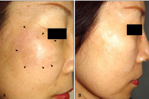
A 52-year-old Korean woman presented with a 3-month history of asymptomatic and edematous plaques on both cheeks. She complained that the lesions had gradually enlarged in size. The physical examination revealed slightly indurated, edematous, and erythematous to brow- nish plaque on the right cheek (Fig. 1A). The lesion of the left cheek was not prominent and no other parts of the body were affected. The patient was otherwise healthy.
The routine hematologic and biochemical tests were within the normal limits. Histopathological examination of a biopsy from the right cheek lesion revealed reticular epithelial degeneration and areas of cavitation within the pilosebaceous units. Lymphocytes, histiocytes and many eosinophils infiltrated around and into the hair follicles and the sebaceous glands (Fig. 2A, B). No atypical lymphocytic infiltrate was observed. Alcian blue pH 2.5 staining revealed the accumulation of acid mucopoly- saccharide in the degenerated areas (Fig. 2C). These clinicopathological findings were considered diagnostic of primary FM.
The patient was initially treated with minocycline, dapsone and topical steroids, but the lesions did not improve. We switched the treatment to methotrexate and intralesional steroids. However, the effect was incomplete
KR Kim, et al
286 Ann Dermatol
Fig. 1. (A) A slightly indurated, edematous, and erythematous to brownish plaque on the right cheek. (B) Complete healing after indomethacin and IFN α treatment.
Fig. 2. (A) Degeneration of the pilosebaceous unit with formation of cystic spaces and inflammation (H&E stain, ×100). (B) An inflammatory infiltrate composed of lymphocytes, histiocytes, and many eosinophils is present around and within the sebaceous gland epithelium (H&E stain, ×200). (C) Positive staining with alcian blue pH 2.5 (original magnification ×200).
and new lesions appeared. Therefore, methotrexate was discontinued and indomethacin was administered at a dose of 25 mg twice daily together with intralesional steroids. Remarkable improvement was observed 3 months after the start of this treatment. Thus, the intralesional steroid injections were stopped and indomethacin was continued at a half dose for a further 4 months, and complete remission of the skin lesions was maintained. At that time, indomethacin was not available because the production of this drug was stopped in Korea. The lesions recurred a month after indomethacin discontinuation. We administered intralesional IFN α-2a 3×10⁶ IU biweekly, and dramatic improvement was observed after 5 injec- tions. The interval between successive IFN α-2a ad- ministrations was progressively increased up to every 4
weeks. Complete remission was achieved after 6 months of treatment with IFN α. The IFN α treatment was well tolerated with only pain during the injection. Furthermore, no signs of any recurrence were observed 4 months after the end of treatment (Fig. 1B).
DISCUSSION
FM is an epithelial reaction pattern that is characterized by the accumulation of mucinous material in the epithelial hair follicle sheaths and the sebaceous glands8. This disorder is divided into two types. The primary type of FM occurs when there is no underlying associated skin disease. The secondary type is associated with a number of inflammatory disorders and malignant conditions,
Follicular Mucinosis Responding to Indomethacin and Intralesional Interferon Alpha
Vol. 21, No. 3, 2009 287 including mycosis fungoides and other lymphomas3. We
consider that our case is the primary type of FM based upon the absence of an atypical lymphocytic infiltrate in the skin lesion and the complete remission after treatment with indomethacin or IFN α.
The infiltrate mainly consisted of lymphocytes, but there were variable numbers of histiocytes and eosinophils1,3. It has been suggested that the keratinocytes that form the affected follicles produce intracellular mucin and they eventually degenerate, which is induced by the T lym- phocytes of the infiltrate9. On the basis of this postulation, we used various anti-inflammatory agents such as steroids, minocycline, and dapsone, but these drugs failed to control the eruption.
The exact mechanism of the effect of indomethacin and IFN α on FM is not well established. Indomethacin is an inhibitor of cyclooxygenase activity that reduces the synthesis of eosinophil chemotactic factor and lympho- kine, resulting in the inhibition of the chemataxis of eosinophils and the inhibited activation of lymphocytes and macrophages10,11. IFN α has imunomodulatory ef- fects, which include enhancement of suppressor T cells and inhibition of the helper T cell function, and a stimulating effect on the phagocytic and metabolic activities of macrophages12,13. The actions of IFN α on the eosinophils are inhibition of chemotaxis and the de- creased production of hydrogen peroxide and peroxidase in response to stimuli14. In addition, IFN α inhibits the release of cytotoxic proteins such as eosinophil cationic protein and eosinophil-derived neurotoxin, as well as the molecules involved in eosinophil differentiation and activation such as IL-515. Based upon the findings of the infiltrate of many eosinophils in the skin lesion and the complete response to agents that inhibit the eosinophil functions, we suggest that various inflammatory mediators released by eosinophils might play an important role in the mucin production and in the degeneration of the follicular and sebaceous glandular epithelium.
There have been some reports showing indomethacin or IFNs are effective to treat the primary type of FM5,7. To the best of our knowledge, the primary type of FM responding to indomethacin and low-dose IFN α, respectively, in a single case has not been reported in the English literature.
In the present report, we describe the complete remission of the primary type of FM that was treated with indo- methacin and low-dose IFN α, respectively. Further
studies are warranted to determine the role of eosinophils in the pathogenesis of FM.
REFERENCES
1. Pinkus H. Alopecia mucinosa; inflammatory plaques with alopecia characterized by root-sheath mucinosis. AMA Arch Derm 1957;76:419-424.
2. Jablonska S, Chorzelski T, Lancucki J. Mucinosis follicularis.
Hautarzt 1959;10:27-33.
3. Emmerson RW. Follicular mucinosis. A study of 47 patients.
Br J Dermatol 1969;81:395-413.
4. Yotsumoto S, Uchimiya H, Kanzaki T. A case of follicular mucinosis treated successfully with minocycline. Br J Dermatol 2000;142:841-842.
5. Kodama H, Umemura S, Nohara N. Follicular mucinosis:
response to indomethacin. J Dermatol 1988;15:72-75.
6. Kubba RK, Stewart TW. Follicular mucinosis responding to dapsone. Br J Dermatol 1974;91:217-220.
7. Meissner K, Weyer U, Kowalzick L, Altenhoff J. Successful treatment of primary progressive follicular mucinosis with interferons. J Am Acad Dermatol 1991;24:848-850.
8. Hempstead RW, Ackerman AB. Follicular mucinosis. A reaction pattern in follicular epithelium. Am J Dermato- pathol 1985;7:245-257.
9. Reed RJ. The T-lymphocyte, the mucinous epithelial inter- stitium, and immunostimulation. Am J Dermatopathol 1981;
3:207-214.
10. Takematsu H, Tagami H. Eosinophilic pustular folliculitis.
Studies on possible chemotactic factors involved in the formation of pustules. Br J Dermatol 1986;114:209-215.
11. Morley J, Beets JL, Bray MA, Paul W. Regulation of allergic responses by prostaglandins: a review. J R Soc Med 1980;73:443-447.
12. Martinez J, de Misa RF, Boixeda P, Arrazola JM, Ledo A.
Long-term results of intralesional interferon alpha-2B in discoid lupus erythematosus. J Dermatol 1993;20:444-446.
13. Yoshida R, Murray HW, Nathan CF. Agonist and antagonist effects of interferon alpha and beta on activation of human macrophages. Two classes of interferon gamma receptors and blockade of the high-affinity sites by interferon alpha or beta. J Exp Med 1988;167:1171-1185.
14. Saito H, Hayakawa T, Yui Y, Shida T. Effect of human interferon on different functions of human neutrophils and eosinophils. Int Arch Allergy Appl Immunol 1987;82:133- 140.
15. Aldebert D, Lamkhioued B, Desaint C, Gounni AS, Gold- man M, Capron A, et al. Eosinophils express a functional receptor for interferon alpha: inhibitory role of interferon alpha on the release of mediators. Blood 1996;87:2354- 2360.