Korean J Hepatobiliary Pancreat Surg 2013;17:131-134
Case Report
Peribiliary cysts developed in normal underlying liver:
report of a case
Hee Joon Kim, Choong Young Kim, Young Hoe Hur, Jung Chul Kim, Chol Kyoon Cho, and Hyun Jong Kim
Department of Surgery, Chonnam National University College of Medicine, Gwangju, Korea
Peribiliary cysts, known as cystic dilatation, of the extramural peribiliary glands of the bile duct are rare, and are usually detectable under conditions of pre-existing hepatobiliary diseases such as liver cirrhosis. Preoperative diagnosis is often difficult, because they are usually asymptomatic. Distinction of peribiliary cysts from premalignant or malignant cystic lesions is mandatory. Herein, we report a case of peribiliary cyst, which was preoperatively assumed as unilobar Caroli's diseases in healthy young patients and briefly discuss the management of the condition. (Korean J Hepatobiliary Pancreat Surg 2013;17:131-134)
Key Words: Cyst; Hepatic neoplasm; Caroli disease
Received: July 24, 2013; Revised: July 30, 2013; Accepted: August 5, 2013 Corresponding author: Chol Kyoon Cho
Department of Surgery, Chonnam National University Hwasun Hospital, 322, Seoyang-ro, Hwasun-eup, Hwasun 519-763, Korea Tel: +82-61-379-7646, Fax: +82-61-379-7661, E-mail: [email protected]
Copyright Ⓒ 2013 by The Korean Association of Hepato-Biliary-Pancreatic Surgery Korean Journal of Hepato-Biliary-Pancreatic Surgery ∙ ISSN: 1738-6349
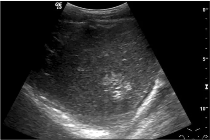
Fig. 1. Ultrasonogram demonstrating a multiseptated cystic mass at the segment VI.
INTRODUCTION
Peribiliary cysts originate from cystic dilatation of the peribiliary glands.1 Almost all the peribiliary cysts are found in patients with advanced liver diseases, and partic- ularly with disturbance of the portal venous systems, as almost all patients have severe portal hypertensions.
Peribilary cysts should be distinguished from premalig- nant conditions, such as Caroli's diseases, localized pri- mary sclerosing cholangitis, mucinous cystic neoplasm (MCN) and intraductal papillary neoplasm of the bile duct (IPNB).
Surgical treatment was indicated for our patient, as the diagnosis of Caroli's disease could not be excluded in the present case of a young patient. Subsequently, he under- went elective liver resection. Herein, we report a case of peribiliary cyst resembling unilobar Caroli's disease for a healthy young patient.
CASE
A 31-year-old man was referred to our hospital for fur- ther evaluations on the presence of hepatic mass, detected by ultrasonogram (US) performed for routine screening
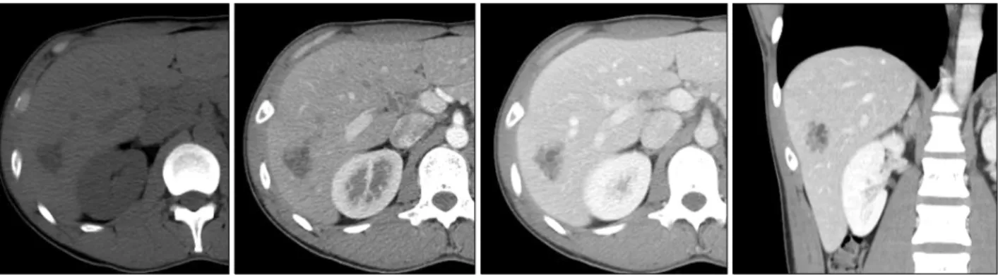
examinations (Fig. 1). The patient was asymptomatic. He had no history of liver diseases or alcohol abuses. All the routine laboratory tests, including liver function tests and tests for tumor markers, yielded normal findings. An ul- trasonogram indicated a 3 cm sized multiseptated cystic mass in the right hepatic lobe. An abdominal computed tomography (CT) scan and a magnetic resonance chol- angiopancreatography (MRCP) demonstrated a 2.8×2.2 cm sized multilocular, irregular cystic mass in segment VI (Fig. 2, 3). The mass was located at a distance from the
132 Korean J Hepatobiliary Pancreat Surg Vol. 17, No. 3, August 2013
Fig. 2. Computed tomography scan showing a multiseptated non-enhancing cystic mass at the segment VI.
Fig. 4. Gross finding of peribiliary cysts. Multiple cysts dis- tributed along the major segmental portal tract of the segment VI.
Fig. 3. Magnetic resonance chol- angiopancreatography demonst- rating a cluster of hyperintense cystic lesions on T2-weighted image involving the segment VI.
hepatic hilum. The findings suggested unilobar Caroli's disease or biliary cystic neoplasm, such as mucinous cyst- ic neoplasm or intraductal papillary neoplasm of the bile duct. Regarding the potential risk of malignancy in the context for suspected Caroli's disease, the mucinous cystic neoplasm and intraductal papillary neoplasm of the bile duct were discussed with the patient, who consequently opted for surgical resection.
A right hemihepatectomy was performed. At lapa- rotomy, multiple cystic lesions were found surrounding the segment VI bile duct. The patient was discharged 2 weeks following surgery without any complications.
Histological examinations of the resected specimen
Hee Joon Kim, et al. Peribiliary cysts in normal liver 133
Fig. 5. Microphotographs of per- ibiliary cysts. (A) Normal appear- ing bile ducts are seen without in- flammation and hyperplasia. (B) The peribiliary cyst is lined by flattened epithelium.
showed multiple cysts distributed along the major seg- mental portal tracts of the segment VI (Fig. 4). On micro- scopic examination, numerous peribiliary cysts lined by cubic or flattened epithelium were seen around normal in- trahepatic bile ducts. There was no evidence of dysplasia (Fig. 5).
DISCUSSION
Peribiliary cysts were first described by Nakanuma et al. in 1984, as an incidental finding in seven autopsy livers.1 The authors suggested that the peribiliary cysts arose from cystic dilatation of the intrahepatic extramural peribiliary glands in the periductal connective tissues.
Terada and Nakanuma have reported the presence of peri- biliary cysts in 202 livers (20.2%) among 1,000 consec- utive autopsy livers.2 However, the degree of cystic dilata- tion varies, and the majority are microscopically recognized. In clinical practices, the conditions are still poorly identified.
Until today, the pathogenesis of peribiliary cysts is unclear. Inflammation and circulatory disturbance in the liver are considered as possible mechanisms of cystic for- mation because the cysts are mainly found in patients with severe liver diseases, such as liver cirrhosis, alcoholic liv- er diseases, portal hypertensions, or thrombosis.1-4 Genetic predisposition may exist as peribiliary cysts have been re-
ported in association with autosomal dominant polycystic kidney diseases.5 However, in our case, peribiliary cysts were developed without any underlying hepatic diseases.
This observation supports a developmental etiology. Fusai et al. have reported the development of peribiliary cysts in normal underlying livers.6
Peribiliary cysts are usually asymptomatic. In some pa- tients, they often increase in size and number, resulting in obstructive jaundice.7,8 However, peribiliary cysts are commonly known as being harmless. Until today and to the best of our knowledge, no cases of malignant degener- ation have been documented so far. Once peribiliary cysts are diagnosed accurately, the hepatectomy is unnecessary and follow-ups without invasive treatments are recom- mended. In our case, preoperatively, the cystic lesion was considered as a unilobar Caroli’s disease. Caroli's disease is associated with cholangiocarcinoma. Cholangiocarcion- ma has been reported in approximately 7% of patients with Caroli's diseases.9 Consequently, hepatectomy was being performed in our patient.
Upon imaging, peribiliary cysts may sometimes be mis- taken for biliary dilatations, especially on US examination.
Drip infusion cholangiographic (DIC) CT is useful for di- agnosis of peribiliary cysts.10 In diagnosing peribiliary cysts, the most important factor demonstrates that the cysts have no communication with the biliary tract. On DIC-CT, peribiliary cysts are not filled with contrast ma-
134 Korean J Hepatobiliary Pancreat Surg Vol. 17, No. 3, August 2013
terials since these cysts do not communicate with the lu- men of the biliary tree. MRCP is also useful in the diag- nosis of peribiliary cysts. Biliary dilatations are demon- strated as tubular structures on one side along the portal tracts. On the other hand, peribiliary cysts usually exist on both sides along the portal tract, which is diagnostic.
Nevertheless, in some cases, distinguishing peribiliary cysts from biliary dilatations is a difficult task.
Histologically, the cyst epithelium is cuboidal or of low columnar, thus, is relatively poor in mucus and rarely shows papillary proliferative features.2 In literature, there are no reported cases of malignant transformations.
Peribilary cysts can occur in the normal liver and can mimic the conditions of unilobar Caroli's disease. Accurate identifications of peribiliary cysts and differentiations from other potentially premalignant or malignant cystic le- sions are mandatory.
REFERENCES
1. Nakanuma Y, Kurumaya H, Ohta G. Multiple cysts in the hepatic
hilum and their pathogenesis. A suggestion of periductal gland origin. Virchows Arch A Pathol Anat Histopathol 1984;404:
341-350.
2. Terada T, Nakanuma Y. Pathological observations of intrahepatic peribiliary glands in 1,000 consecutive autopsy livers. II. A pos- sible source of cholangiocarcinoma. Hepatology 1990;12:92-97.
3. Nakanuma Y. Peribiliary cysts: a hitherto poorly recognized disease. J Gastroenterol Hepatol 2001;16:1081-1083.
4. Nakanuma Y. Peribiliary cysts have at least two different pathogeneses. J Gastroenterol 2004;39:407-408.
5. Kida T, Nakanuma Y, Terada T. Cystic dilatation of peribiliary glands in livers with adult polycystic disease and livers with soli- tary nonparasitic cysts: an autopsy study. Hepatology 1992;16:
334-340.
6. Fusai G, Tucker O, Nik Sulaiman NM, et al. Peribiliary cysts can mimic Caroli's disease: a case report. Int J Surg Pathol 2005;13:379-382.
7. Pang T, Kuo S, Hugh TJ, et al. The role of peribiliary cysts in biliary obstruction. ANZ J Surg 2010;80:699-702.
8. Ikenaga N, Chijiiwa K, Otani K, et al. A case of peribiliary cyst presenting with obstructive jaundice. J Gastrointest Surg 2009;
13:174-176.
9. Dayton MT, Longmire WP Jr, Tompkins RK. Caroli's Disease:
a premalignant condition? Am J Surg 1983;145:41-48.
10. Tohma T, Miura F, Cho A, et al. Usefulness of computed tomog- raphy during cholangiography for the diagnosis of multiple hep- atic peribiliary cysts: a report of three cases with chronic liver disease. J Hepatobiliary Pancreat Surg 2009;16:372-375.