http://dx.doi.org/10.4174/astr.2015.89.5.240 Annals of Surgical Treatment and Research
Choledochoduodenal fistula in Mainland China: a review of epidemiology, etiology, diagnosis and management
Ming-Bing Wu, Wen-Feng Zhang1, Ying-Lin Zhang1, Di Mu2, Jian-Ping Gong1
Department of Surgery, the Second Hospital of Chongqing New North Zone, Chongqing, 1Chongqing Key Laboratory of Hepatobiliary Surgery and Department of Hepatobiliary Surgery, Second Affiliated Hospital, Chongqing Medical University, Chongqing, 2Department of Infectious Diseases, Institute for Viral Hepatitis, Key Laboratory of Molecular Biology for Infectious Diseases, Ministry of Education, The Second Affiliated Hospital of Chongqing Medical University, Chongqing, China
INTRODUCTION
Biliary-enteric fistula was first described by Bartholin in 1654, but so far biliary-enteric fistulas are still rarely reported, which are thought to be one or multiple pathological perfora- tions between biliary tree and gastrointestinal tract [1,2].
Choledochoduodenal fistula (CDF), the special type of biliary- enteric fistulas, is nearly 90% caused by cholecystolithiasis [3].
Increasing cases have been reported in the last 30 years since the progress in hepatobiliary techniques, such as endoscopic retrograde cholangiography (ERCP), magnetic resonance cho- langiopancreatography (MRCP), which have been applied to extensively reevaluate hepatobiliary diseases in the clinic, especially in Mainland China. However, the preoperative diag- nosis of CDF is still difficult because of the nonspecific and/
or minimal clinical symptoms [4]. Hence, CDFs are resulting in Purpose: Choledochoduodenal fistula (CDF) is an extremely rare condition even in the most populous nations. However, diagnostic tools are inadequate for the young surgeon to be made aware of such a rare condition before surgery. Hence, basic understanding of the epidemiology, etiology, and management for this unusual but discoverable condition are necessary and essential.
Methods: The exclusive case reports of CDF, which were published from 1983 to 2014 concerning mainland Chinese people, were performed to review the epidemiology, etiology, and management.
Results: A total of 728 cases were incorporated into this review among 48 papers. More than half of the CDF cases were female (416) with an average age of 57.3 years. CDF was usually caused by cholelithiasis (573 of 728). Epigastric pain (589 of 728) and cholangitis (395 of 728) were the most common symptoms of CDF. CDF was usually detected and confirmed by endoscopic retrograde cholangiopancreatography (ERCP) (475 of 728) in Mainland China. The fistulas larger than 1 cm (82 of 654) were recommended for surgical biliary reconstruction. Fistulas between 0.5 cm and 1.0 cm (467 of 654) which were followed frequently by cholangitis attacks also required surgery; the rest were recommended to have stone removal and/
or the application of an effective biliary drainage. Fistulas less than 0.5 cm (105 of 654) were usually received conservative therapy.
Conclusion: CDF should be considered in differential diagnosis of recurrent epigastric pain and cholangitis. A possible ERCP should be arranged to investigate carefully. Depending on the size of fistula and clinical presentation, different programs for CDF are indicated, ranging from drug therapy to choledochojejunostomy.
[Ann Surg Treat Res 2015;89(5):240-246]
Key Words: Biliary fistula, Epidemiology, Disease management
Reviewed January February March April May June July August September October November December
Received April 13, 2015, Reviewed May 22, 2015, Accepted May 29, 2015 Corresponding Author: Jian-Ping Gong
Chongqing Key Laboratory of Hepatobiliary Surgery and Department of Hepatobiliary Surgery, Second Affiliated Hospital, Chongqing Medical University, 76 Linjiang road, Chongqing 400010, China
Tel: +86-13996286589, Fax: +82-6-23-63693532 E-mail: [email protected]
Copyright ⓒ 2015, the Korean Surgical Society
cc Annals of Surgical Treatment and Research is an Open Access Journal. All articles are distributed under the terms of the Creative Commons Attribution Non- Commercial License (http://creativecommons.org/licenses/by-nc/4.0/) which permits unrestricted non-commercial use, distribution, and reproduction in any medium, provided the original work is properly cited.
tough challenges for surgeons, especially for young surgeons.
In this review, we have mentioned exclusive cases reports of CDF in Chinese populations concerning epidemiology, etiology, diagnosis and management.
METHODS
The MeSH terms biliary fistula and intestinal fistula with the subheadings of China were searched in the English databases such as PubMed, Embase, ISI Web of Science and the Chinese databases such as SinoMed, CQVIP, CNKI, WANFANG data. A total of 1,379 papers have been researched. Cholecystoduodenal fistula was not encountered in discussions. Data on the papers were extracted from the full text. Papers were reviewed elabo- rately, except for those without specific reference or already reported. A total of 48 case reports from 1983 to 2014 con- cerning 728 cases of CDF were finally accorded with the inclusion criteria [5-52]. Statistical calculations have not been built as they are lacking in the heterogeneity of data.
RESULTS
Epidemiology
Large-scale studies of ERCP have reported that the incidence of CDF ranges from 2.53% to 5.3% [22-24,27,45]. More than half of the cases are found in females (57.14%). The ratio of male/
female is 0.75. The mean age of the 728 patients is 48.9 ranging from 18 to 82. The case reports of CDF, which have accurate age data, totals 24 papers concerning 33 cases. In these papers, the mean age of male (n = 13) and female (n = 20) are respectively 48 and 57.3. The distribution of CDF in different districts of Mainland China is shown in Fig. 1.
Etiology
CDF is a complication of cholelithiasis, including gallstone, choledocholithiasis, hepatolith, and biliary stenosisin, which account for 73.76% of CDF in all cases. The remainder of cases is rarely caused by iatrogenic injury (9.20%), spontaneity (8.93%), penetrating peptic ulcer (3.16%), adjacent organs tumors (3.58%), and abdominal tuberculosis (0.27%) and so on (Table 1).
Clinical presentation
Generally, no typical symptoms of CDF are presented. The most common presenting symptoms of nonobstructing biliary- enteric fistulas are epigastric pain (80.91%) and cholangitis such as jaundice (54.26%) and fever (50.69%), nausea and/or vomiting (10.30%), abdominal distension (0.39%), asymptomatic (0.11%), and diarrhea (0.11%). A case of melena and a case of kaolin stools have been respectively reported by Cao et al. [5]
and Shen and Zhao [18]. Cachexia such as anorexia, ascites and weight loss commonly present in the advanced tumor patient [5,19,22,27,30,41,52] (Table 1).
Diagnostic methods
ERCP is the most common and effective method for the diagnosis of CDF. In the 728 cases of CDF, 475 cases were confirmed by ERCP (65.25%), the rest were detected by opera- tion (23.21%), X-barium meal examination (4.40%), T-tube cholangiography (3.43%), MRCP (1.79%), gastroscope/duode- noscope (1.37%), percutaneous cholangiography (0.41%), autopsy (0.14%) and so on. Under the detection of ERCP, surgery, gastro- scope/duodenoscope, and the fistula size are classified as less than 0.5-cm group, more than 1.0-cm group and 0.5- to 1.0-cm group, respectively. The number of above groups is respectively 105 cases, 82 cases, and 467 cases. Ultrasound and CT have rarely been useful in CDF diagnosis (Table 1).
Northeast 15
10
5
0
No.ofarticles
Northwest Centra l
Nort h
Souther n
SoutheastSouthwest 1
5 9
10
14
6
2
Northeast 400
300
200
100
0
No.ofpublishedcases
Northwest Centra l
Nort h
Souther n
SoutheastSouthwest 5
57
113 64
364
107
18
Fig. 1. The distribution of choledochoduodenal fistula (CDF) in different districts of Mainland Chinese. Distribution in 48 arti- cles regarding 728 cases of CDF fistula published from 1983 to 2014.
Management strategies for CDF
At present, treatments of CDF are controversial. In the 728 cases of CDF, patients treated by biliary reconstruction including resection of hepatic bile duct stricture or stricturoplasty side-to- side choledochojejunostomy, intrahepatic cholangiojejunostomy and transection of common bile duct (CBD) account for 46.84%.
The rest were nonoperative treatment (14.01%), endoscopic sphincterotomy (EST) (13.74%), fistula repair surgery (11.95%), drainage of CBD (9.89%), subtotal gastrectomy (2.34%). Four cases were under vater papilla reconstruction [34]. Two cases suffered from fistulation [46-47]. One case was implanted with a self-expandable metal stent in the CBD to heal the fistula [52]
(Table 1).
DISCUSSION
In the West, the most common communication was cholecys- toduodenal, but in Asia, choledochoduodenal was the most common type [53]. This may be related to physical endurance.
Asians are prone to endure pain when they suffer from cho- langitis or CBD stone, so CDF is more prevalent in Asian patients [54]. Especially, Southwest China’s population more easily suffers from gallstone, choledocholithiasis, hepatolith, and biliary stenosisin than other regions of Mainland China [23,24,27]. We also know from these reports above that women are prone to suffer from CDF, which may be due to a higher prevalence of cholelithiasis in women. In contrast, CDF historically has been a complication of duodenal ulcer disease and has been seen more often in men.
CDF is a well-known but relatively rare complication of duodenal ulcer in recent years, because the development of antacid drugs helps us control the disease more easily than before [55]. The major cause of duodeno-biliary fistula is inflammation of the bile duct due to gallstones, and minor
causes include duodenal ulcer, pancreatic neoplasm, and inflam mation of neighboring organs [56]. As we know, there are two parts of narrow in CBD, one part is of the duodenal papilla, and the other is the site of the bile duct in entering the duode- num. When stones impact on either of the two narrow parts and press against the wall of CBD, inflammation would occur repeatedly and necrosis will form eventually [57].
Perampullary adenocarcinoma or pancreatic carcinoma could lead to CDF when a tumor presses against the CBD directly, making it obstructive [58]. Then CBD enlarges as the pressure in bile duct increases, causing CBD to crack and finally a fistula formation [59]. The carcinoma could penetrate the local tissues and cause necrosis and fistula formation as well [60].
Duodenal ulcer could penetrate the intestinal and biliary walls into CBD and ultimately cause a fistula, which would usually occur in the site of the duodenal bulb. The final result is the iatrogenic factor, which can not be neglected. For example, when one patient undergoes exploration of CBD, ducts around the periampullary region could be injured during passage of a rigid choledochal bougie in the CBD. Furthermore, iatrogenic CDF could occur while performing sphincteroplasty or EST carelessly [61,62]. In addition, previous studies also reported CDF formation in a patient with duodenal tuberculosis, liver transplantation and metallic biliary stent placement.
Moreover, perapapillary choledochoduodenal fistula (PCDF) is mainly caused by a spontaneous migration of a CBD stone into the duodenum. Karincaoglu et al. [63] reviewed 841 patients who underwent ERCP between 1993 and 2002 for evaluation of PCDF. They found that 16 had a PCDF at the papilla Vater and none of these 16 patients had a history of pancreatitis, duodenal ulcer, nor had they undergone biliary surgery or ERCP previously. This study indicated that PCDF was more frequently associated with CBD stones than with biliary surgery and bougienage.
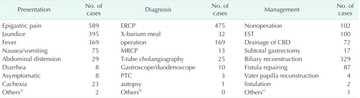
Table 1. The general conditions of cases
Presentation No. of
cases Diagnosis No. of
cases Management No. of
cases
Epigastric pain 589 ERCP 475 Nonoperation 102
Jaundice 395 X-barium meal 32 EST 100
Fever 369 operation 169 Drainage of CBD 72
Nausea/vomiting 75 MRCP 13 Subtotal gastrectomy 17
Abdominal distension 29 T-tube cholangiography 25 Biliary reconstruction 329
Diarrhea 8 Gastroscope/duodenoscope 10 Fistula repairing 87
Asymptomatic 8 PTC 3 Vater papilla reconstruction 4
Cachexia 23 autopsy 1 fistulation 2
Othersa) 2 Othersb) 0 Othersc) 1
ERCP, endoscopic retrograde cholangiopancreatography; EST, endoscopic sphincterotomy; CBD, common bile duct; MRCP, magnetic resonance cholangiopancreatography; PTC, percutaneous transhepatic cholangiography.
a)A case of melena and a case of kaolin stools. b)Ultrasound and CT mainly indicated that air is present within the biliary system. c)One case is implanted a selfexpandable metal stent in the CBD to heal the fistula.
CDF has been categorized by Ikeda and Okada [64] and Gong et al. [23,24] separately. According to the location of the fistula, Ikeda divided CDFs into two types. Type I is located on the longitudinal fold of the papilla, while type II is on the posterior wall of the duodenal bulb (Fig. 2A). However, Gong et al [23, 24] have divided them into three types, the first type is type A, which is an orifice of CDF located more than 2 cm away from the papilla. The second is type B, characterized by an orifice of CDF located less than 2 cm away from the papilla. Lastly, type C, also called PCDF, is an orifice of CDF located on the papilla fold (Fig. 2B). The classification of CDF is important for the diagnosis and treatment in the clinic, and the position of fistula can suggest what the possible cause of CDF is.
The clinical manifestations of CDF are various and nonspeci- fic. To determine the typical presentations of CDF, more specific clinical datum are needed. What is more, assistant examinations are necessary in the diagnosis. There are some signs related to these diseases. The presence of an internal biliary fistula was suggested in some cases by the findings of pneumobilia, atrophic gallbladder, and biliary stones [65].
Furthermore, the bile sample collected from the CBD showed high levels of pancreatic enzymes, including amylase and phospholipase-A2 [66]. High levels of these enzymes may indicate an abnormal connection between the duodenum and the CBD. Despite the increased rate of CDF diagnosis, treatment and management strategies for CDF have not been established yet, and more investigation is needed in this area.
Some studies suggest that patients with no symptoms of illness probably should not receive treatment, especially surgical. Treatment can sometimes be accomplished through nonsurgical (endoscopic) or percutaneous interventions. Even in patients with symptoms conservative treatment of H2 antagonist or proton pump inhibitors and drugs for the eradica- tion of Helicobacter pylori infection (when abundant), which often happens, is advised. This therapy can also lead to less
CDF [67-70]. Surgery is recommended when there are surgical complications, duodenal stenosis, severe symptoms of peptic ulcer disease in those who have not responded to conservative treatment, especially bleeding and recurrent cholangitis.
On the other hand, Choi et al. [71] have regarded CDF as a biliary abnormality that was prone to ascending biliary infec- tion. To avoid recurrence of bile tract infection and the risk of biliary stricture existing after healing of the fistula, aggressive therapy to correct CDF was mandatory. In addition, recurrent gallstone ileus caused by CDF was considered as a definitive indication for surgical managements. What is more, CDF presented a high risk for biliary tract carcinoma, and surgical management to be necessary for CDF, even if the patient had no significant clinical symptoms [72,73].
Agarwal et al. [74] have reported that most of their patients were operated on with biliary enteric anastomosis, and the remaining patients underwent other therapeutic procedures, such as sphincterotomy, biliary stenting, and nasobiliary drainage. According to the sizes of fistula, Li et al. [35] proposed the following strategies for fistula treatments: for fistula orifices larger than 1 cm, CBDs larger than 2 cm and with complications in the biliary tree, after removing the stones, Roux-en-Y choledochojejunostomy or Roux-en-Y intrahepatic cholangiojejunostomy to build an effective drainage, a tran- section of the CBD was applied to prevent the reflux of duode- nal juice; for fistula orifices between 0.5 and 1.0 cm, CBDs greater than 2 cm without any biliary complications and reflux of duodenal juice, a side-to-side choledochojejunostomy without transecting the duct and an effective biliary drainage was applied; for fistula orifices larger than 0.5 cm, CBDs larger than 1.2 cm and CDF caused by secondary stones from the gallbladder, the surgery included stone removal, cholecystectomy and CBD examination followed by drainage;
for fistula orifices less than 0.5 cm, without complications in the bile duct tree non-surgical treatments were applied. This
A B
1
2
3
4 5
1 2
3
4 5
5
4 5
4
5 4
Type I Type II Type A
Type B
Type C
Fig. 2. The classification of cho- ledochoduodenal fistula (CDF).
(A) The Ikeda’s classification. Type I was located on the longitudinal fold of the papilla, while type II was on the posterior wall of the duodenal bulb. (B) The Gong’s classification. Type A is an orifice of CDF located more than 2 cm away from the papilla. Type B is an orifice of CDF located less than 2 cm away from papilla.
Type C, or perapapillary CDF, is an orifice of CDF located on the papilla fold. 1, duodenum;
2, CBD; 3, pancreatic duct; 4, major duodenal papilla; 5, CDF.
study suggested that larger fistula increased the frequency of cholangitis episodes and needed surgical treatment for fistula itself. In many patients, when there was stenosis of the duodenum, it was recommended gastric resection.
Biliary-enteric fistula including CDF is one of the reasons for converting from laparoscopic cholecystectomy (LC) to open surgery. Thus, surgery is preferred over laparoscopic operation for treating CDF. However, as the skill of LC improves, LC would expand its indication [56]. Periselneris and Bong [75] have reported that no patient suffered from death or intraoperative complications when the laparoscopic approach to the CDF was performed. And Lee et al. [76] also reported three cases of biliary-enteric fistula including CDF treated successfully by laparoscopic surgery without any postoperative complication, eventually. We can know that laparoscopic surgery is an ad- vanced technique and the treatment of choice for CDF irrespec- tive of the preoperative diagnosis from these studies.
In a word, there are several treatment options for CDF.
Management of CDF depends on their type and etiological severity of the disease and the general condition of each patient. In fistulas with complicating duodenal ulcers, medical management or surgery can be used. Surgery must be reserved for patients with poorly controlled or recurrent ulcer symptoms, major ulcer complications, such as perforation, hemorrhage, or obstruction, or exceptional cases with cholangitis or biliary obstruction. Endoscopic management and the extraction of a bile duct stone (if present) may be needed. There is a justified
trend towards conservative management of iatrogenic bile duct perforations. The current case was also managed conservatively, by reducing the pressure gradient with T-tube drainage and antibiotics.
In conclusion, patients with CDF may have nonspecific symptoms, which make the diagnosis difficult. To assist in diagnosing CDF, imaging procedures are needed. And ERCP can increase the rate of CDF diagnosis significantly. Although the rate of diagnosis has increased, treatment and management plans for CDF have not been established. Managements of CDF depend on the types, etiology, severity of the diseases, and the general condi tion of each patient. Despite open surgery being preferred over laparoscopic surgery for treating CDF, laparoscopic surgery was reported effective for cases with CDF irrespective of the pre operative diagnosis. And to prove the effectiveness of lapar oscopic surgery, more data are needed to be collected.
CONFLICTS OF INTEREST
No potential conflict of interest relevant to this article was reported.
ACKNOWLEDGEMENTS
This study was supported by National Natural Science Foundation of China (No. 81071339).
1. Marshall T, Kamalvand K, Cairns SR.
Endoscopic treatment of biliary enteric fistula. BMJ 1990;300:1176.
2. Liu TM, Chiu HH. Images in clinical med- icine. Gallstone ileus. N Engl J Med 2010;
362:345.
3. Lin CT, Hsu KF, Yu JC, Chu HC, Hsieh CB, Fu CY, et al. Choledochoduodenal fistula caused by cholangiocarcinoma of the distal common bile duct. Endoscopy 2009;41 Suppl 2:E319-20.
4. Luu MB, Deziel DJ. Unusual complications of gallstones. Surg Clin North Am 2014;
94:377-94.
5. Cao JY, Fang Z, Guo WT, Dai LH. A case report of choledochoduodenal fistula.
Tianjin Yi Yao 1983;6:978-80.
6. Xu RN. Parapapillary choledochoduodenal
fistula: diagnosis and treatment. Zhong- hua Wai Ke Za Zhi 1984;22:672-3, 701.
7. He SL, Tang RS. A case report of chole- dochoduodenal fistula. Lichuang Fang- shexue Za Zhi 1984;3:120-1.
8. Jiao TY. A case report of choledochoduode- nal fistula. Shanghai Med J 1985;5:420-1.
9. Dai XZ, Chen MZ. Clinical assessment of para-ampulla choledocho-duodenal fistulae in 27 cases. Zhonghua Nei Ke Za Zhi 1986;25:655-7, 700-1.
10. Li XC. Two case reports of choledochoduo- denal fistula. Beijing Med J 1987;4:251-2.
11. Gu GH. Choledochoduodenal fistula: a long-term complication of T tuber drain- age. J Third Mil Med Univ 1987;4:443.
12. Li Z. A case report of choledochoduodenal fistula after T tuber drainage. J Chin Pract
Surg 1991;4:201.
13. Wang ZL, Lu Qide. A case report of choledochoduodenal fistula secondary penetrated duodenal peptic ulcer. J Pract Radiol 1993;3:507.
14. Fei X. Pedical A case report of omentum repairing the choledochoduodenal fistula.
J Hepatobiliary Surg 1995;4:216.
15. Li YC. 2 Cases of choledochoduodenal fistula. Hainan Yi Xue 1995;2:135.
16. Zhu JR, Kai GW, Zhang YC. X-ray analysis for choledochoduodenal fistula. J Pract Radiol 1995;3:183-4.
17. Hu MX. 13 Cases of choledochoduodenal fistula after T tuber drainage. Curr Physi- cian 1996;12:34.
18. Shen WS, Zhao P. 4 Cases of choledocho- duodenal fistula. J Chin Pract Surg 1996;
REFERENCES
8:486-7.
19. Zhao DL, You L. Cardiac cancer implicated with choledochoduodenal fistula. Chin J Clin Gastroenterol 1996;2:82.
20. Li WM. Surgical treatment for choledocho- duodenal fistula: 11 cases report. J Mod Oper Surg 1997;4:286.
21. Fan XJ, Geng XQ. 4 Rare cases of choledo- choduodenal fistula. Yunnan Yi Yao 1997;
5:398-9.
22. Li NF, Zhang YD, Liu S. 69 Cases of choledochoduodenal fistula: a retrospec- tive study of ERCP. Chin J Endosc 1999;4:
34-5.
23. Gong JP, Zhou YB, Han BL. Diagnosis and classification of choledochoduodenal fistula. Chin J Digest Endosc 1999;2:42-3.
24. Gong JP, Zhou YB, Han BL. Diagnosis and surgical management of choledochoduo- denal fistula. J Third Mil Med Univ 2000;
22:287-9.
25. Wang XX, Tao F. Choledochoduodenal fistula induced by the partially prolapsed T tube: two cases report. Zhejiang Pract Med 2001;6:57-8.
26. Tian ZL, Wu DX. A case report of choledo- choduodenal fistula. J Hebei Med Uni 2001;22:106.
27. Li ZH, Zhou YB, Chen M, Liu JK, Chen K. Clinical analysis of juxtapapillary choledochoduodenal fistula: report of 47 cases. Chin J Gen Surg 2001;16:331-3.
28. Ku RX, Chen, F, Liu SL, Wei NR, Ge XH, Ablet. Diagnosis of choledochoduodenal fistula by barium meal analysis: a case report. Xinjiang Med J 2001;1:65.
29. Xie J. A case report of choledochoduodenal fistula after T tuber drainage. J Clin Surg 2002;10:10.
30. Yu HZ, Li FY, Du Y, Geng XP, Xiong QR.
Diagnosis and treatment of choledocho- duodenal fistul. J Clin Surg 2002;10:88-9.
31. Gu J, Li JS, Ren J AN. Discussion of the suitable management and preventive methods for distal choledochoduodenal fistula. Chin J Gen Surg 2002;17:725-6.
32. Hou YL, Chen ZW, Cheng H, Fu G.
Analysis of 7 cases of postoperative distal choleo-duodonum fistulas. Fu Bu Wai Ke 2004;17:230-1.
33. Hao RR, Yu L, Wang HJ. A case report of
gallstone ileus induced by choledocho- duodenal fistul. Beijing Med 2005;27:719.
34. Lan XH, Xu L, Wang LY, Wang YZ. Diag- nosis and treatment for peripapillary choledo-choduodenal fistula. Clin Med Chin 2005;1:71-2.
35. Li ZH, Ding J, Ye Y, Cai L, Liu X, Liu J, et al. New strategy to prevent ascending cholangitis in larger choledochoduodenal fistula. ANZ J Surg 2006;76:796-800.
36. Zhao DP, Sun W, Xu HM, Ma YW. 7 Cases of choledochoduodenal fistula after T tuber drainage. Acta Acad Med Weifang 2006;28:76.
37. Lin D, Zhang LH, Jiang TR, Lan SQ, Yang LK. 16 Cases reports of choledochoduo- denal fistula diagnosed by ERCP. Fujian Med J 2006;28:118-9.
38. Peng B, Du JP, Deng B, Cun BY, Jiang LL, Zhang SF. A case report of pri mary pleomorphic intrahepatic cholangio- carcinoma complicated with choledocho- duodenal fistula. J Sichuan Univ 2006;
3:455.
39. Sheng W, Zhao J, Luo HY, Liu ZB. A case report of choledochoduodenal fistula. J Clin Radiol 2007;26:87.
40. Chen HP, Bai L, Li WB, Liang C, Han SX. A case report of choledochoduodenal fistula induced by foreign bodies on the border of duodenum ball drop. Chin J Endosc 2007;13:1341-2.
41. Xu ZY, Liu L, Zhang CQ, Li F, Li QQ. Clinic analysis 0f 9 cases with choledochoduo- denal fistula associated with pancreatic carcinoma. Chin J Mod Med 2007;17:1386- 8, 92.
42. Wang An G. 5 Cases of choledochoduo- denal fistula after T tuber drainage. J Liaoning Med Univ 2010;31:73-4.
43. Chen RB. A case report of choledochoduo- denal fistula. J Yangtze Univ 2010;1:107-8.
44. Wang WZ, He CH, Yi HF. 2 Cases of spon- taneous chronic choledochoduodenal fistula. J Pract Med 2010;26:4050.
45. Zong KC, You HB, Gong JP, Tu B. Diagnosis and management of choledochoduodenal fistula. Am Surg 2011;77:348-50.
46. Li SW, Tian CP, Feng MH. A case re port of choledochoduodenal fistula compli- cated cholecystolithiasis and choledocho-
lithiasis. Public Med Forum Mag 2012;34:
4632.
47. Yi XL, Li XH, Wang J. Management of cho- ledochoduodenal fistula. For All Health 2012;18:66-7.
48. Ding CL, Ma D. Management of choledo- choduodenal fistula. Chin J Aesthet Med 2012;21:84-5.
49. Liu Q, Lu MX, Zhang TH, Feng CL. 78 Cases report of choledochoduodenal fistula. Chin J Gerontol 2012;32:3534-5.
50. Li CS, Yuan D, Zhang J, Yuan YS. A case report of choledochoduodenal fistula diagnosed by ERCP. J Chin Physician 2012;
14:718.
51. Zhao S, Wang J, Ge J, Zhang X, Liu J, Zhang A, et al. Implantation of covered self- expandable metal stent in the com mon bile duct for the treatment of choledocho- duodenal fistula. J Clin Gastroenterol 2014;48:383-4.
52. Zhang YL, Xu JG, Zhu YY. Analysis of diagnosis and surgical treatment of the choledochoduodenal fistula. Chin J Mod Med 2014;29:103-5.
53. Halabi WJ, Kang CY, Ketana N, Lafaro KJ, Nguyen VQ, Stamos MJ, et al. Surgery for gallstone ileus: a nationwide comparison of trends and outcomes. Ann Surg 2014;
259:329-35.
54. Chang WT, Lin NC, Hsia CY, Liu CS, Tsai HL, Loong CC. Liver transplantation for a renal transplantation recipient with secon dary sclerosing cholangitis by chole- dochoduodenal fistula. Asian J Surg 2012;
35:49-52.
55. Neumann H, Nagel A, Bernatik T, Wickles N, Neurath MF, Raithel M. Endo scopic closure of large, spontaneous, choledo- choduodenal fistula by using an over- the-scope clip. Gastrointest Endosc 2011;
74:200-2.
56. Hong T, Xu XQ, He XD, Qu Q, Li BL, Zheng CJ. Choledochoduodenal fistula caused by migration of endoclip after lapar oscopic cholecystectomy. World J Gastroenterol 2014;20:4827-9.
57. Moghaddam JA, Amini M, Adibnejad S. Development of bile duct bezoars following cholecystectomy caused by choledochoduodenal fistula formation: a
case report. BMC Gastroenterol 2006;6:1.
58. Williamson JB, Draganov PV. Choledo- choduodenal fistula after biliary metallic stent placement for pancreatic cancer. Dig Endosc 2014;26:292.
59. Chaudhari D, Saleem A, Murthy R, Baron T, Young M. Choledochoduodenal fistula after biliary placement of a self- expanding metallic stent for palliation of pancreatic cancer. Endoscopy 2013;45 Suppl 2 UCTN:E77.
60. Okabe Y, Kaji R, Ishida Y, Noda T, Sasaki Y, Tsuruta O, et al. Successful endo scopic extraction of a large impacted choledo- cholithiasis in the ampulla of vater: two interesting cases. Dig Endosc 2010;22 Suppl 1:S103-6.
61. Song TJ, Hyun YS, Lee SS, Park do H, Seo DW, Lee SK, et al. Endoscopic ultrasound- guided choledochoduodenostomies with fully covered self-expandable metallic stents. World J Gastroenterol 2012;
18:4435-40.
62. Chikamori F, Okumiya K, Inoue A, Kuniyoshi N. Laparoscopic cholecysto- fistulectomy for preoperatively diag nosed cholecystoduodenal fistula. J Gastro- enterol 2001;36:125-8.
63. Karincaoglu M, Yildirim B, Kantarceken B, Aladag M, Hilmioglu F. Association of peripapillary fistula with common bile
duct stones and cholangitis. ANZ J Surg 2003;73:884-6.
64. Ikeda S, Okada Y. Classification of chole- dochoduodenal fistula diagnosed by duo- denal fiberscopy and its etiological signi- ficance. Gastroenterology 1975;69:130-7.
65. Yang D, Wang Z, Duan ZJ, Jin S. Laparo- scopic treatment of an upper gastro- intestinal obstruction due to Bouveret's syndrome. World J Gastroenterol 2013;
19:6943-6.
66. Mohammed N, Godfrey EM, Subramanian V. Cholecysto-duodenal fistula as the source of upper gastrointestinal bleeding.
Endoscopy 2013;45 Suppl 2 UCTN:E250-1.
67. Miyamoto S, Furuse J, Maru Y, Tajiri H, Muto M, Yoshino M. Duodenal tubercu- losis with a choledocho-duodenal fistula.
J Gastroenterol Hepatol 2001;16:235-8.
68. Jaballah S, Sabri Y, Karim S. Choledocho- duodenal fistula due to duodenal peptic ulcer. Dig Dis Sci 2001;46:2475-9.
69. Mueller XM, Tevaearai HT, Stumpe F, Hurni M, Ruchat P, Fischer AP, et al. Gas- trointestinal disease following heart trans- plantation. World J Surg 1999;23:650-5.
70. Povoski S, Shamma'a J. Fistula involving portal vein and duodenum at the site of a duodenal ulcer in a patient after previous extrahepatic bile duct resection and
brachytherapy. Dig Surg 2003;20:53-5.
71. Choi D, Lim HK, Kim MJ, Kim SJ, Kim SH, Lee WJ, et al. Liver abscess after per- cutaneous radiofrequency ablation for hepatocellular carcinomas: frequency and risk factors. AJR Am J Roentgenol 2005;
184:1860-7.
72. Ohtsuka T, Tanaka M, Inoue K, Nabae T, Takahata S, Yokohata K, et al. Is peri- papillary choledochoduodenal fistula an indication for endoscopic sphincterotomy?
Gastrointest Endosc 2001;53:313-7.
73. Dwivedi AN, Kumar S, Rana S, Maurya B.
Transmural invasion of hepatic flexure of colon causing cholecystocolic fistula by aggressive gallbladder carcinoma. World J Surg Oncol 2013;11:86.
74. Agarwal N, Sharma BC, Garg S, Kumar R, Sarin SK. Endoscopic management of postoperative bile leaks. Hepatobiliary Pancreat Dis Int 2006;5:273-7.
75. Periselneris N, Bong JJ. Choledocho-duo- denal fistula encountered during emer- gency laparotomy for upper gastro-inte- stinal haemorrhage: what should be the surgical strategy? Clin Ter 2011;162:547-8.
76. Lee JH, Han HS, Min SK, Lee HK. Lapar- oscopic repair of various types of biliary- enteric fistula: three cases. Surg Endosc 2004;18:349.