ORIGINAL ARTICLE
DOI: 10.4174/jkss.2011.80.6.390
JKSS
Journal of the Korean Surgical Society pISSN 2233-7903ㆍeISSN 2093-0488
Received October 31, 2010, Accepted February 22, 2011 Correspondence to: Suk-Kyung Hong
Division of Trauma and Surgical Critical Care, Department of Surgery, Asan Medical Center, University of Ulsan College of Medicine, 388-1 Pungnap 2-dong, Songpa-gu, Seoul 138-736, Korea
Tel: +82-2-3010-3510, Fax: +82-2-3010-4709, E-mail: [email protected]
cc Journal of the Korean Surgical Society is an Open Access Journal. All articles are distributed under the terms of the Creative Commons Attribution Non-Commercial License (http://creativecommons.org/licenses/by-nc/3.0/) which permits unrestricted non-commercial use, distribution, and reproduction in any medium, provided the original work is properly cited.
Non-invasive ventilation for surgical patients with acute respiratory failure
Byoung Chul Lee, Kyu Hyouck Kyoung, Young Hwan Kim, Suk-Kyung Hong
Division of Trauma and Surgical Critical Care, Department of Surgery, Asan Medical Center, University of Ulsan College of Medicine, Seoul, Korea
Purpose: Acute respiratory failure is a relatively common complication in surgical patients, especially after abdominal surgery. Non-invasive ventilation (NIV) is increasingly used in the treatment of acute respiratory failure. We have assessed the usefulness of NIV in surgical patients with acute respiratory failure. Methods: We retrospectively reviewed the medical charts of patients who were admitted to a surgical intensive care unit between March 2007 and February 2008 with acute res- piratory failure. The patients who have got respiratory care for secondary reason such as sepsis and encephalopathy were ex- cluded from this study. Results: Of the 74 patients who were treated with mechanical ventilation, 15 underwent NIV and 59 underwent invasive ventilation. The causes of acute respiratory failure in the NIV group were atelectasis in 5 patients, pneu- monia in 5, acute lung injury in 4, and pulmonary edema in 1, this group included 3 patients with acute respiratory failure af- ter extubation. Overall success rate of NIV was 66.7%. Conclusion: NIV may be an alternative to conventional ventilation in surgical patients with acute respiratory failure. Use of NIV may avoid re-intubation in patients who develop respiratory fail- ure after intubation.
Key Words: Non-invasive ventilation, Acute respiratory failure, Pulmonary atelectasis, Pneumonia, Post-operative complica- tions
INTRODUCTION
Acute respiratory failure (ARF) is a complication in sur- gical patients, especially after abdominal surgery. ARF af- ter abdominal surgery is closely associated with a reduc- tion in lung volume caused by alveolar collapse and dys- function of the diaphragm, increased intraabdominal pressure after surgery, and decreased respiration caused by pain [1-4]. Treatment of post-operative ARF usually in- cludes endotracheal tube insertion and mechanical ven-
tilation.
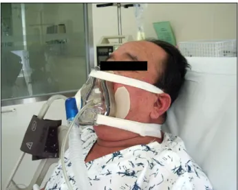
More recently, however, patients with post-operative ARF have been treated by non-invasive ventilation (NIV) [5], consisting of attachment of a mask to the face without endotracheal tube insertion (Fig. 1), followed by deliveries of positive air pressure by pressure support ventilation (PSV) and positive end expiratory pressure (PEEP) or con- tinuous positive airway pressure (CPAP) [6,7]. NIV with positive pressure ventilation can relieve a patient’s in- spiratory efforts and improve gas exchange without the
Fig. 1. Non-invasive ventilation.
need for endotracheal intubation. This may benefit pa- tients since endotracheal intubation has been associated with numerous complications. NIV decreases the devel- opment of ventilator associated pneumonia, allows the patient to eat and drink while receiving positive pressure ventilation and decreases the use of sedative drugs and neuromuscular blockers required to endure mechanical ventilation. Consequently, NIV shortens weaning time from mechanical ventilators and can avoid hypotension caused by the use of sedatives [6].
NIV was initially applied to treat patients with chronic obstructive pulmonary disease. In addition to successfully treating ARF, NIV reduced invasive intubation and mor- tality rates, and shortened treatment periods [8-10]. NIV has been utilized primarily to treat patients with hyper- carbic respiratory failure, and its therapeutic role in treat- ing psychogenic ARF and ARF after abdominal or thoracic surgery is expanding [11-15]. We therefore assessed the usefulness of NIV in surgical patients with ARF.
METHODS
We retrospectively reviewed the medical records of the medical charts of patients admitted to the surgical in- tensive care unit in our institution for ARF between March 2007 and February 2008.
ARF was defined as a tachypnea (respiration rate >35),
hypercapnea (PaCO2 >45 mmHg and PH <7.35), hypo- xemia (PaO2/FiO2 <150), severe dyspnea, and the use of accessory respiratory muscles. Patients were excluded if they had hemodynamic instability, decreased mental sta- tus, inability to expectorate airway secretions or facial trauma.
NIV was performed using Performa Trak facial masks (Philips Respironics, Woerden, Netherland) either pres- sure control ventilation or PSV. Treatment goals included a volume per respiration of 6 to 8 mL/kg, with PEEP ad- justed to 5 to 15 cmH2O according to the conditions of each patient’s lung. Arterial blood gas analysis (ABGA), respi- ration rate and patient compliance were monitored continuously. NIV was converted to invasive ventilation with endotracheal intubation if respiratory status was worsened despite NIV support.
Weaning was performed using the optimal method that reduced the time of mechanical ventilation, including al- ternation of mechanical ventilation with venturi masks, reservoir bags. Patients were extubated if they were con- scious, could breathe on their own, could readily ex- pectorate, and could maintain smooth tidal volume under a positive pressure respiration <8 cm H2O.
ARF patients were divided into two groups, according to whether they underwent NIV or invasive ventilation.
Their clinical features were compared including patient age, gender, acute physiology and chronic health evalua- tion (APACHE) II score at the time of the application of mechanical ventilation, PaO2/FiO2 ratio, whether or not immune suppressors were used, and the cause of respira- tory failure. Causes of respiratory failure were diagnosed by clinical symptoms, ABGA, and chest radiographs.
Success of treatment was defined as weaning from me- chanical ventilation. Multivariate logistic regression anal- ysis was used to determine factors predictive of the suc- cess of NIV.
RESULTS
Classification of patient groups according to un- derlying diseases
We identified 74 patients who were treated in the surgi-
Table 2.Characteristics of the patients with acute respiratory failure Non-invasive Invasive
ventilation ventilation P-value (n = 15) (n = 59)
Age (yr) 56 ± 14 54 ± 18 0.13
Male 10 (66.7) 49 (83.0) 0.64
APACHE II score 14 ± 6 17 ± 6 0.2
PaO2/FiO2 115 ± 71 137 ± 69 0.4
Immunosuppressant 5 (33.3) 26 (44.1) 0.18
Etiology 0.032
Atelectasis 5 (33.3) 5 (8.5)
Pneumonia 5 (33.3) 36 (61.0)
Acute lung injury 4 (26.7) 14 (23.7) Pulmonary edema 1 (6.6) 3 (5.0) Pulmonary embolism 0 (0) 1 (1.7)
Day of mechanical 6.0 ± 6.0 13.9 ± 18.0 0.62 ventilation (day)
Day of ICU stay (day) 9.4 ± 7.6 17.6 ± 19.6 0.58
Mortality 2 (13.3) 13 (22.0) 0.32
Values are presented as mean ± SD or number (%).
APACHE, acute physiology and chronic health evaluation; ICU, intensive care unit.
Table 1. Underlying condition of the patients with acute respiratory failure (n = 74)
Non-invasive Invasive ventilation ventilation
Use of immunosuppressant 5 12
Multiple trauma 1 5
Post-operation
Elective surgery 9 38
Emergency surgery 0 4
cal intensive care unit for ARF, including 45 patients who developed ARF immediately after elective surgery includ- ing transplantation, 17 patients taking immune suppres- sors after transplantation, 6 patients with multiple trau- mas, 4 patients who developed ARF immediately after emergency surgery, and 2 patients who developed ARF during follow-up observation after surgery other than transplantation. Of these 74 patients, 52 (77%) developed acute respiratory failure within 1 month after abdominal surgery or multiple traumas, with ARF directly associated with surgery and trauma. The 17 patients who developed ARF while taking immune suppressors were being treated with the latter after organ transplantation, including liver, kidney, pancreas, and other organs (Table 1).
Comparison of invasive ventilation and NIV pa- tient groups
Of the 74 ARF patients, 59 patients (79.7%) received in- vasive ventilation, and 15 (20.3%) received NIV (Table 2).
Mean age in the invasive ventilation and NIV groups were 54 ± 18 years and 56 ± 14 years, respectively. The invasive ventilation group consisted of 39 males (66.2%) and 29 fe- males (33.8%), whereas the NIV group consisted of 5 males (33.3%) and 10 females (66.7%). Causes of ARF in the in- vasive ventilation group were pneumonia 36 patients (61.0%), acute lung injury 14 patients (23.7%), atelectasis 3 patients (5.0%), pulmonary edema 3 patients (5.0%), and pulmonary embolism 1 patient (1.7%). Causes of ARF in the NIV group were atelectasis 5 patients (33.3%), pneu- monia 5 patients (33.3%), acute lung injury 4 patients (26.7%), and patient with psychogenic pulmonary edema 1 patient (6.6%).
Of the 59 patients in the invasive ventilation group, 26 (44.1%) were immunosuppressed. Their mean APACHE II
score at the time of ARF development was 17 ± 6 points, and their mean PaO2/FiO2 was 137± 69. Of the 15 patients in the NIV group, 5 (33.3%) were immunosuppresed.
Their mean APACHE II score was 14 ± 6 points, and their mean PaO2/FiO2 was 115 ± 71 points.
The mean duration of artificial respirator application in the invasive ventilation group was 13.9 ± 18.0 days, their mean stay in the intensive care unit was 17.6 ± 19.6 days, and 13 of these 59 patients (22.0%) died. In comparison, the mean duration of artificial respirator application in the NIV group was 6.0 ± 6.0 days, their mean stay in the in- tensive care unit was 9.4 ± 7.6 days, and 2 of these 15 pa- tients (13.3%) died.
Although the mean days of mechanical ventilation and stay in the intensive care unit differed in the two group, these differences were not statistically significant (P = 0.62 and 0.58 respectively). This may have been due to the se- verity of illness in several patients who received invasive ventilation requiring treatment for a long time.
Conversion from NIV to invasive ventilation
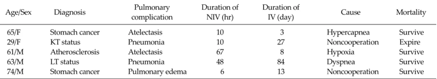
Among the 15 patients in the NIV group, 10 (66.7%) were treated successfully without endotracheal intuba- tion, whereas 5 (33.3%) were converted to invasive ven-Table 3. The characteristics of the patients who were converted from non-invasive ventilation to invasive ventilation
Pulmonary Duration of Duration of
Age/Sex Diagnosis Cause Mortality
complication NIV (hr) IV (day)
65/F Stomach cancer Atelectasis 10 3 Hypercapnea Survive
29/F KT status Pneumonia 10 27 Noncooperation Expire
61/M Atherosclerosis Atelectasis 67 8 Hypoxia Survive
63/M LT status Pneumonia 48 84 Dyspnea Survive
74/M Stomach cancer Pulmonary edema 6 13 Noncooperation Survive
NIV, non-invasive ventilation; IV, invasive ventilation; KT, kidney transplantation; LT, liver transplantation.
Table 4. The patients with respiratory failure after extubation who were successfully weaned with non-invasive ventilation
Age/Sex Diagnosis Pulmonary
complication
Duration of IV (day)
Duration of NIV (day)
Extubation-NIV (hr)
Cause of conversion
63/F Gastric cancer Atelectasis 7 1 7 Dyspnea
70/M Hepatocellular carcinoma Atelectasis 2 2 12 Dyspnea
38/F Multiple trauma Acute lung injury 8 2 3 Hypoxia
IV, invasive ventilation; NIV, non-invasive ventilation.
tilation during treatment. The causes of conversion in- cluded patient noncooperation in 2 patients, the deterio- ration of hypercapnea in 1, deterioration of hypoxia in 1, and dyspnea symptoms in 1 (Table 3). Causative diseases in these 5 patients included atelectasis in 2, pneumonia in 2 and psychogenic pulmonary edema in 1. The mean time to conversion in these 5 patients was 28.2 hours after the start of NIV. Particularly, in patients whose causative dis- ease was pneumonia, there was a trend toward a longer treatment period even after the conversion to invasive ventilation.
Application of NIV to ARF patients after extuba- tion
Of the 59 patients who received invasive ventilation, 3 were treated by NIV for respiratory failure after ex- tubation, thus avoiding re-intubation (Table 4). Respira- tory failure after extubation was defined as development of dyspnea symptoms within 12 hours after weaning. Of these 3 patients, 2 had post-operative atelectasis and 1 had acute lung injury due to multiple trauma. All were treated successfully by NIV without re-intubation.
Factors predictive of the success of NIV
NIV was successful in 10 of 15 patients. Univariate and
multivariate analysis showed that success was related to the etiology of ARF, especially atelectasis (P = 0.032). NIV was more likely to be successful in patients with post- operative atelectasis, whereas invasive ventilation was more likely to be successful in patients with pneumonia. In contrast, patient age, gender, treatment with or immune suppressors, and the severity of respiratory function were not predictive of success (Table 2).
DISCUSSION
We have shown here that surgical patients with ARF can be treated effectively with NIV. The likelihood of NIV suc- cess is determined by the disease causing ARF not by the severity at the time of ARF or level of hypoxia. In partic- ular, NIV was more likely to be successful in patients with postoperative atelectasis, whereas invasive ventilation was more likely to be successful in patients with pneumonia.
Although, hypoxia develops in 30 to 50% of patients af- ter surgery, it improves slowly over several weeks in most patients without symptoms. In some patients, however, hypoxia can progress to dyspnea. Impaired gas exchange can be induced by the reduction in muscle tension caused
by anesthesia, decreases in respiratory capacity, tidal vol- ume, functional residual capacity and diaphragm dys- function caused by surgery elevated abdominal pressure and pain. In particular, post-operative fluid therapy, trans- fusion, inflammatory reaction, and sepsis are factors that aggravate respiratory failure. Positive pressure ven- tilation may be the mainstay of ARF treatment. A random- ized prospective study of patients who underwent thor- aco-abdominal surgery for aortic aneurysm and were treated with CPAP or oxygen showed that CPAP de- creased the rate of intubation, increased the level of oxy- gen saturation and shortened hospital stay [16]. Other studies have also shown that positive pressure respiration after surgery is effective prophylactically in assisting pul- monary function lost during surgery, and therapeutically in treating patients with post-operative respiratory fail- ure, with NIV being successful in most patients [17-19].
During the initial period of NIV use in our institution, the mask did not adhere closely to the face, resulting in air leakage, skin compression by the mask and receipt of ex- cessive pressure. Thus, the skin became necrotized and pa- tients were quite uncomfortable, contributing to the NIV failure. Due to improvements of mask materials, espe- cially the use of helmet masks that are not in direct contact with the patient’s face, patient discomfort and complica- tions have been reduced, increasing patient compliance with NIV [11]. Moreover, nurses and physicians have gained more experience with NIV including both manipu- lation methods and patient education. Thus, of our 15 pa- tients treated with NIV therapy, only 5 (33.3%) were con- verted to invasive ventilation during treatment, whereas the remaining 10 (66.7%) were treated successfully with NIV without endotracheal intubation. Others have also re- ported that 30 to 50% of patients are converted to invasive ventilation requiring intubation. Factors predicting fail- ure of NIV include pneumonia, disease severity, older age and lack of improvement 1 hour after application of NIV [20,21]. Among our 5 patients who failed NIV and were converted to invasive ventilation, compliance with NIV was determined within 3 days of its application and mean period of invasive ventilation after conversion was 27 days, longer than in other patients who received mechan- ical ventilation. Our findings suggest that the selection of
appropriate indications for NIV is very important, and that the delay of appropriate treatments could prolong the treatment period.
In NIV, positive pressure ventilation is applied with the cooperation of patients, thus preventing hypotension caused by the use of sedatives, shortening treatment peri- ods, reducing complications associated with intubation and reducing the incidence of pneumonia. Nonetheless, NIV may be associated with complications such as gastric distention and pulmonary aspiration, and NIV may im- pede the ability of patients to cough and expectorate par- ticularly in patients later converted to invasive ventilation.
Of ARF patients weaned from mechanical ventilation as planned, 6 to 23% require re-intubation with 48 to 72 hours [22-24]. This is due primarily to obstruction of the upper airways, insufficient coughing or expectoration, deterio- ration of consciousness, or myocardial failure. Although re-intubation reflects disease severity, intubation itself is an independent factor associated with increases in rates of pneumonia and death and duration of hospitalization [23,25]. Therefore, prevention of re-intubation is very im- portant in the treatment of critically ill patients. In patients who developed respiratory failure after weaning, NIV was unable to lower mortality or re-intubation rate [25].
However high risk patients who received NIV soon after extubation, without re-intubation, could be successfully weaned from mechanical ventilation with a reduced mor- tality rate within the intensive care unit [26]. Similarly, all 3 of our patients who developed ARF after extubation were treated successfully with NIV without re-intubation.
Due to the retrospective design of our study, we could not control for factors such as the disease causing ARF at the time of therapeutic intervention, disease severity and clinical symptoms. This study, however, is to compare NIV with invasive ventilation in surgical patients in Korea. Prospective analysis of the prophylactic and ther- apeutic effects of NIV are therefore required.
In conclusion, we found that NIV may be successful in surgical patients who develop ARF, particularly in se- lected patients with post-operative atelectasis. The se- verity of ARF or hypoxia at the time of intervention was not associated with the effects of NIV. Moreover, NIV can successfully prevent re-intubation in patients who devel-
op respiratory failure after extubation.
CONFLICTS OF INTEREST
No potential conflict of interest relevant to this article was reported.
REFERENCES
1. Warner DO. Preventing postoperative pulmonary compli- cations: the role of the anesthesiologist. Anesthesiology 2000;92:1467-72.
2. Vassilakopoulos T, Mastora Z, Katsaounou P, Doukas G, Klimopoulos S, Roussos C, et al. Contribution of pain to in- spiratory muscle dysfunction after upper abdominal sur- gery: a randomized controlled trial. Am J Respir Crit Care Med 2000;161(4 Pt 1):1372-5.
3. Lawrence VA, Dhanda R, Hilsenbeck SG, Page CP. Risk of pulmonary complications after elective abdominal sur- gery. Chest 1996;110:744-50.
4. Warner DO. Preventing postoperative pulmonary compli- cations: the role of the anesthesiologist. Anesthesiology 2000;92:1467-72.
5. Jaber S, Chanques G, Jung B. Postoperative Non-invasive ventilation. Anesthesiology 2010;112:453-61.
6. Mehta S, Hill NS. Non-invasive ventilation. Am J Respir Crit Care Med 2001;163:540-77.
7. Pennock BE, Kaplan PD, Carlin BW, Sabangan JS, Magovern JA. Pressure support ventilation with a sim- plified ventilatory support system administered with a na- sal mask in patients with respiratory failure. Chest 1991;100:1371-6.
8. Ambrosino N, Foglio K, Rubini F, Clini E, Nava S, Vitacca M. Non-invasive mechanical ventilation in acute respira- tory failure due to chronic obstructive pulmonary disease:
correlates for success. Thorax 1995;50:755-7.
9. Räsänen J, Heikkilä J, Downs J, Nikki P, Väisänen I, Viitanen A. Continuous positive airway pressure by face mask in acute cardiogenic pulmonary edema. Am J Cardiol 1985;55:296-300.
10. Kindgen-Milles D, Müller E, Buhl R, Böhner H, Ritter D, Sandmann W, et al. Nasal-continuous positive airway pres- sure reduces pulmonary morbidity and length of hospital stay following thoracoabdominal aortic surgery. Chest 2005;128:821-8.
11. Conti G, Cavaliere F, Costa R, Craba A, Catarci S, Festa V, et al. Non-invasive positive-pressure ventilation with differ- ent interfaces in patients with respiratory failure after ab- dominal surgery: a matched-control study. Respir Care 2007;52:1463-71.
12. Jaber S, Delay JM, Chanques G, Sebbane M, Jacquet E,
Souche B, et al. Outcomes of patients with acute respira- tory failure after abdominal surgery treated with Non-in- vasive positive pressure ventilation. Chest 2005;128:
2688-95.
13. Antonelli M, Conti G, Bufi M, Costa MG, Lappa A, Rocco M, et al. Non-invasive ventilation for treatment of acute respiratory failure in patients undergoing solid organ transplantation: a randomized trial. JAMA 2000;283:
235-41.
14. Hilbert G, Gruson D, Vargas F, Valentino R, Gbikpi- Benissan G, Dupon M, et al. Non-invasive ventilation in immunosuppressed patients with pulmonary infiltrates, fever, and acute respiratory failure. N Engl J Med 2001;344:
481-7.
15. Feltracco P, Serra E, Barbieri S, Milevoj M, Furnari M, Rizzi S, et al. Non-invasive ventilation in postoperative care of lung transplant recipients. Transplant Proc 2009;41:1339- 44.
16. Kindgen-Milles D, Müller E, Buhl R, Böhner H, Ritter D, Sandmann W, et al. Nasal-continuous positive airway pres- sure reduces pulmonary morbidity and length of hospital stay following thoracoabdominal aortic surgery. Chest 2005;128:821-8.
17. Squadrone V, Coha M, Cerutti E, Schellino MM, Biolino P, Occella P, et al. Continuous positive airway pressure for treatment of postoperative hypoxemia: a randomized con- trolled trial. JAMA 2005;293:589-95.
18. Antonelli M, Conti G, Rocco M, Bufi M, De Blasi RA, Vivino G, et al. A comparison of Non-invasive positive- pressure ventilation and conventional mechanical ven- tilation in patients with acute respiratory failure. N Engl J Med 1998;339:429-35.
19. Antonelli M, Conti G, Esquinas A, Montini L, Maggiore SM, Bello G, et al. A multiple-center survey on the use in clinical practice of Non-invasive ventilation as a first-line intervention for acute respiratory distress syndrome. Crit Care Med 2007;35:18-25.
20. Wallet F, Schoeffler M, Reynaud M, Duperret S, Workineh S, Viale JP. Factors associated with Non-invasive ven- tilation failure in postoperative acute respiratory in- sufficiency: an observational study. Eur J Anaesthesiol 2010;27:270-4.
21. Antonelli M, Conti G, Moro ML, Esquinas A, Gonzalez- Diaz G, Confalonieri M, et al. Predictors of failure of Non-invasive positive pressure ventilation in patients with acute hypoxemic respiratory failure: a multi-center study.
Intensive Care Med 2001;27:1718-28.
22. Epstein SK, Ciubotaru RL, Wong JB. Effect of failed ex- tubation on the outcome of mechanical ventilation. Chest 1997;112:186-92.
23. Esteban A, Alía I, Tobin MJ, Gil A, Gordo F, Vallverdú I, et al. Effect of spontaneous breathing trial duration on out- come of attempts to discontinue mechanical ventilation.
Spanish Lung Failure Collaborative Group. Am J Respir Crit Care Med 1999;159:512-8.
24. Torres A, Gatell JM, Aznar E, el-Ebiary M, Puig de la Bellacasa J, González J, et al. Re-intubation increases the
risk of nosocomial pneumonia in patients needing me- chanical ventilation. Am J Respir Crit Care Med 1995;152:
137-41.
25. Esteban A, Frutos-Vivar F, Ferguson ND, Arabi Y, Apezteguía C, González M, et al. Non-invasive positive- pressure ventilation for respiratory failure after extuba-
tion. N Engl J Med 2004;350:2452-60.
26. Ferrer M, Valencia M, Nicolas JM, Bernadich O, Badia JR, Torres A. Early Non-invasive ventilation averts extubation failure in patients at risk: a randomized trial. Am J Respir Crit Care Med 2006;173:164-70.