INTRODUCTION
Jrais a high-frequency antigen that has been included in the 901 series (antigen number, 901005) of the Interna- tional Society of Blood Transfusion blood group classifi- cation [1]. The frequency of occurrence of this antigen is known to be over 99% in most ethnic groups [2]. Although the Japanese population is known to have the highest
number of Jr(a-) individuals, the incidence of the Jr(a-) phenotype is estimated to be only 0.03-0.12% even in the Japanese population [3], and this percentage is thought to be lower in Caucasians, Mexicans, and Arabs [4]. The incidence of the Jr(a-) phenotype in the Korean popula- tion has not yet been reported.
Jrawas first reported in 1970 by Stroup and MacIlroy [5], who identified 5 cases in which anti-Jra antibody was detected during the assessment of a high frequency antigen in the Caucasian population. Although case reports regarding anti-Jraantibody have been published occasionally over the past 40 yr, its clinical significance has not been well established. This is mainly because of the diversity of patient outcomes and scarcity of report- ed data. The 2 main clinical scenarios associated with
511 511
Hemolytic Disease of the Newborn Associated with Anti-Jr
aAlloimmunization in a Twin Pregnancy: The First Case Report in Korea
Hyungsuk Kim, M.D.
1, Min-Jeong Park, M.D.
2, Tae-Jung Sung, M.D.
3, Ji Seon Choi, M.D.
4, Jungwon Hyun, M.D.
1, Kyoung Un Park, M.D.
1,5, and Kyou-Sup Han, M.D.
1Department of Laboratory Medicine1, Seoul National University College of Medicine, Seoul; Departments of Laboratory Medicine2and Pediatrics3, Hallym University College of Medicine, Seoul; Department of Laboratory Medicine4, National Cancer Center, Goyang;
Department of Laboratory Medicine5, Seoul National University Bundang Hospital, Seongnam, Korea
511 511
Jrais a high-frequency antigen found in all ethnic groups. However, the clinical significance of the anti-Jraantibody has remained controversial. Most studies have reported mild hemolytic dis- ease of the newborn and fetus (HDNF) in Jra-positive patients. Recently, fatal cases of HDNF have also been reported. We report the first case of HDNF caused by anti-Jraalloimmunization in twins in Korea. A 33-yr-old nulliparous woman with no history of transfusion or amniocentesis was admitted at the 32nd week of gestation because of vaginal bleeding caused by placenta previa. Anti-Jraanti- bodies were detected in a routine laboratory examination. An emergency cesarean section was performed at the 34th week of gestation, and 2 premature infant twins were delivered. Laboratory examination showed positive direct antiglobulin test and Jr(a+) phenotype in the red blood cells and the presence of anti-Jraantibodies in the serum in both neonates. The infants underwent pho- totherapy for neonatal jaundice; this was followed by conservative management. They showed no further complications and were discharged on the 19th postpartum day. Preparative management to ensure the availability of Jr(a-) blood, via autologous donation, and close fetal monitoring must be performed even in cases of first pregnancy in Jr(a-) women. (Korean J Lab Med 2010;30:511-5)
Key Words : Anti-Jra, Neonatal jaundice, Fetal erythroblastosis, Hematologic pregnancy compli- cations
Received :June 10, 2010 Manuscript No :KJLM10-108 Revision received :July 6, 2010
Accepted :July 19, 2010
Corresponding author :Kyou-Sup Han, M.D.
Department of Laboratory Medicine, Blood Bank, Seoul National University Hospital, 28 Yeongeon-dong, Jongno-gu, Seoul 110-744, Korea
Tel : +82-2-2072-3519, Fax : +82-2-762-9411 E-mail : [email protected]
ISSN 1598-6535 The Korean Society for Laboratory Medicine
anti-Jra positivity are incompatible transfusions and hemolytic disease of the newborn and fetus (HDNF). In the former scenario, no biologic or clinical evidence of hemolysis due to mild, acute, or delayed transfusion reac- tions has been reported for individuals with anti-Jraanti- bodies [6]. In the latter scenario, limited availability of data has led to conflicting results [6]. We present a case of neonatal jaundice associated with anti-Jraalloimmu- nization in premature twins; this case occurred during the first pregnancy of a Korean woman. This is the first report documenting HDNF related to anti-Jraalloimmu- nization in Korea.
CASE REPORT
A 33-yr-old nulliparous woman in her 32nd gesta- tional week visited the emergency room because of vagi- nal bleeding. Her medical history was unremarkable except for the fact that she was a hepatitis B carrier.
She had no history of blood transfusion or amniocente- sis. She had a twin pregnancy after in vitro fertilization and embryo transfer (IVF-ET). A routine obstetric exam- ination revealed that vaginal bleeding was caused by pla- centa previa, and the patient was admitted to undergo conservative perinatal management.
Initial laboratory tests showed a low hemoglobin level (10.9 g/dL). The antibody-screening test (Search-Cyte;
Medion Diagnostics AG, Du_dingen, Switzerland) yielded weakly positive, irregular results in both the reagent cells only in the antiglobulin phase. The patient’s ABO and RhD blood type was O+. Antibody identification test (Data-Cyte; Medion Diagnostics AG) revealed the pres- ence of a pan-reactive antibody that showed weak posi- tivity against all the 11 reagent panel cells in the antiglob- ulin phase. The results of the antibody identification tests conducted at room temperature and at 37℃ with albu- min enhancement were negative. The indirect antiglob- ulin test (IAT) with autologous control and direct antiglob- ulin test (DAT) also yielded negative results. The anti- body characteristics indicated that the corresponding antigen was a high-frequency red blood cell (RBC) anti-
gen. Screening of the patient’s RBC phenotype with a panel of antisera showed that the RBCs were positive for k, Dib, and Lub. A negative reaction against Jraantiserum prepared in-house confirmed the patient’s Jr(a-) phe- notype. Crossmatching of the patient’s serum and previ- ously confirmed Jr(a-) O+ RBCs yielded negative results.
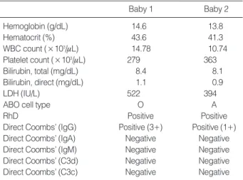
Because of frequent uterine contraction and vaginal spotting, an emergency cesarean section was performed at the 34th week of gestation. The patient delivered 2 premature male infants. Their body weight and Apgar scores at 1 and 5 min after birth were as follows: baby 1, 1.91 kg, 4, and 7; baby 2, 2.2 kg, 5, and 8. Both babies underwent phototherapy for the treatment of neonatal jaundice. The results of the laboratory tests conducted on the 4th day of birth are shown in Table 1. The ABO/
RhD blood type of baby 1 and baby 2 were O+ and A+, respectively. The antibody screening and identification test results of the 2 infants were similar to those of their mother. In both tests, the serum of the 2 infants weakly reacted with all the reagent-panel RBCs only in the antiglobulin phase. IAT with autologous control yielded positive results in both infants. The RBCs of both infants showed reactivity with Jraantiserum; this confirmed the Jr(a+) phenotype (the infants’RBCs were treated with chloroquine to dissociate the antibodies on the RBC sur- face before testing). Crossmatching with the infants’
Abbreviations: WBC, white blood cell; LDH, lactate dehydrogenase.
Baby 1 Baby 2
Hemoglobin (g/dL) 14.6 13.8
Hematocrit (%) 43.6 41.3
WBC count (×103/mL) 14.78 10.74
Platelet count (×103/mL) 279 363
Bilirubin, total (mg/dL) 8.4 8.1
Bilirubin, direct (mg/dL) 1.1 0.9
LDH (IU/L) 522 394
ABO cell type O A
RhD Positive Positive
Direct Coombs’ (IgG) Positive (3+) Positive (1+)
Direct Coombs’ (IgA) Negative Negative
Direct Coombs’ (IgM) Negative Negative
Direct Coombs’ (C3d) Negative Negative
Direct Coombs’ (C3c) Negative Negative
Table 1. Results of laboratory tests conducted on the neonates on the 4th day of birth
serum and previously confirmed Jr(a-) O+ RBCs showed negative results. Antibody-identification test with the elu- ate of the infants’RBCs was not performed because of the inadequate quantity of the referred blood sample.
Taken together, these data indicated the presence of anti-Jraantibodies of maternal origin in the twins’serum and the role of these antibodies in the positive DAT results in the neonates. The infants were discharged after con- servative management on the 19th postpartum day, with- out further complications.
DISCUSSION
Although the anti-Jra antibody was identified nearly half a decade ago, its clinical significance with regard to hemolytic reactions has not been clearly established. This is mainly because of the paucity of reported data. In most of the cases described in the literature, HDNF did not occur, and when it did occur, its intensity was mild to moderate, and hence, no treatment beyond phototherapy was required [3, 4, 7-13]. A few cases of incompatible Jr(a+) RBC transfusion with no or delayed hemolytic transfusion reactions have also been reported [14-16].
However, recent evidence has shown that anti-Jra antibodies have the potential for causing fatal HDNF and acute hemolytic reactions. Acute hemolytic transfu- sion reaction caused by anti-Jra antibodies has been reported in a 45-yr-old Japanese woman [15]. Peyrard et al. [6] reported the first case of fatal HDNF in a 28- yr-old Caucasian woman of Spanish Gypsy origin. She had an obstetric history of 2 abortions and 1 full-term pregnancy (G4P1). At the time of the first abortion, she developed massive hemorrhage that required transfusion of 10 units of RBCs. Fetal cardiomegaly associated with intrauterine growth retardation was confirmed at the 35th week of her 4th pregnancy. An emergency cesare- an section was performed at the 36th week. The newborn was hydropic, had severe anemia, and died 30 hr after birth. Anti-Jraantibodies, which were later identified in the mother’s serum, were thought to be the cause of severe fetal anemia. Arriaga et al. [17] also reported a similar
fatal case of hydrops fetalis associated with anti-Jraanti- bodies in a 39-yr-old Caucasian woman with a history of multiple pregnancies. An earlier case reported in 2006 also described severe fetal anemia in a 40-yr-old Japanese woman (G4P3), but the infant was born healthy after 4 intravascular transfusions via the umbilical cord [18].
The common feature of the 3 cases of HDNF described above is that the mothers had multiple pregnancies with or without transfusion history. In many previously pub- lished cases reporting no or mild to moderate clinical HDNF associated with anti-Jraantibody positivity, the Jr(a-) pregnant women had no transfusion history [6].
Thus, it can be presumed that the clinical significance of the anti-Jra antibody may depend on the frequency of exposure to the Jraantigen. Previous studies have shown that anti-Jra antibodies may develop during pregnancy and that they can cross the placenta [3, 9]. The Jraanti- gen has also been reported to be fully developed at birth [7]. Although anti-Jraantibodies can be stimulated by a first pregnancy, HDNF associated with anti-Jraantibody was thought to occur in subsequent pregnancies [6]. How- ever, in our case, the mother was nulliparous and had no history of transfusion or amniocentesis. The present pregnancy was the only identifiable cause of alloimmu- nization. Although confirmative findings such as nega- tive antibody screening test results before the present pregnancy were not available, it should be noted that the findings in this case suggest that HDNF can occur in a first pregnancy. This may probably be the reason for the low severity of HDNF. Another interesting finding was that the DAT results for both neonates were posi- tive and that the positivity in the DAT result of baby 1, whose ABO/Rh blood type was O+, was greater than that in the DAT result of baby 2. The reasons for the differences in DAT results are not clear. The effect of having twins on anti-Jraalloimmunization requires fur- ther investigation. Although we could not evaluate the eluates of the twin’s RBCs because of lack of specimens, the presence of anti-Jraantibodies in the twin’s serum and the Jr(a+) phenotype of the twin’s RBCs indicated that anti-Jraantibodies were the cause of the positive
DAT results.
Several in vivo and in vitro studies have been performed to assess the clinical significance of anti-Jraantibodies.
Kendall [14] performed the traditional 51Cr RBC survival test and showed the moderately rapid destruction of incompatible Jr(a+) RBCs after transfusion to a Jr(a-) patient. Ogasawara and Mazuda [19] reported that the Jraantigen density was lower than the D-antigen den- sity in RBCs with a weak D phenotype and was signifi- cantly lower than the density of high-incidence antigens such as Dib. They concluded that the small number of antigens on the RBC membrane may account for the general lack of clinical significance of anti-Jraantibod- ies. Miyazaki et al. [20] described that the fluorescence intensity of Jr(a+) RBCs was significantly lower than that of E+ RBCs from E homozygotes; this finding sug- gested that the number of Jraantigen sites is substan- tially lower than that of Rh antigen sites. The monocyte monolayer assay (MMA) has also been used in some stud- ies to predict the potential clinical significance of anti- bodies to high-frequency antigens [15, 21, 22]. However, the exact mechanism of the toxicity of anti-Jraantibod- ies remains unclear.
One of the difficulties in identifying anti-Jraantibod- ies is the lack of commercially available antisera. A human monoclonal anti-Jraantibody has been produced from a human-mouse heterohybridoma (HMR0921) [20]. How- ever, the biochemical structure of the Jraantigen and the molecular basis of the Jr(a-) phenotype are currently unknown [6]. Our laboratory has been identifying anti- Jra antibodies since 1995 after donation of Jra antisera and Jr(a-) type O RBCs obtained from Osaka Red Cross Center. We are currently using in-house Jra antisera and Jr(a-) RBCs prepared by constantly storing con- firmed patient samples. Although we have been identi- fying a few cases of anti-Jraalloantibodies annually, this is the first case of HDNF related to anti-Jraalloimmu- nization in Korea. The first case of anti-Jraalloimmu- nization in Korea was reported in 1995 in a 35-yr-old pregnant woman with a history of multiple pregnancies and abortions (G6P4L1D3A1) [23]. Anti-Jraantibodies were
identified after an anamnestic response following the transfusion of 1 RBC unit. However, the infant did not show any evidence of HDNF. Because a large percentage of Jr(a-) individuals appear to be of Asian origin [18], further studies on the incidence of the Jr(a-) phenotype in the Korean population may reveal interesting findings.
In conclusion, the potential of the anti-Jraantibody as a clinically significant alloantibody should be considered even in cases of first pregnancy. Preventive management and close fetal monitoring are recommended, especially if the mother has a high antibody titer and a history of multiple pregnancies and/or transfusion. Because Jr(a-) RBC units are very difficult to obtain, measures must be taken to ensure the availability of appropriate blood types.
Autologous donations should always be considered; sib- lings are also potential donors of Jr(a-) RBCs. In emer- gent situations where Jr(a-) RBCs are not available, transfusion of the least incompatible units is inevitable.
Nevertheless, close monitoring of the patient is neces- sary while performing subsequent transfusions.
REFERENCES
1. Daniels GL, Fletcher A, Garratty G, Henry S, Jorgensen J, Judd WJ, et al. Blood group terminology 2004: from the International Society of Blood Transfusion committee on terminology for red cell surface antigens. Vox Sang 2004;87:304-16.
2. Roback JD, Combs MR, et al. eds. Technical manual. 16th ed. Bethes- da: American Association of Blood Banks, 2008:411-36.
3. Nakajima H and Ito K. An example of anti-Jracausing hemolytic disease of the newborn and frequency of Jraantigen in the Japanese population. Vox Sang 1978;35:265-7.
4. Vedo M and Reid ME. Anti-Jrain a Mexican American. Transfusion 1978;18:569.
5. Stroup M and MacIlroy M. Jra-five examples of an antibody defining an antigen of high-frequency in the Caucasian population [abstract].
Proceedings of the 23rd Annual Meeting of the American Associa- tion of Blood Banks. 1970:86.
6. Peyrard T, Pham BN, Arnaud L, Fleutiaux S, Brossard Y, Guerin B, et al. Fatal hemolytic disease of the fetus and newborn associated with anti-Jra. Transfusion 2008;48:1906-11.
7. Tritchler JE. An example of anti-Jra. Transfusion 1977;17:177-8.
8. Orrick LR and Golde SH. Jra-mediated hemolytic disease of the new- born infant. Am J Obstet Gynecol 1980;137:135-6.
9. Toy P, Reid M, Lewis T, Ellisor S, Avoy DR. Does anti-Jracause hemolytic disease of the newborn? Vox Sang 1981;41:40-4.
10. Takabayashi T, Murakami M, Yajima H, Tsujiei M, Ozawa N, Yaji- ma A. Influence of maternal antibody anti-Jraon the baby: a case report and pedigree chart. Tohoku J Exp Med 1985;145:97-101.
11. Bacon J, Sherrin D, Wright RG. Case report, anti-Jra. Transfusion 1986;26:543-4.
12. Levene C, Sela R, Dvilansky A, Yermiahu T, Daniels G. The Jr(a-) phenotype and anti-Jrain two Beduin Arab women in Israel. Trans- fusion 1986;26:119-20.
13. Bellver-Pradas J, Arriaga-Chafer F, Perales-Marin A, Maiques-Mon- tesinos V, Serra-Serra V. Obstetric significance of anti-Jr(a) antibody.
Am J Obstet Gynecol 2001;184:75-6.
14. Kendall AG. Clinical importance of the rare erythrocyte antibody anti-Jra. Transfusion 1976;16:646-7.
15. Kwon MY, Su L, Arndt PA, Garratty G, Blackall DP. Clinical signif- icance of anti-Jra: report of two cases and review of the literature.
Transfusion 2004;44:197-201.
16. Chung HJ, Lim JH, Park HJ, Kwon SW. Transfusion of Jra-positive
red blood cells to a Jra-negative patient with anti-Jra. Korean J Blood Transfus 2007;18:111-5.
17. Arriaga F, Gomez I, Linares MD, Gascon A, Carpio N, Perales A.
Fatal hemolytic disease of the fetus and newborn possibly due to anti-Jra. Transfusion 2009;49:813.
18. Ishihara Y, Miyata S, Chiba Y, Kawai T. Successful treatment of extremely severe fetal anemia due to anti-Jraalloimmunization.
Fetal Diagn Ther 2006;21:269-71.
19. Ogasawara K and Mazuda T. Characterization of Jraantibodies by monocyte phagocytosis assays and flow cytometry analysis. Acta Haematol Jpn 1990;53:1131-7.
20. Miyazaki T, Kwon KW, Yamamoto K, Tone Y, Ihara H, Kato T, et al. A human monoclonal antibody to high-frequency red cell anti- gen Jra. Vox Sang 1994;66:51-4.
21. Nance SJ, Arndt P, Garratty G. Predicting the clinical significance of red cell alloantibodies using a monocyte monolayer assay. Trans- fusion 1987;27:449-52.
22. Garratty G. Predicting the clinical significance of red cell antibodies with in vitro cellular assays. Transfus Med Rev 1990;4:297-312.
23. Kim HK, Park Q, Nahm CH, Kwon OH, Park YW. A case report of anti-Jrain pregnant woman. Korean J Blood Transfus 1995;6:185-8.