Vol. 23, Suppl. 1, 2011 S57
Received July 27, 2010, Revised September 10, 2010, Accepted for publication September 10, 2010
Corresponding author: Jung Soo Kim, M.D., Ph.D., Department of Radiation Oncology, Chonbuk National University Medical School, 634-18, Keumam-dong, Jeonju 561-712, Korea. Tel: 82-63-250-1196, Fax: 82-63-250-1192, E-mail: jskim@jbnu.ac.kr
This is an Open Access article distributed under the terms of the Creative Commons Attribution Non-Commercial License (http://
creativecommons.org/licenses/by-nc/3.0) which permits unrestricted non-commercial use, distribution, and reproduction in any medium, provided the original work is properly cited.
Ann Dermatol Vol. 23, Suppl. 1, 2011 http://dx.doi.org/10.5021/ad.2011.23.S1.S57
CASE REPORT
The Three Dimensional Conformal Radiotherapy for Hyperkeratotic Plantar Mycosis Fungoides
Sun Young Lee, M.D., Hyoung Cheol Kwon, M.D., Yong-Sun Cho, M.D.
1, Kyung-Hwa Nam, M.D.
1, Chull-Wan Ihm, M.D.
1, Jung Soo Kim, M.D., Ph.D.
Departments of Radiation Oncology and 1Dermatology, Chonbuk National University Medical School, Jeonju, Korea
The localized early-stage of Mycosis fungoides (MF) (stage IA-IIA) is usually treated with topical agents, such as nitrogen mustard, steroids, and phototherapy (UVB/PUVA) as first line therapy; response to these initial treatments is usually good. However, hyperkeratotic plantar lesions are clinically rare and have decreased responsiveness to topical agents.
For such cases, physicians may consider local radiotherapy.
Here, a case of an 18-year-old Korean woman who was treated with three-dimensional conformal radiotherapy (3D-CRT) for hyperkeratotic plantar lesions that were refrac- tory to UVA-1, methotrexate, and topical steroids is reported.
Complete remission was attained after radiotherapy. During the one-year follow-up period, there has been no evidence of disease recurrence and no chronic complications have been observed. (Ann Dermatol 23(S1) S57∼S60, 2011) -Keywords-
Hyperkeratotic plantar lesions, Mycosis fungoides, Three- dimensional conformal radiotherapy (3D-CRT)
INTRODUCTION
Mycosis fungoides (MF) is a primary cutaneous T-cell lymphoma, which usually starts as “flat patches”. Common
locations include the lower abdomen, buttocks, upper thighs, and breasts of women1-4. Patches vary in size, with a round, oval, serpiginous, or polycyclic shape, and they have sharp margins1,2. A case of mycosis fungoides on both soles of the feet was recently treated at our center.
The lesion was not easily recognized as a flat patch and the diagnosis of MF was delayed. After the correct diagnosis, the plantar lesion required special treatment due to its interdigital involvement and plantar furrows, which were difficult to irradiate with the therapeutic wavelength of electron beam commonly used for this condition3. The patient was successfully treated with three-dimensional conformal radiotherapy (3D-CRT).
Here, a case of an 18-year-old Korean woman with MF on the soles of the feet, which was refractory to the usual conventional local treatments, is reported.
CASE REPORT
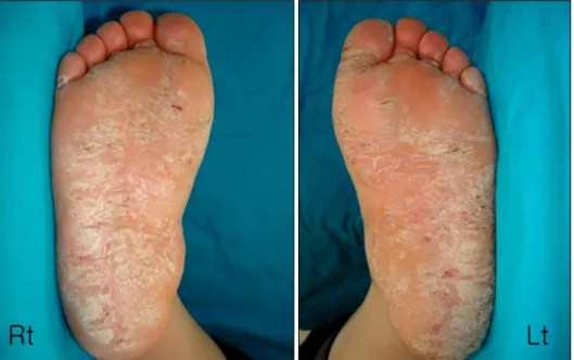
An 18-year-old Korean woman developed hyperkeratotic soles three years prior to presentation to the hospital. For two years before examination, she was treated with oral and topical steroids at local clinics, which caused weight gain and menstrual irregularities, with little improvement of skin lesions. On examination, the soles of both feet showed thick hyperkeratosis with scales and mild ery- thema. The hyperkeratotic plantar surfaces had shallow furrows. The lesion involved almost the entire plantar surface, including the digital furrows and lateral borders of the soles (Fig. 1). There was no family history of palmo- plantar keratoderma.
Biopsy findings of the keratotic plaque were as follows:
the epidermis showed compact hyperparakeratosis, acan- thosis, spongiosis, and mononuclear cell exocytosis (Fig.
2A). Higher power microscopy revealed intraepidermal collections of mononuclear cells showing hyperchromatic
SY Lee, et al
S58 Ann Dermatol
Fig. 1. Thick keratotic scales and furrows with mild erythema involves the entire plantar surface bilaterally, including its lateral borders and in- terdigital spaces.
Fig. 2. (A) A low magnification view shows compact hyper- karatois, acanthosis, and exocytosis with intraepidermal collec- tion of mononuclear cells. The hyperkeratotic horny layer also shows many nuclear remnants (H&E, ×100). (B) The higher magnification of intraepidermal collections of mononuclear cells showed atypical hyperchromatic nuclei of cells com- patible with Pautrier’s microabscesses (H&E, ×400). (C) Papil- lary dermal and intraepidermal exocytic cells were CD4 positive (immunoperoxidase, ×100).
atypical cells that were compatible with Pautrier’s micro- abscesses (Fig. 2B). Infiltrated cells were mostly CD4 posi- tive (Fig. 2C). These findings were consistent with the
diagnosis of patchy stage IA of MF.
UVA-1 phototherapy was started immediately with 20 J/cm2 two times weekly (a total of 20 times, total cumulative
The Three Dimensional Conformal Radiotherapy for Hyperkeratotic Plantar Mycosis Fungoides
Vol. 23, Suppl. 1, 2011 S59 Fig. 4. (A) The plantar skin on the day of completion of treatment shows di- sappearance of thick keratotic scales.
(B) A follow-up picture taken one- year after treatment shows complete resolution of plantar skin lesions.
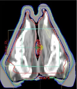
Fig. 3. The image is a three-dimensional conformal radiation treatment field. With the patient in a frog leg position, a 1 cm- thick silicon bolus was placed on the sole, as a tissue com- pensator. The photon beam could reach the entire lesions.
dose: 400 J/cm2). The disease showed only a slight response. Then, methotrexate 15 mg/week and topical di- flucortolone valerate ointment were tried for the next four months, without improvement. Finally, radiation treatment with 3D-CRT using a 6 MV photon beam (Varian, Palo Alto, Ca, USA) was applied to the resistant lesion. To avoid radiation injury to the adjoining joint structures and the surrounding normal tissue, 3D-CRT was applied to plantar lesions and this included interdigital furrows.
The 3D-CRT procedure was as follows: The patient’s legs were placed in a frog leg position. To ensure application of an adequate radiation dose to the skin tissue, a 1 cm- thick silicon bolus was placed on the sole as a tissue compensator. The area for radiation therapy encompassed the normal skin area 2 cm from the margin of the lesion
(Fig. 3). A total dose of 40 Gy was administered with a fractionated daily dose of 2 Gy, five days/week for a total of four weeks.
At the time of completion of treatment, the plantar skin had recovered to normal soft smooth skin with mild erythema (Fig. 4A). The post-treatment biopsy examina- tion, performed one month after the end of treatment, sho- wed absence of the previously-present lymphoid infil- trates. All of the lesions were resolved and the patient has been free of disease for one-year of follow up (Fig. 4B).
DISCUSSION
For early stage MF, the goal of therapy is to achieve a complete remission. The choice of treatment method for early stage localized lesions is dependent on the local nature of the lesions or the plaque thickness and the extent of the disease. For early stage MF (stage IA-IIA), Whittaker et al1. recommended that skin-directed therapy (SDT), including topical emollients, steroids, bexarotene gel, UVB/PUVA, and superficial radiotherapy, such as with an electron beam, should be tried as first line therapy.
By nature, plantar skin has a very thick keratinized layer, and an irregular contour, including digital furrows. There- fore, plantar MF is less responsive to skin-directed therapy, compared to other areas of the cutaneous with MF5. My- cosis fungoides and other variants of cutaneous T-cell lymphomas are very radiosensitive; local thick plaque skin lesions can be treated successfully with local radio- therapy6-10.
Technical modifications of local radiotherapy have allo- wed for optimization of dose distribution, resulting in improved clinical outcome and reduction of chronic com- plications. The intensity of radiation must be chosen to provide an adequate dose to the bottom of the lesion. An
SY Lee, et al
S60 Ann Dermatol
electron beam can be used successfully for treatment of superficial and flat cutaneous lesions of MF; however, for deeper and irregular contoured lesions, electron beam irradiation provides an indistinct dose distribution and ineffective dose delivery to deeper lesions10. A photon beam is usually used for treatment of deep and irregular lesions10. However, for dermatological conditions, this is not commonly used because of the untoward effects on deep regions of the body. Therefore, 3D-CRT using a photon beam was considered for treatment of this patient.
The 3D-CRT protocol begins with individualized, 3D digital data sets, which are used in generation of 3D computer images of the patient’s lesions and the normal adjacent tissue anatomy. With 3D-CRT, an adequate amount of therapeutic radiation can be delivered to lesions while significantly reducing radiation to the su- rrounding normal tissues. 3D-CRT increases the effective treatment dose to malignant cells while avoiding harmful effects on normal tissue10. In the present case, a 1 cm- thick silicon bolus was used to provide an adequate radiation dose to the superficial skin layer. A silicon bolus is a tissue equivalent material that is placed directly on the skin surface in order to even out the irregular contours of a patient, which results in presentation of a flat normal surface to the photon beam10.
For successful local radiotherapy, the extent of radiation on the skin is important. Inclusion of the adjoining normal skin, up to 2∼3 cm away from the margin of the visible lesions, is recommended, with a total dose of at least 40 Gy9. Otherwise, or with a tight field size without including adjoining normal skin, recurrence at the margins has been commonly reported7. In the present case, radiation treatment with a total dose of 40 Gy, including a 2 cm width of normal skin, was performed.
In this case, satisfactory local control without severe com- plications was obtained for hyperkeratotic plantar MF.
3D-CRT should be considered for treatment of early stage MF in patients who are refractory to topical agents. How- ever, as local relapse has been commonly reported for
many skin-directed therapies1,7, regular follow-up is need- ed in order to carefully monitor these patients.
REFERENCES
1. Whittaker SJ, Marsden JR, Spittle M, Russell Jones R; British Association of Dermatologists; U.K. Cutaneous Lymphoma Group. Joint British Association of Dermatologists and U.K.
Cutaneous Lymphoma Group guidelines for the manage- ment of primary cutaneous T-cell lymphomas. Br J Dermatol 2003;149:1095-1107.
2. Siegel RS, Pandolfino T, Guitart J, Rosen S, Kuzel TM.
Primary cutaneous T-cell lymphoma: review and current concepts. J Clin Oncol 2000;18:2908-2925.
3. Kneitz H, Bröcker EB, Becker JC. Mycosis fungoides bullosa:
a case report and review of the literature. J Med Case Reports 2010;4:78.
4. Duvic M, Cather J. Emerging new therapies for cutaneous T-cell lymphoma. Dermatol Clin 2000;18:147-156.
5. Pandya MJ, Rawal RC, Bilimoria FE. Unusual presentation of T-cell lymphoma. Indian J Dermatol Venereol Leprol 1997;
63:370-372.
6. Jacob R, Scala M, Fung MA. A case of syringotropic cutaneous T-cell lymphoma treated with local radiotherapy. J Am Acad Dermatol 2009;60:152-154.
7. Curt H, Sushil B, Luther WB, Eric CV. Cutaneous T-cell lymphoma. In: Halperin EC, Perez CA, Brady LW, editors.
Perez and Brady's principles and practice of radiation oncology. 5th ed. Philadelphia: Lippincott Williams &
Wilkins, 2007:1766-1776.
8. Samant RS, Fox GW, Gerig LH, Montgomery LA, Allan DS.
Total scalp radiation using image-guided IMRT for pro- gressive cutaneous T-cell lymphoma. Br J Radiol 2009;
82:e122-e125.
9. Eich HT, Eich D, Micke O, Süttzer H, Casper C, Krieg T, et al. Long-term efficacy, curative potential, and prognostic factors of radiotherapy in primary cutaneous B-cell lym- phoma. Int J Radiat Oncol Biol Phys 2003;55:899-906.
10. Purdy JA. Three-dimensional conformal radiation therapy:
physics, treatment planning, and clinical aspects. In: Hal- perin EC, Perez CA, Brady LW, editors. Perez and Brady's principles and practice of radiation oncology. 5th ed. Phila- delphia: Lippincott Williams & Wilkins, 2007:218-238.