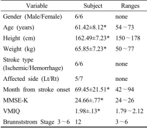
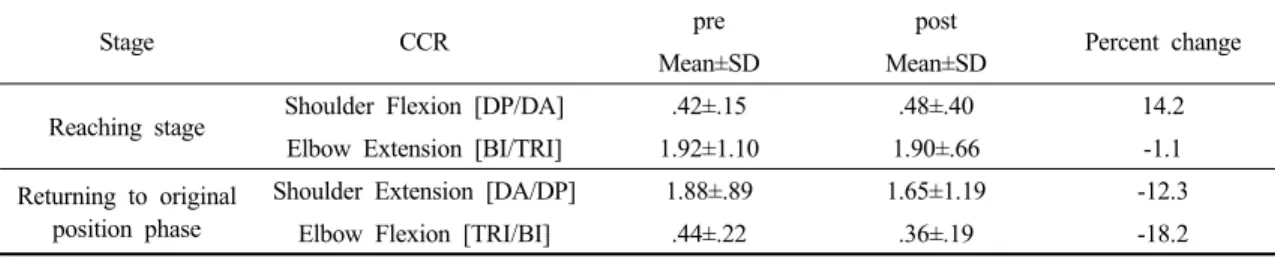
Effects of Mental Practice on Function and Muscle Activation of Upper Extremity in Stroke Patients
7
0
0
전체 글
(2)
(3)
(4)
(5)
(6)
(7)
수치
관련 문서
Changes in the level of physical fitness of obese women who performed complex exercise were significantly different in muscle endurance,
Sixth, there was no significant correlation between lower extremity muscle thickness, static stability, and dynamic stability of excellent athletes in college
It was summarized as follows that group participating in after-school basketball activities for 12 weeks showed decrease of the body fat(%) and increase
In exercise group, change in physical strength after measurement showed significant increase in muscular strength, muscle endurance, cardiovascular endurance, agility,
This study was to do a comparative analysis on kinematic differences and differences in muscle activity between the skilled and the unskilled in windsurfing
In this study, two experiments were conducted to understand the effects of additional charge on the detailed growth mechanism of Alq 3 and to determine the effect of
The purpose of this study was to investigate the lower extremity muscle strength and student health of male middle school students. Twenty middle school
The improvement factors of the performance showed significant increase in muscle strength, muscle endurance, cardiovascular endurance, power, agility and
