*한양대학교 의과대학 한양대학교병원 흉부외과학교실
Department of Thoracic and Cardiovascular Surgery, Hanyang University Seoul Hospital, College of Medicine, Hanyang University
**한양대학교 의과대학 영상의학교실
Department of Diagnostic Radiology, College of Medicine, Hanyang University
논문접수일:2010년 6월 7일, 논문수정일:2010년 7월 26일, 심사통과일:2010년 8월 17일 책임저자:정원상 (133-792) 서울시 성동구 행당동 17번지, 한양대학교병원 흉부외과
(Tel) 02-2290-8461, (Fax) 02-2290-8462, E-mail: [email protected] 본 논문의 저작권 및 전자매체의 지적소유권은 대한흉부외과학회에 있다.
CC
This is an open access article distributed under the terms of the Creative Commons Attribution Non-Commercial License (http://creative- commons.org/licenses/by-nc/3.0) which permits unrestricted non-commercial use, distribution, and reproduction in any medium, provided the original work is properly cited.
양전자단층촬영 /전산화단층촬영(integrated PET/CT)을 이용한 비소세포폐암의 림프절 병기판정
김지훈*ㆍ정원상*ㆍ김영학*ㆍ김 혁*ㆍ전석철**
Accuracy of Nodal Staging with Integrated PET/CT Scanning in Non-small Cell Lung Cancer
Ji-Hoon Kim, M.D.*, Won Sang Chung, M.D.*, Young-Hak Kim, M.D.*, Hyuck Kim, M.D.*, Seok-Chol Jeon, M.D.**
Background: For staging primary lung cancer, integrated positron emission tomography/computed tomography (PET/
CT) imaging is popular. The purpose of this study was to evaluate the accuracy of PET/CT scanning in lymph no- dal staging of lung cancer. Material and Method: We studied 48 patients who had received CT, PET/CT and pul- monary resections due to primary non-small cell lung cancer in our hospital between January 2006 and August 2009. Mediastinal lymph nodes were classified as superior mediastinal nodes, aortic nodes, inferior mediastinal no- des, or N1 nodes. We compared the power of CT and PET/CT for diagnosing pulmonary lymph nodes for each of the four types of nodes. Result: PET/CT was more sensitive than CT for all groups except inferior mediastinal nodes. However, the differences were not significant (McNemar’s test: superior mediastinal nodes, p=0.109; aortic nodes, p=1.000; inferior mediastinal nodes, p=0.625, N1 nodes, p=0.424). Conclusion: The accuracy of PET/CT is similar to that of CT alone for staging lymph nodes. The two imaging modalities might be used as complementary, cooperative tools. We expect that integrated PET/CT will be found to be significantly mmore sensitive after more trials are done and more data is accumulated.
(Korean J Thorac Cardiovasc Surg 2010;43:700-704) Key words: 1. Carcinoma, non-small cell, lung
2. Lymphatic metastasis 3. Computed tomography 4. Positron emission tomography
서 론
원발성 폐암의 진단 및 임상적 병기 결정에 있어서 흉 부전산화단층촬영(Chest computed tomography, Chest CT) 은 필수적인 검사로 자리잡았다. 하지만 CT의 특성상 림
프절 전이 여부를 판단할 수 있는 근거는 림프절의 크기 밖에 없어서 실제 병기와는 차이를 보이는 경우가 많이 있다[1].
이를 보완할 수 있는 검사로 대두된 것이 바로 F-18 flu-
oro-2-deoxyglucose (F-18 FDG)를 이용한 양전자단층촬영
Table 1. Demographics of the patients
Male Female Total
Number Age mean (yrs)*
Age max (yrs) Age min (yrs)
34 63.82±8.17
79 50
14 60.36±11.52
74 34
39 62.81±9.28
79 34
*=No difference between male and female (p=0.104).
(Positron emission tomography, PET)으로, 이는 정상 조직보 다 암 조직에서 현저히 증가되어 있는 당대사(glucose me- tabolism)의 활성도를 측정하여 암의 존재여부를 알 수 있 다[2]. 하지만 PET 영상은 암조직의 정확한 위치를 구분할 수 없어서, CT와의 비교, 상관 분석 없이는 한정된 정보밖 에 얻을 수 없다.
이러한 필요성으로 최근 개발된 F-18 FDG 양전자단층촬 영/전산화단층촬영 결합영상(PET/CT fusion imaging, integrat- ed PET/CT)은 여러 암의 진단에 활발히 이용되고 있다.
이 연구에서는 원발성 폐암의 림프절 병기 결정에 있어 서, F-18 FDG PET/CT의 효용성에 대해 CT와 비교 평가하 고자 한다.
대상 및 방법
2006년 1월부터 2009년 8월까지 본원 흉부외과에서 악 성종양으로 폐절제술을 받은 110명의 환자 중, 술 전 본원 에서 조영증강 흉부전산화단층촬영(CT)과 전신 양전자단 층촬영/전산화단층촬영 결합영상(PET/CT fusion imaging, integrated PET/CT) 검사를 모두 시행 받고, 술 후 원발성 비소세포폐암을 진단 받은 48명의 환자들을 대상으로 하 였다. 대상환자들의 임상기록과 방사선 판독소견, PET/CT 판독소견, 수술 기록 및 조직학적 검사기록을 비교하여 조사하였다.
대상환자들의 남녀비는 약 2.4:1이었고(남자 34명, 여 자 14명), 진단 당시 평균나이는 62.81±9.28세로, 최고령은 79세, 최저령은 34세이었다(Table 1).
검사에 사용한 CT기기는 Sensation 16 (Siemens Medical System, Erlangen, Germany)와 Brilliance CT-64 (Philips, Cleveland, USA)이었으며, 조영제 (Ultravist
Ⓡ300
Ⓡ, 370
Ⓡ, Schering Korea, Seoul, Korea) 80∼100 mL를 초당 2∼2.5 mL 속도로 정주한 후 절편 간격(slice interval)이나 절편의 중복(overlap) 없이 연속적인 5 mm 절편두께로 조영증강 영상을 얻었다. 흡기 상태에서 콩팥의 상부까지 촬영하였 으며, 양팔을 머리 위로 올려놓고 앙와위로 촬영하였다.
검사에 사용한 integrated PET-CT 기기는 Biograph 6
®(Siemens Medical Solutions USA, Inc.)로, 6시간 이상 환자 를 금식 시키고 F-18 fluoro-2-deoxyglucose (F-18 FDG)를 정주 후, 차렷 자세의 앙와위에서 얕은 호흡을 하며 전신 CT 및 PET 영상을 얻어 합성하였다.
모든 림프절들은 American Thoracic Society (ATS)의 분 류에 따라 나뉘어, CT에서는 단축의 길이가 10 mm 이상
인 경우, PET-CT에서는 주변 조직에 비해 F-18 FDG 섭취 (uptake)가 현저히 증가한 경우를 악성판단의 기준으로 하 였다. 단, 림프절에 석회화가 동반되어 있는 경우는 양성 (benign) 병변으로 간주하였다.
수술은 모두 후측방개흉술을 이용하여 폐절제술 및 종 격동림프절절제술을 시행하였고, 이들은 모두 ATS 분류 에 따라 나뉘어 동결절편(frozen section) 및 영구절편(per- manent section) 검사를 시행하였다. 이 결과를 바탕으로 술 전 CT 및 PET-CT에서의 림프절 소견과 술 후 조직학 적 림프절 소견을 비교하여 진양성(true positive), 진음성 (true negative), 위양성(false positive), 위음성(false negative) 으로 구분하고, 이를 이용하여 민감도(sensitivity), 특이도 (specificity), 양성예측도(positive predictive index), 음성예측 도(negative predictive index) 등을 계산하였다. CT와 PET- CT의 정확도 정도의 비교는 McNemar test로 통계적 유의 성을 검증하였다. 통계에는 Microsoft
®Excel 2007 및 SPSS Statistics version 17.0 프로그램을 사용하였다.
결 과
48명의 대상환자 중 폐암의 조직학적 진단은 편평세포 암 20예, 선암 25예, 기관세지폐포암(bronchioloalveolar cell carcinoma) 2예, 그리고 대세포암종(large cell carcinoma)가 1예 있었다. 수술은 폐엽절제술(lobectomy)이 38예, 이엽절 제술(bilobectomy)이 5예, 전폐절제술(pneumonectomy)이 3 예, 기타 폐절제술이 2예가 시행되었다. 기타 폐절제술로 는 폐기능이 심각하게 저하되어있어 폐구역절제술(seg- mentectomy) 및 이구역절제술(bisegmentectomy)을 각각 1 예씩 시행하였다.
검사에서 수술까지 걸린 일수는 CT가 평균 20±11.66일 이었으며, PET/CT는 평균 13±29.53일이었다.
최종 병기는 Stage Ia가 17예(35.42%), Ib가 8예(16.33%),
IIa가 4예(8.16%), IIb가 8예(16.33%), IIIa가 6예(12.24%),
IIIb가 5예(10.20%)를 차지하였다. 이 중 18예에서는 술전
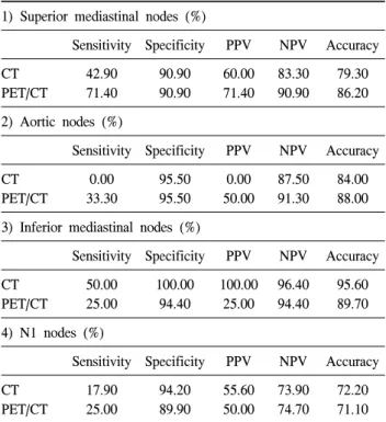
Table 2. Diagnostic performances of CT and PET/CT according to the each nodal groups
1) Superior mediastinal nodes (%)
Sensitivity Specificity PPV NPV Accuracy CT
PET/CT
42.90 71.40
90.90 90.90
60.00 71.40
83.30 90.90
79.30 86.20 2) Aortic nodes (%)
Sensitivity Specificity PPV NPV Accuracy CT
PET/CT
0.00 33.30
95.50 95.50
0.00 50.00
87.50 91.30
84.00 88.00 3) Inferior mediastinal nodes (%)
Sensitivity Specificity PPV NPV Accuracy CT
PET/CT
50.00 25.00
100.00 94.40
100.00 25.00
96.40 94.40
95.60 89.70 4) N1 nodes (%)
Sensitivity Specificity PPV NPV Accuracy CT
PET/CT
17.90 25.00
94.20 89.90
55.60 50.00
73.90 74.70
72.20 71.10 PPV=Positive predictive value; NPV=Negative predictive value;
CT=Computed tomography; PET=Positron emission tomography.
병기와 술후 병기에 변화가 있었고, 수술전의 병기보다 술후의 병기가 더 상향된 경우가 10예로 20.83%였고, 병 기가 하향 조정된 경우가 8예로 16.67%이었다.
수술 중 생검한 림프절은 총 917개이었고, nodal station 수로는 209개이었다. 본 연구에서는 표본수의 제한으로 ATS의 분류에 따라 각 림프절을 superior mediastinal no- des, aortic nodes, inferior mediastinal nodes, 그리고 N1 no- des의 4군으로 분류하여 CT와 PET/CT의 진단력에 대한 통계분석을 실시하였다. Superior mediastinal nodes에는 highest mediastinal, upper paratracheal, prevascular and retro- tracheal, 그리고 lower paratracheal including azygos nodes가 포함되고, aortic nodes에는 subaortic (AP window), para- aortic (ascending aorta 혹은 phrenic)가 들어간다. 또한 in- ferior mediastinal nodes에는 subcarinal, paraesophageal, pul- monary ligament nodes가, N1 nodes에는 hilar, interlobar, lo- bar, segmental, 그리고 subsegmental nodes가 포함된다.
4개의 군 가운데, inferior mediastinal nodes를 제외한 나 머지 군들은 CT보다 PET/CT에서 민감도가 향상되는 소 견을 보였다. 하지만 두 검사의 진단력 차이를 각각의 군 에 대해 McNemar’s test를 사용하여 분석한 결과 통계적으
로 유의한 차이는 보이지 않았다(p-values; superior media- stinal nodes=0.109, aortic nodes=1.000, inferior mediastinal nodes=0.625, N1 nodes=0.424) (Table 2).
고 찰
폐암의 비침습적 진단에는 단순방사선촬영, 전산화단층 촬영(CT), 자기공명영상(MRI)과 같은 영상의학검사들과 PET와 같은 핵의학 검사가 이용되고 있다.
그 중 병기결정에 아직까지 가장 중요한 역할을 하고 있는 필수 검사는 바로 CT로, 림프절의 전이여부를 결정 하는 데에는 그 크기를 기준으로 하게 된다. 대개는 림프 절의 크기가 단경이 10 mm 이상이면 악성 전이를 진단한 다[3]. 하지만 그 동안 이루어진 여러 연구들에 의하면, 정 상 크기의 림프절이라도 악성 전이가 되어 있는 경우가 상당수 있어서 림프절 병기 결정에 있어서 CT의 효용에 대해서는 부정적인 견해가 많다[4-6].
Arita 등은 40명의 폐암 수술 대상환자들을 대상으로 한 전향적 연구에서 CT만으로는 N stage를 진단하는 것은 불 가능하다고 결론지었다[7]. 같은 맥락으로, Erdogan 등은 술전 진단적 종격동경검사를 모든 폐암 환자에서 시행해 야 한다고 주장했다[8]. 또한 Robert 등은 50명의 폐암 진 단 환자들을 대상으로 한 연구에서, CT 보다는 흉강경을 이용한 병기 진단이 더욱 정확하여 CT만으로 수술을 진 행하기 보다는 흉강경 검사를 먼저 시행하면 불필요한 수 술을 막을 수 있을 것이라고 결론지었다[9].
림프절 전이 진단에 대한 회의적 시각은 MRI에 대해서 도 비슷하여서, Webb 등이 170명의 폐암 환자에 대해서 시행한 전향적 연구에서 MRI와 CT의 종격동 림프절 전이 여부 진단에 있어서는 차이가 없다고 발표하였다[6]. Bo- nomo 등이 발표한 연구에서도 역시 같은 내용으로 결론 지어졌다[1]. 이는 CT나 MRI나 모두 해부학적인 것을 보 는 검사 방법으로, 크기 이외에는 악성 여부를 판단할 수 있는 지표가 없는 것에 기인한다.
PET는 그와는 대조적으로, 조직의 구조 보다는 기능을
평가하는 검사로, 암조직에서는 일반조직에서 보다 활발
한 대사가 일어난다는 점을 이용한다. 현재 가장 많이 이
용되는 추적자는 포도당 유도체인 F-18-2-deoxy-2-fluoro-
D-glucose로, 이를 정주하면 악성 종양세포뿐만 아니라, 대
사작용이 활발한 인체 조직에 많이 섭취된다[2]. 많은 연
구들에서 종격동 림프절 평가에 있어서 PET가 CT보다는
정확도가 높음을 보고하고 있다[10-13]. 하지만 PET는 공
간해상력이 좋지 않다는 점 때문에 PET 단독으로는 정확 한 병기 결정이 불가능하다[9]. 따라서 CT와 PET를 모두 시행하여 두 검사를 비교하면, 각 검사를 단독으로 실시 했을 때 보다 더욱 상세하고 정확한 정보를 얻을 수 있다 [14-16]. 그렇지만 두 검사를 단독으로 각각 시행한 경우, 환자의 상태의 변화, 촬영 당시의 조건 등으로 인해 완벽 한 합성을 하는 것은 사실상 불가능하다. 그래서 최근 개 발된 기기가 바로 integrated PET/CT이다. 이 기기는 CT와 PET를 거의 동시에 촬영하여 컴퓨터로 두 검사의 합성 영 상을 얻을 수 있는 것으로, PET 영상에 있는 병소를 국소 화할 수 있으며, CT영상에 보이는 병소의 활성도를 평가 할 수 있게 해준다[17].
Shim 등이 106명의 환자들을 대상으로 연구한 PET/ CT 의 민감도, 특이도, 정확도는 각각 85%, 84%, 84%로, 이는 CT 단독으로만 병기 진단을 했을 때의 수치보다 유의하 게 높은 것들이었다[18]. PET/CT는 T 병기에서 가장 좋은 성적을 보이며, N과 M 병기에서도 역시 훌륭한 결과를 나타낸다[19,20]. 하지만, PET이 가지고 있는 고유의 위양 성, 위음성 문제는 PET/CT에서도 여전히 문제가 될 수 밖 에 없다.
본 연구에서는 기존의 진단법인 CT와 PET/CT를 비교하 였으나, 통계적으로 유의한 이득을 발견하지는 못하였다.
이는 수술을 시행한 환자들만을 연구의 대상으로 하여 병 기별 환자 수를 충분히 확보할 수 없었던 것에 기인한 것 으로 사료된다. 즉, 종격동 림프절에 전이가 되어있지 않 은 기수(stage)가 상대적으로 많은 비율을 차지하여 두 진 단법간의 차이를 규명하기에는 어려움이 있었다.
하지만 잠정적으로 수술이 가능한 폐암 환자들에 있어 서 PET/CT의 효용은 종격동 림프절의 전이 여부의 판단 뿐만 아니라, 원격전이의 발견에도 있다[20]. 즉, 흉부 단 순 방사선 촬영에서 폐암이 의심될 경우, 흉부 CT를 시행 하여 병기를 결정하고, 몸의 다른 부분에 원격전이가 있 는지 확인하는 의미로 PET/CT를 시행하는 것이다. 단순 PET만을 사용할 경우, 병변의 유무는 추정할 수는 있어 도, 정확한 병변의 위치를 지정하기에는 무리가 있지만, PET/CT의 경우에는 한 번의 촬영으로 병변의 국소화까지 비교적 정확하게 할 수 있어 많은 도움을 준다.
결 론
본 연구에서 종격동 림프절에 대한 PET/CT의 진단력은 조영증강 CT와 비슷한 정도로 나타났다. 현재 PET/CT는
CT와 상호보완적인 개념으로써 사용되어야 할 것으로 사 료되며, 앞으로 촬영 기술 및 판독 수준이 더욱 발전하고 결과들이 많이 축적되었을 때에는 보다 좋은 성적을 보일 것으로 생각된다.
참 고 문 헌
1. Bonomo L, Ciccotosto C, Guidotti A, Storto ML. Lung can-
cer staging: the role of computed tomography and magnetic resonance imaging. Eur J Radiol 1996;23:35-45.
2. Kubota K. From tumor biology to clinical PET: a review of
positron emission tomography (PET) in oncology. Ann Nucl
Med 2001;15:471-86.3. Glazer GM, Orringer MB, Gross BH, et al. The mediastinum
in non-small cell lung cancer: CT-surgical correlation. Am J
Radiol 1984;142:1101-5.4. Beadsmoore CJ, Screaton NJ. Classification, staging and
prognosis of lung cancer. Eur J Radiol 2003;45:8-17.
5. McLoud TC, Bourgouin PM, Greenberg RW, et al. Bron-
chogenic carcinoma: analysis of staging in the mediastinum with CT by correlative lymph node mapping and sampling.
Radiology 1992;182:319-23.
6. Webb WR, Gatsonis C, Zerhouni EA, et al. CT and MR
imaging in staging non-small cell bronchogenic carcinoma:
report of the Radiologic Diagnostic Oncology Group. Radio-
logy 1991;178:705-13.7. Arita T, Matsumoto T, Kuramitsu T, et al. Is it possible to
differentiate malignant mediastinal nodes from benign nodes by size? Chest 1996;110:1004-8.
8. Cetinkaya E, Turna A, Yildiz P, et al. Comparison of
clinical and surgical-pathologic staging of the patients with non-small cell lung carcinoma. Eur J Cardiothorac Surg
2002;22:1000-5.9. Roberts JR, Blum MG, Arildsen R, et al. Prospective com-
parison of radiologic, thoracoscopic, and pathologic staging in patients with early non-small cell lung cancer. Ann Tho-
rac Surg 1999;68:1154-8.10. Steinert HC, Hauser M, Alleman F, et al. Non-small cell
lung cancer: nodal staging with FDG PET vs CT with correlative lymph node mapping and sampling. Radiology
1997;202:441-6.11. Vansteenkiste JF, Stroobants SG, De Leyn PR, et al.
Mediastinal lymph node staging with FDG PET scan in patients with potentially operable non-small cell lung cancer;
a prospective analysis of 50 cases. Leuven Lung Cancer
Group. Chest 1997;112:1480-6.12. Guhlmann A, Storck M, Kotzerke J, Moog F, Sunder- Plassmann L, Reske SN. Lymph node staging in non-small
cell lung cancer: evaluation by [18F]FDG positron emission
tomography (PET). Thorax 1997;52:438-41.
13. Pieterman RM, van Putten JW, Meuzelaar JJ. Preoperative
staging of non-small cell lung carcinoma with positron emi- ssion tomography. N Engl J Med 2000;343:290-2.
14. Kim OG, Choh JH, Sung SH. Efficacy of positron emission
tomography in diagnosing pulmonary tumor and staging of lung cancer: comparing to computed tomography. Korean J
Thorac Cardiovasc Surg 2003;36:79-85.15. Vansteenkiste JF, Stroobants SG, De Leyn PR, et al. Lymph
node staging in non-small cell lung cancer with FDG-PET scan: a prospective study on 690 lymph node stations from 68 pations. J Clin Oncol 1998;16:2142-9.
16. Verschakelen JA, Bogaert J, De Wever W. Computed tomog-
raphy in staging for lung cancer. Eur Respir J 2002;19(suppl
35):40s-8s.17. Beyer T, Townsend DW, Brun T, et al. A combined PET/CT
scanner for clinical oncology. J Nucl Med 2000;41:1369-79.
18. Shim SS, Lee KS, Kim BT, et al. Non-small cell lung
cancer: prospective comparison of integrated FDG PET/CT and CT alone for preoperative staging. Radiology 2005;236:
1011-9.
19. Lardinois D, Weder W, Hany TF, et al. Staging of non-
small-cell lung cancer with integrated positron-emission to- mography and computed tomography. N Engl J Med 2003;
348:2500-7.
20. Devaraj A, Cook GJR, Hansell DM. PET/CT in non-small