Euptelea pleiosperma 에탄올 추출물의 항산화 및 항염증 활성
진경숙1, 박정애1, 이지영1, 강지숙1, 권현주1,2, 김병우1,2*
1동의대학교블루바이오소재개발및실용화지원센터
2동의대학교생명응용학과
Received: March 7, 2014 / Revised: April 7, 2014 / Accepted: April 7, 2014
서 론
생명체의대사과정에서끊임없이발생하는활성산소종 (Reactive Oxygen Species, ROS)은산화적스트레스의유발 을통해세포조직에치명적인손상을유발하며암, 뇌질환, 심장질환, 동맥경화, 당뇨병, 천식등생체내에서발생하는 수많은질환의원인이될뿐아니라, 노화를일으키는직·
간접적인원인물질로작용하는것으로알려져있다[3, 7, 12,
24, 25]. 또한염증은외부자극에대한생체조직의방어기
전의하나로지속적인염증반응은조직의손상을일으켜암
을비롯한각종질병을유발하는것으로알려져있다[5, 8].
이에따라최근많은연구들이항산화및항염증활성을바 탕으로한생리활성보유신소재개발및그활성기전의규 명에주력하고있으며특히천연유래소재로부터유용성분 을추출하고생리활성을규명하여기능성소재로서의가능 성을타진하는연구가활발히이루어지고있다[16, 17, 26].
대표적인 cellular defensive phase 2 detoxifying antioxidant enzyme으로 알려진 heme oxygenase (HO)-1, NAD(P)H dehydrogenase 1 (NQO1), thioredoxin reductase 1 (TrxR1)의 유도는산화적스트레스를방어하는중요한기전중하나로 다양한 carcinogen으로부터세포를보호하는 chemoprevention
에중요한역할을담당하는것으로알려져있다[6, 33]. 특히
천연에서유래한다양한 dietary phytochemical은 nuclear factor E2-related factor 2 (Nrf2)에의해조절되는 phase 2 detoxifying antioxidant enzyme의 발현 증가를 통해 chemopreventive function을나타내며 extracellular signal- regulated kinase (ERK), c-jun N-terminal kinase (JNK), and Anti-Oxidative and Anti-Inflammatory Activities of Euptelea Pleiosperma Ethanol Extract
Kyong-Suk Jin1, Jung Ae Park1, Ji Young Lee1, Ji Sook Kang1, Hyun Ju Kwon1,2, and Byung Woo Kim1,2*
1Blue-Bio Industry Regional Innovation Center, 2Department of Life Science and Biotechnology, College of Natural Science, Dong-Eui University, Busan 614-714, Republic of Korea
In this study, the anti-oxidative and anti-inflammatory activities of Euptelea pleiosperma ethanol extract (EPEE) were evaluated using in vitro assays and cell culture model systems. EPEE possessed a more potent scavenging activity against 1,1-diphenyl- 2-picryl hydrazyl than the ascorbic acid used as a positive control. EPEE effectively suppressed lipopolysaccharide (LPS), in addition to hydrogen peroxide induced reactive oxygen species on RAW 264.7 cells. Furthermore, EPEE induced the expression of the anti-oxidative enzyme heme oxygenase 1 (HO-1) and its upstream transcription factor, nuclear factor-E2- related factor 2 (Nrf2), dose and time dependently. The modulation of HO-1 and Nrf2 expression might be regulated by mitogen-activated protein kinases and phosphatidyl inositol 3 kinase/Akt as their upstream signaling pathways. On the other hand, EPEE inhibited LPS induced nitric oxide (NO) formation without cytotoxicity. Suppression of NO formation was the result of the down regulation of inducible NO synthase (iNOS) by EPEE. Suppression of NO and iNOS by EPEE may be modulated by their upstream transcription factor, nuclear factor κB, and AP-1 pathways. Taken together, these results provide important new insights into E. pleiosperma, namely that it possesses anti-oxidative and anti-inflammatory activities, indicating that it could be utilized as a promising material in the field of nutraceuticals.
Keywords: Euptelea pleiosperma, anti-oxidative activity, anti-inflammatory activity, upstream transcription factor
*Corresponding author
Tel: +82-51-890-2900, Fax: +82-51-890-2914 E-mail: [email protected]
© 2014, The Korean Society for Microbiology and Biotechnology
p38과 같은 mitogen-activated protein kinases (MAPKs)와 phosphatidylinositol 3-kinase (PI3K)/Akt와같은상위신호 전달기전의 영향을 받는다[13, 18, 20, 28]. 이러한 chemo-
prevention은항산화활성을기초로하여함암뿐만아니라
염증, 뇌및심혈관계질환, 노화등의예방및치료기전과 도상호작용하는것으로알려져있어그중요성이더욱커 지고있다[4, 14, 27, 31].
생체내염증반응은대식세포(macrophage)에서과량생
산되는염증매개인자(inflammatory mediators)로부터유 래되는데 inducible nitric oxide synthase (iNOS)로 부터 생산되는 nitric oxide (NO)와 cyclooxygenase 2 (COX-2)로 부터생산되는 prostaglandin E2 (PGE2) 등이대표적이다. 외부 자극에 의해 과량 생산된 염증 매개인자는 tumor necrosis factor α (TNF-α), interleukin 1β (IL-1β) 등과같 은사이토카인을생산하여다양한염증반응을일으킨다[14,
15]. 염증 반응의 대표적인 세포 실험계 중 하나인 RAW
264.7 murine macrophage에 lipopolysaccharide (LPS) 등 의염증유발인자를처리하면 iNOS 및 COX-2의발현유도
에의해 NO와 PGE2 등염증매개인자의생성및이를통
한사이토카인분비량증가를확인할수있으며이러한일 련의 반응은 대표적인 염증 상위신호전달기전인 nuclear factor (NF)-κB와 activator protein (AP)-1에의해조절되는 것으로알려져있다[20, 22, 28]. 그러므로이러한염증매개인 자와그상위신호전달기전을효과적으로제어할수있는물질 들이염증의예방및치료를위한소재로서각광받고있다.
Euptelea pleiosperma는 Eupteleaceae과에속하는낙엽수 이며영춘목으로도불린다. 동아시아지역, 주로중국및히 말라야일대에분포한다. 잎은식용가능하며, 꽃과가지의 껍질은약용으로알려져있으나그구체적인효능에대해서 는알려진바가없으며, 특히항산화및항염증효과에대해 서는전혀알려진바없다. 이에본연구에서는천연에서유
래한생리활성보유신소재개발의일환으로E. pleiosperma
95% 에탄올추출물(EPEE)이보유한항산화및항염증활
성을 in vitro assay system 및 cell culture model system 을이용하여분석함으로써기능성소재로서의활용가능성 을확인해보고자하였다.
재료 및 방법
E. pleiosperma 추출물의 제조
본연구에서사용한E. pleiosperma 95% 에탄올추출물
(EPEE)은한국생명공학연구원, 해외생물소재허브센터에서
구입(분양번호 FBM123-004)하여사용하였다. 건조및분쇄 한시료를 95% 에탄올을용매로하여 45oC에서 3일간초음 파추출을수행하였다. 추출이끝난시료를 filter로여과하
여고형물을없애고감압농축(N-1000SW, EYELA, Japan) 및동결건조(FDU2100, EYELA, Japan)하여사용전까지 4oC에보관하였다.
DPPH radical 소거 활성 측정을 통한 EPEE의 항산화능 분석
항산화작용의주요기전중하나인전자공여능은인체내 에서 생성되는 free radical의 전자를 공여함으로써 free
radical에의한노화와질병을억제한다. 항산화작용의주
요지표로서의전자공여능은특히천연물이보유한항산화 능의측정에많이사용되고있으며[7], 1,1-diphenyl-2-picryl hydrazyl (DPPH) radical 소거능분석을이용하여측정하였 다. DPPH는비교적안정한 free radical로써, ascorbic acid, tocopherol, polyhydroxy 방향족화합물, 방향족아민류에의 해환원되어짙은자색이탈색되는원리를이용하여항산화 활성을간단히측정할수있는동시에식물체의항산화활 성과도연관성이매우높기때문에많이이용되고있는방 법이다[11].
DPPH radical scavenging activity 측정을위해 EPEE를 농도별(0.1024-12.8 μg/ml)로 메탄올에 녹여 준비하고 96 well plate에메탄올에용해된 1.5×10⁻⁴ M DPPH 40 μl와 각시료 160 μl를분주한혼합액을실온에서 30분간반응시 킨 후, multi-plate reader (Paradigm, Beckman, CA, USA)를이용하여 520 nm에서흡광도를측정하였다. 시료 를첨가하지않은음성대조군과비교하여 free radical 소거
정도를 백분율로 나타내고, 50% 저해 농도(Inhibitory
Concentration, IC50)를계산하였다. 대표적인항산화제로
DPPH radical 소거활성측정시양성대조군으로주로사
용되는 ascorbic acid를 함께 비교 분석하였다. DPPH radical 소거능의백분율공식은다음과같다.
DPPH radical scavenging activity (%) = {1−(A−B)/C}×100
A: sample absorbance at 520 nm B: color control absorbance at 520 nm C: control absorbance at 520 nm
RAW 264.7 murine macrophage의 배양
항산화 및 항염증 활성의 세포 실험 모델계로 murine
macrophage cell line인 RAW 264.7을 American Type Tissue Collection (ATCC®, TIB-71TM, Manassas, VA, USA)로부터 구입하여 10% fetal bovine serum (FBS) 및 penicillin/streptomycin (Pen/Strep)이포함된 DMEM 배지 에서배양하였다[23].
EPEE의 세포생존율 분석
활성분석수행전시료가세포생존율에미치는영향을확 인함과동시에세포독성을유발하지않는시료의처리농
도를결정하기위해 EPEE에의한세포독성유발유무를
WST assay를 통해 수행하였다. 1.0×10⁵ cell을 24-well tissue culture plate에분주하여 24시간동안 부착시키고, EPEE 처리 24시간후 WST 시약이든배지로교체하여한 시간 동안 반응시킨 다음 multi-plate reader를 이용하여
450 nm에서흡광도를측정하였다. 이후독성을유발하지
않는농도범위에서실험을수행하였다.
EPEE의 reactive oxygen species (ROS) scavenging activity 분석
ROS는과량생산시 DNA, protein, lipid를포함한생체 내분자에산화적인변형을유발하여다양한질병의원인이
되므로 ROS 소거능은항산화능의중요한지표로활용된다
[19]. Hydrogen peroxide (H2O2)는대표적인 ROS 중하나 로소재의항산화능의규명하기위한많은연구에서 ROS 유 도제로사용되고있다[29, 30, 32]. 또한그람음성세균의세 포외벽을구성하는주요인자인 lipopolysaccharide (LPS) 는대표적인염증유발인자로산화적스트레스또한유발 하는것으로알려져있어항산화능및항염증활성을규명
하기위한많은연구에사용되고있다[1, 21]. 본연구에서는
EPEE가보유한항산화능을 H2O2및 LPS로유도한 ROS 생 성에시료가미치는영향을통해분석하였다. 이를위해 RAW 264.7 cell에 cell permeable fluorescent dye인 50 μM의 dichlorofluorescin diacetate (DCFH-DA)를 2시간동안 전 처리한후제거하고 500 μM의 H2O2혹은 1 μg/ml의 LPS를
농도별시료와함께처리한후시료에의한 ROS 생성억제
능을 multiplate reader를이용한 fluorescence 측정을통해 분석하였다.
항산화 효소 HO-1, TrxR1, NQO1 및 그 전사인자인 Nrf2 의 발현 조절능 분석
EPEE의항산화활성기전을알아보기위해대표적인항
산화효소인 HO-1, TrxR1, NQO1과그전사인자인 Nrf2의
시료 처리에 의한 단백질 발현 변화를 Western blot
hybridization으로 분석하였다. HO-1과 p-p38, p-JNK, p- ERK, 그리고 p-Akt의일차항체는 Cell Signaling Technology
(MA, USA)로부터 구입하였고, p-Nrf2의 일차항체는
Calbiochem (CA, USA)로부터구입하였으며, TrxR1, NQO1, Nrf2, Actin의 일차항체와 anti-goat, anti-rabbit, 그리고 anti-mouse 등의이차항체는 Santa Cruz Biotechnology (CA, USA)에서구입하여사용하였다. 시료처리가끝난배양세 포에서단백질을추출하여 bradford assay로단백질농도를
결정한후 50 μg의단백질을 10% sodium dodecyl sulfate- polyacrylamide gel electrophoresis (SDS-PAGE)로전기영 동하고 nitrocellulose membrane에 blotting한후 1:1,000- 5,000으로희석한대상단백질의일차항체와 hybridization 하였다. Membrane washing 후 horse radish peroxidase (HRP)가부착된이차항체(1:1,000)로한시간동안반응시 킨 후 chemiluminescence detection system (FluoChem® FC2, AlphaInnotech, USA)을이용하여단백질발현을분석 하였다.
EPEE의 NO 생성 억제능 분석
대표적인 free radical 중하나인 NO는생체내에서중요 한세포신호전달물질로서작용하나과잉생산시산화적스
트레스의유발을통해염증및세포손상의원인이된다[10].
이러한 NO 생성억제능의분석은 Park 등[23]의방법을변 형하여 수행하였다. RAW 264.7 cell을 24-well tissue culture plate에 well 당 1.0×10⁵ cell을 seeding하여부착 시킨후 1μg/ml의 LPS를처리하여 NO 생성을유도하고식 물추출물에의한 NO 생성저해능을 Griess reaction을통 해분석하였다.
EPEE의 항염증 활성 기전 분석
EPEE가보유한 NO 생성억제능의기전을밝히기위해
NO 생성의핵심단백질인 iNOS의단백질발현을분석하였
다. 또한 EPEE에의한 NO 생성및 iNOS의발현저해능이 NF-κB 및 AP-1에의해조절될가능성을알아보기위해 LPS 로 유도된 NF-κB p65와 inhibitory κBα (IκBα), 그리고 AP-1의 subunit인 c-Jun의인산화에 EPEE가미치는영향 을분석하였다. Western blot hybridization을위한 iNOS, p-p65, p-IκBα, 그리고 p-c-Jun의일차항체는 Cell Signaling Technology (MA, USA)로부터구입하여사용하였다. 시료
처리가 끝난 배양 세포에서 단백질을 추출하여 bradford
assay로단백질 농도를결정한후 50 μg의단백질을 10%
SDS-PAGE로 전기영동하고 nitrocellulose membrane에 blotting한후대상단백질의항체와 hybridization하였다. Membrane washing 후 HRP가 tagging된이차항체로한시 간동안반응시킨후 chemiluminescence detection system 을이용하여단백질발현을분석하였다.
데이터 처리 및 통계 분석
모든실험결과는최소 3회이상의반복실험을통하여얻 은 데이터를 평균(mean) ± 표준편차(standard deviation, SD)로나타내었고, 필요시대표적인그림이나데이터를제 시하였다. 각데이터의통계분석은 SPSS 20.0 software를 이용한 unpaired Student’s t-test를통해p 값이 0.05 미만
(p < 0.05)인경우유의성이있는것으로판단하였다.
결과 및 고찰
EPEE의 항산화능 분석
EPEE의항산화능보유유무및그정도를알아보기위해
먼저 항산화능의 주요 지표 중 하나인 DPPH radical scavenging activity를분석하였다. 그결과 Table 1에제시 된바와같이 EPEE의농도증가에따라강한 radical 소거 능을 보여 0.1024, 0.512, 2.56, 12.8 μg/ml의 시료처리에 의해 DPPH radical 소거능이 각각 26.11, 40.49, 93.83, 98.00%로 나타나 50% 소거 농도를 나타내는 IC50 값이 0.88μg/ml로 양성 대조군으로 사용한 ascrobic acid, 즉 vitamin C의 IC50값인 1.53 μg/ml 보다높은활성을보여 매우강한항산화능을보유함을확인하였다. 이에 EPEE가 보유한항산화능의정도및기전을세포수준에서확인할필 요성이제기되었다.
EPEE의 ROS scavenging activity 분석
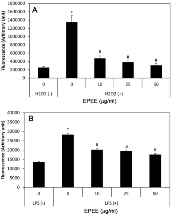
DPPH radical scavenging activity 분석을통해 EPEE가 보유한높은항산화능이확인됨에따라그작용기전을좀 더자세히알아보기위해먼저 RAW 264.7 cell에대표적인 산화적스트레스유도인자인 H2O2와 LPS를각각처리하여 EPEE에의한 ROS scavenging activity를분석하였다. 그 결과 Fig. 1에제시한바와같이 H2O2와 LPS에의해각각유
도된 ROS 생성이 EPEE의처리에의해효과적으로저해되
는것으로나타나 EPEE가 DPPH radical 뿐만아니라세포 실험계에서 H2O2와 LPS에의해유도된산화적스트레스또 한효과적으로감소시킴을확인하였다.
EPEE가 항산화 효소 HO-1, TrxR1, NQO1 및 상위 전사 인자 Nrf2의 발현에 미치는 영향
강한항산화능을보유한천연유래소재들이 Nrf2에의한 항산화효소계의발현유도를통해활성을나타낸다는것이
Table 1. DPPH radical scavenging activity of EPEE.
Reagent Concentration (μg/ml)
Inhibition rate (%)
EPEE 0.1024
0.512
26.11 ± 0.89 40.49 ± 0.93
2.56 93.83 ± 0.40
12.8 98.00 ± 0.61
Ascorbic acid (Positive control)
0.512 31.90 ± 0.02
2.56 12.8
96.47 ± 0.13 98.41 ± 0.17
Fig. 1. Effect of EPEE on H2O2- (A) and LPS- (B) induced ROS scavenging activity in RAW 264.7 cells. Values are represented as the mean ± SD (n = 6). *, #Significantly different from the vehi- cle control [0, H2O2 or LPS (−)] and H2O2- or LPS- induced control [0, H2O2 or LPS (+)], respectively (p < 0.05).
Fig. 2. Modulation of anti-oxidative enzymes, HO-1, NQO1, TrxR1, and their upstream transcription factor, Nrf2 protein expression and phosphorylation in RAW 264.7 cells by EPEE.
(A) Dose dependency and (B) time course study. Actin was used as an internal control.
여러연구를통해밝혀짐에따라 EPEE가보유한항산화능 의작용기작을알아보고자하였다[9, 27]. 이를위해대표적 인항산화효소들로천연물에의한항산화활성에의해주 로발현이유도되는세효소인 HO-1, TrxR1, NQO1의단백 질발현과그상위전사인자인 Nrf2의단백질발현및인산 화에 EPEE가미치는영향을분석하였다. 그결과 Fig. 2A 에제시된바와같이 6시간동안 10-50 μg/ml의시료처리 에의해세효소중 HO-1의발현이강한증가를보였다. 뿐 만아니라 HO-1의상위전사인자인 Nrf2의단백질발현과
인산화또한증가되는것으로나타나 EPEE에의한 HO-1의
발현증가가 Nrf2의발현증가및인산화에의해나타날가
능성을보였다. 이러한결과는시간의변화에따른발현형 태를관찰한 Fig. 2B에서도유사한양상을보여 50 μg/ml의
EPEE를시간별로처리한결과세효소중 HO-1의발현이
증가하는것으로나타났으며이는 Nrf2의발현증가와인산 화에서기인할것으로판단되었다[2, 6].
EPEE가 상위신호전달인자인 MAPKs와 PI3K/Akt의 인산 화에 미치는 영향
EPEE가보유한항산화능의상위신호전달기전을알아보 기위해 p38, JNK, 그리고 ERK 등의 MAPKs와 Akt의인 산화에 EPEE가미치는영향을분석한결과 Fig. 3에제시 된바와같이 6시간동안 10-50 μg/ml의시료처리에의해 4종모두인산화가증가되는것으로나타났으며그정도및
경향은다소차이를보여 p-38과 JNK는농도의존적인인산
화의증가를보였으나 ERK와 Akt는처리농도에상관없이 유사한정도의인산화를나타내었다. 이러한결과를통해 EPEE의항산화능이 HO-1과그상위전사인자인 Nrf2의발
현증가를통해나타나며그러한일련의과정이 MAPKs와
PI3K/Akt와같은상위신호전달인자에의해나타날가능성
을확인하였다.
EPEE의 NO 생성 저해능 분석
EPEE가항산화능뿐만아니라항염증활성또한보유하
는지를알아보기위해먼저 NO 생성에미치는효과를알아
보았다. LPS로자극을 유도한쥐대식세포주 RAW 264.7
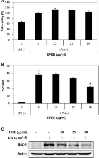
cell에서농도별 EPEE의처리에따른 NO 생성양의변화를 분석한결과 Fig. 4에제시된바와같이 10-50 μg/ml의시료 처리에의해세포독성의유발없이농도의존적인 NO 생성
저해능을보였으며이는 NO 생성단백질인 iNOS의발현저
해에서기인하는것으로나타났다. 이러한결과를통해 EPEE
가산화적스트레스뿐만아니라염증성자극에대한방어능 또한보유함을확인하였다.
EPEE가 NF-κB 및 AP-1의 인산화에 미치는 영향 분석 EPEE가 iNOS의발현저해에따른 NO 생성억제능을보
Fig. 3. Effect of EPEE on the phosphorylation of MAPKs and PI3K/Akt signaling molecules in RAW 264.7 cells. Actin was used as an internal control.
Fig. 4. Effect of EPEE on cell viability (A), LPS-induced NO formation (B), and iNOS protein expression (C) in RAW 264.7 cells. (A, B) Values are represented as the mean ± SD (n = 3). *,
#Significantly different from the vehicle control [0, LPS (−)] and LPS-induced control [0, LPS (+)], respectively (p < 0.05). (C) Actin was used as an internal control.
유함을확인함에따라항염증활성의상위신호전달기전인 NF-κB와 AP-1의연관성을알아보기위해 EPEE가 LPS에 의해 유도된 NF-κB p65와 IκBα, 그리고 AP-1의 subunit
중하나인 c-Jun의인산화에미치는영향을분석하였다. 그
결과 Fig. 5에제시된바와같이 2시간동안의 LPS 처리에 의해유도된세전사인자의인산화가 EPEE 농도의증가에 따라유의적으로억제되는것으로나타났다. 이러한결과는 EPEE의항염증활성이항염증상위신호전달인자들의일련 의조절기작을통해이루어질가능성을시사하였다.
이상의결과를통해 EPEE가높은항산화능과항염증활
성을보유함을처음으로확인하였으며, 이러한결과는신규 소재에대한새로운기능성데이터를구축함과동시에향후 생리활성보유기능성소재로서의활용을위한근거자료로 이용될수있을것으로판단된다.
요 약
본 연구에서는 Euptelea pleiosperma 에탄올 추출물 (EPEE)의 항산화능과 항염증 생리활성을 in vitro assay system 및 cell culture model system을이용하여분석하였 다. 먼저 EPEE의 항산화능을 DPPH radical scavenging activity로분석한결과 radical 소거능의정도가양성대조 군으로사용한 ascorbic acid 보다높은활성을보여매우강 한항산화능을보유함을확인하였다. 또한 RAW 264.7 세포 주를이용한 H2O2 및 LPS의유도에의해생성된 ROS에대 한소거능을분석한결과에서도농도의존적인강한소거능 을보였다. 뿐만아니라대표적인항산화효소중하나로천 연물에의한항산화능활성에의해 주로발현이유도되는 hemeoxygenase 1 (HO-1) 및 그 전사 인자인 nuclear factor-E2-related factor 2 (Nrf2)의단백질발현이 EPEE의 처리에의해유의적으로증가됨을보였으며이러한 HO-1 및 Nrf2의발현변화는상위신호전달계인 MAPKs 및 PI3K/Akt 에의해조절될가능성을보였다. 한편 EPEE가 LPS에의
해유도된 NO 생성에미치는영향을분석한결과농도의존 적인 NO 생성저해능을보였으며이는 NO 생성단백질인 iNOS의 발현 저해에서 기인함을 확인하였다. 이와 같은 EPEE의 NO 생성억제효과는염증상위신호전달계인 NF- κB 및 AP-1의조절을통해일어날가능성을보였다. 이러한
결과를통해 EPEE의높은항산화능과항염증활성을처음
으로확인하였으며향후기능성소재로서유용하게활용될 수있을것으로판단된다.
Acknowledgments
This work was supported by Blue-Bio Industry Regional Innova- tion Center (RIC08-06-07) at Dong-Eui University as a RIC pro- gram under Ministry of Trade, Industry and Energy (MOTIE) and Busan city.
References
1. Awad N, Khatib N, Ginsberg Y, Weiner Z, Maravi N, Thaler I, et al. 2011. N-acetyl-cysteine (NAC) attenuates LPS-induced maternal and amniotic fluid oxidative stress and inflammatory responses in the preterm gestation. Am. J. Obstet. Gynecol.
450: e15-20.
2. Bryan HK, Olayanju A, Goldring CE, Park BK. 2013. The Nrf2 cell defence pathway: Keap1-dependent and -independent mechanisms of regulation. Biochem. Pharmacol. 85: 705-717.
3. Cencioni C, Spallotta F, Martelli F, Valente S, Mai A, Zeiher AM, et al. 2013. Oxidative stress and epigenetic regulation in ageing and age-related diseases. Int. J. Mol. Sci. 14: 17643- 17663.
4. Chapple SJ, Siow RC, Mann GE. 2012. Crosstalk between Nrf2 and the proteasome: therapeutic potential of Nrf2 induc- ers in vascular disease and aging. Int. J. Biochem. Cell Biol.
44: 1315-1320.
5. Chawla A, Nguyen KD, Goh YP. 2011. Macrophage-mediated inflammation in metabolic disease. Nat. Rev. Immunol. 11:
738-749.
6. Giudice A, Arra C, Turco MC. 2010. Review of molecular mechanisms involved in the activation of the Nrf2-ARE signal- ing pathway by chemopreventive agents. Meth. Mol. Biol. 647:
37-74.
7. Gonzalez-Burgos E, Gomez-Serranillos MP. 2012. Terpene compounds in nature: a review of their potential antioxidant activity. Curr. Med. Chem. 19: 5319-5341.
8. Grivennikov SI, Greten FR, Karin M. 2010. Immunity, inflam- mation, and cancer. Cell. 140: 883-899.
9. Hu R, Saw CL, Yu R, Kong AN. 2010. Regulation of NF-E2- related factor 2 signaling for cancer chemoprevention: antioxi- dant coupled with antiinflammatory. Antioxid. Redox. Signal.
13: 1679-1698.
10. Kalyanaraman B. 2013. Teaching the basics of redox biology Fig. 5. Modulation of EPEE on the upstream signaling path-
way for anti-inflammatory activity in RAW 264.7 cells. Actin was used as an internal control.
to medical and graduate students: Oxidants, antioxidants and disease mechanisms. Redox. Biol. 1: 244-257.
11. Kedare SB, Singh RP. 2011. Genesis and development of DPPH method of antioxidant assay. J. Food Sci. Technol. 48:
412-422.
12. Khansari N, Shakiba Y, Mahmoudi M. 2009. Chronic inflam- mation and oxidative stress as a major cause of age-related diseases and cancer. Recent Pat. Inflamm. Allergy Drug Dis- cov. 3: 73-80.
13. Kocanova S, Buytaert E, Matroule JY, Piette J, Golab J, de Witte P, et al. 2007. Induction of heme-oxygenase 1 requires the p38MAPK and PI3K pathways and suppresses apoptotic cell death following hypericin-mediated photodynamic ther- apy. Apoptosis. 12: 731-741.
14. Kundu JK, Surh YJ. 2008. Inflammation: gearing the journey to cancer. Mutat. Res. 659: 15-30.
15. Kundu JK, Surh YJ. 2012. Emerging avenues linking inflam- mation and cancer. Free Radic. Biol. Med. 52: 2013-2037.
16. Lee JC, Hou MF, Huang HW, Chang FR, Yeh CC, Tang JY, et al. 2013. Marine algal natural products with anti-oxidative, anti-inflammatory, and anti-cancer properties. Cancer Cell Int.
13: 55.
17. Li J, Zhang H, Huang W, Qian H, Li Y. 2012. TNF-α inhibitors with anti-oxidative stress activity from natural products. Curr.
Top Med. Chem. 12: 1408-1421.
18. Li L, Dong H, Song E, Xu X, Liu L, Song Y. 2014. Nrf2/ARE pathway activation, HO-1 and NQO1 induction by polychlori- nated biphenyl quinone is associated with reactive oxygen species and PI3K/AKT signaling. Chem. Biol. Interact. 209:
56-67.
19. Liochev SI. 2013. Reactive oxygen species and the free radi- cal theory of aging. Free Radic. Biol. Med. 60: 1-4.
20. Lu Y, Suh SJ, Kwak CH, Kwon KM, Seo CS, Li Y, et al. 2012.
Saucerneol F, a new lignan, inhibits iNOS expression via MAPKs, NF-κB and AP-1 inactivation in LPS-induced RAW264.7 cells. Int. Immunopharmacol. 12: 175-181.
21. Noworyta-Sokolowska K, Gorska A, Golembiowska K. 2013.
LPS-induced oxidative stress and inflammatory reaction in the rat striatum. Pharmacol. Rep. 65: 863-869.
22. Park CM, Jin KS, Lee YW, Song YS. 2011. Luteolin and chicoric acid synergistically inhibited inflammatory responses via inac- tivation of PI3K-Akt pathway and impairment of NF-κB trans- location in LPS stimulated RAW 264.7 cells. Eur. J.
Pharmacol. 660: 454-459.
23. Park CM, Park JY, Noh KH, Shin JH, Song YS. 2011. Taraxa- cum officinale Weber extracts inhibit LPS-induced oxidative stress and nitric oxide production via the NF-κB modulation in RAW 264.7 cells. J. Ethnopharmacol. 133: 834-842.
24. Pillai S, Oresajo C, Hayward J. 2005. Ultraviolet radiation and skin aging: roles of reactive oxygen species, inflammation and protease activation, and strategies for prevention of inflamma- tion-induced matrix degradation - a review. Int. J. Cosmet. Sci.
27: 17-34.
25. Reuter S, Gupta SC, Chaturvedi MM, Aggarwal BB. 2010.
Oxidative stress, inflammation, and cancer: how are they linked? Free Radic. Biol. Med. 49: 1603-1616.
26. Saw CL, Wu Q, Su ZY, Wang H, Yang Y, Xu X, et al. 2013.
Effects of natural phytochemicals in Angelica sinensis (Danggui) on Nrf2-mediated gene expression of phase II drug metaboliz- ing enzymes and anti-inflammation. Biopharm. Drug Dispos.
34: 303-311.
27. Su ZY, Shu L, Khor TO, Lee JH, Fuentes F, Kong AN. 2013. A perspective on dietary phytochemicals and cancer chemopre- vention: oxidative stress, nrf2, and epigenomics. Top Curr.
Chem. 329: 133-162.
28. Tsai HH, Lee WR, Wang PH, Cheng KT, Chen YC, Shen SC.
2013. Propionibacterium acnes-induced iNOS and COX-2 protein expression via ROS-dependent NF-κB and AP-1 acti- vation in macrophages. J. Dermatol. Sci. 69: 122-131.
29. Wang FW, Wang Z, Zhang YM, Du ZX, Zhang XL, Liu Q, et al.
2013. Protective effect of melatonin on bone marrow mesen- chymal stem cells against hydrogen peroxide-induced apop- tosis in vitro. J. Cell Biochem. 114: 2346-2355.
30. Yagi H, Tan J, Tuan RS. 2013. Polyphenols suppress hydro- gen peroxide-induced oxidative stress in human bone-marrow derived mesenchymal stem cells. J. Cell Biochem. 114: 1163- 1173.
31. Zhang M, An C, Gao Y, Leak RK, Chen J, Zhang F. 2013.
Emerging roles of Nrf2 and phase II antioxidant enzymes in neuroprotection. Prog. Neurobiol. 100: 30-47.
32. Zhang R, Kang KA, Piao MJ, Maeng YH, Lee KH, Chang WY, et al. 2009. Cellular protection of morin against the oxidative stress induced by hydrogen peroxide. Chem. Biol. Interact.
177: 21-27.
33. Zhao CR, Gao ZH, Qu XJ. 2010. Nrf2-ARE signaling pathway and natural products for cancer chemoprevention. Cancer Epidemiol. 34: 523-533.