Phylogenetics, Safety and In Vitro Functional Properties of Bacillus Species Isolated from Iru, a Nigerian Fermented Condiment
Gbenga Adedeji Adewumi
1,2, Sunita Grover
2, Chukwuemeka Isanbor
3, and Folarin Anthony Oguntoyinbo
1*
1
Department of Microbiology, Faculty of Science, University of Lagos, Akoka, Lagos 03005, Nigeria
2
Molecular Biology Unit, Dairy Microbiology Division, ICAR-National Dairy Research Institute, Karnal-132001 (Haryana), India
3
Department of Chemistry, Faculty of Science, University of Lagos, Akoka, Lagos 03005, Nigeria
Received: March 8, 2019 / Revised: June 1, 2019 / Accepted: June 3, 2019
Introduction
Parkia biglobosa (Jacq. Benth) cotyledons are naturally fermented to produce iru or daddawa, a traditional alkaline food condiment, consumed by over ca. 150 million human populations in West Africa [1]. Similar alkaline fermented vegetable protein foods in Asia, produced mainly from soybeans [Glycine max (L.)] include natto
[2], thua nao [3], kinema [4] and tungrymbai [5]. Produc- tion process of iru as well as similar products has been previously described [6], it involves enzymatic and bio- chemical changes, enhanced by the environmental microbes, raw materials, fermentation vessel and pro- cessors, resulting into competitive adaptation and activities of autochthonous, spoilage and pathogenic microorganisms.
Bacillus subtilis has been repeatedly reported as the dominant bacterium responsible for the fermentation of leguminous vegetable protein seeds during condiments production in Africa [7 −9]. Recently, we used culture- Bacillus species were isolated from iru, a traditional fermented condiment in Nigeria. Polyphasic approach was used to evaluate the phylogenetic relationship and strain sub-type of the isolated species. Additionally, the phylogenetic profiles of the species isolated from iru were compared with those of bacilli isolated from different continents. The phylogenetic diversity analysis was performed using the combination of 16S rRNA gene sequencing, ITS-PCR, ITS-PCR-RFLP, and M13 RAPD-PCR. The analysis revealed that Bacillus subtilis U170B and B. subtilis U146A isolated from iru were the closest relatives of strains belonging to the phylogeny of B. subtilis sensu stricto and were related to other bacilli isolated from different continents that had functional benefits. The two isolated species exhibited resistance to acidic pH (pH 2.0). The sur- vival rates of B. subtilis U170B, B. subtilis U146A, and B. clausii UBBC-07 (commercial probiotic strain) cul- tured at pH 2.0 for 3 h were 33.45, 12.44, and 9.53%, respectively. The strains were highly tolerant to bile salts [0.3% (w/v)]. B. subtilis U170B exhibited the highest cell viability (43.45%) when cultured for 3 h in the presence of bile salts, followed by B. subtilis U146A (25%) and B. clausii UBBC-07 (18.94%). B. subtilis U170B and B. subtilis U146A did not exhibit haemolytic activity and were susceptible to different antibiotics. Addi- tionally, these two strains exhibited weak antagonistic activity against B. cereus. The diverse wild strains of B. subtilis can be used as a safe multifunctional starter culture for the industrial production of condi- ments with health benefits.
Keywords: Beneficial, Bacillus subtilis, fermentation, antimicrobial, phylogenetic
*Corresponding author
Tel: +234 805 47 48166, Fax: +234 805 47 48166 E-mail: [email protected]
© 2019, The Korean Society for Microbiology and Biotechnology
independent molecular method to support this informa- tion and confirmed the presence of potential food-borne bacterial pathogens and contaminants, such as Staphy- lococcus saprophyticus, B. thuringiensis, Morganella morganii, Salinicoccus jeotgali and Tetragenococcus halophilus during natural fermentation of P. biglobosa to produce iru [10]. This information further reinforced the need to develop a controlled and sustainable fermentation system, by using multifunctional starter cultures that can initiate fermentation, dominate the process and rapidly ferment the substrate, for improved nutritional benefits, safety and quality, as well as determination of functions that can impart human gut health, especially when fer- mentation bacteria are consumed in large populations.
Furthermore, Bacillus strains used as probiotics for human consumption are allochthonous; they are not normal microbiota of the human gastrointestinal tract (GIT), but have been described as bacteria with frequent transient in the gut, and their endospores when con- sumed in large populations in fermented vegetal protein seeds could germinate and proliferate to establish adhe- sion and colonization for probiotic functions [11]. Germi- nation of Bacillus spores in the GIT of chickens, mice and pigs, and adaptation to intestinal ecosystems as part of their natural life cycle is well documented [12 − 14]. B. coagulans MTCC 5856 in LactoSpore
®, a probi- otic product, strongly adhered to human colonic cells HT-29 and LS174T, for colonization of the intestinal mucosal surfaces [15].
Bacilli exist as both vegetative and stable endospores after fermentation of the legumes during iru production.
They are transmitted with iru to the GIT, and the bene- ficial functional properties of bacilli strains in the gut has been shown in other studies [16]. Here, we deter- mined the genetic diversity of bacilli strains in fer- mented vegetable protein legume seeds, P. biglobosa, and preliminarily demonstrated their potential proper- ties that can enable their consideration for selection as multifunctional strains both as starter cultures and pos- sible gut health beneficial candidates for use in resource limited countries.
Materials and Methods
Bacterial strains
B. subtilis U170B and B. subtilis U146A (NCBI acces-
sion numbers: JN255720 and JN255713 respectively) were previously isolated from iru in Nigeria [17], and deposited in the culture collection of the Department of Microbiology, University of Lagos, Nigeria. B. clausii UBBC-07 (MTCC 5472), a probiotic reference strain was kindly provided by Unique Biotechnology Limited, Hyderabad, India. B. subtilis MTCC 2451 and B. cereus MTCC 430 (Microbial Type Culture Collection and Gene Bank, India), Lactobacillus sakei DSM 20017
T, L. casei DSM 20011
T(Deutsche Sammlung von Mikroorganismen und Zellkulturen, DSMZ, Germany). S. aureus subsp.
aureus ATCC 11632, Escherichia coli ATCC 11229, Listeria monocytogenes ATCC 19118 and Enterococcus faecium ATCC 35667 were obtained from American Type Culture Collection (ATCC), USA. B. cereus MBU 1011, S. aureus MBU 1023, E. coli MBU 1035 and Sal- monella enterica serovar Typhimurium MBU 1047 were from in-house culture collection of National Dairy Research Institute (NDRI), Karnal, India. Bacillus strains were routinely maintained on nutrient agar (HiMedia, India); lactobacilli and E. faecium ATCC 35667 were cultured on MRS agar (Merck, Germany).
Other bacterial cultures were grown on BHI agar (HiMedia).
Genomic DNA extraction
Genomic DNA of Bacillus strains were extracted according to the method previously described by Jeyaram et al. [18], with some modifications by over- night suspension of extracted DNA in 10 mM Tris at pH 8.0. DNA quantity and purity were determined at 260 nm using 2000ND NanoDrop spectrophotometer (Thermo Scientific, USA).
Bacillus species characterization and strains sub-typing
Wild bacilli isolated from iru, type and reference
strains were included in the internal transcribed spacer
(ITS) PCR, ITS-PCR-restriction fragment length poly-
morphism (ITS-PCR-RFLP) and randomly amplified
polymorphic DNA (RAPD-PCR) analysis. The procedure
involved using DNA extracted from the bacteria for PCR
amplification and conditions [19, 20] were based on
16Sf-R2 and 23Sr-R10 primers for ITS-PCR and M13
primer for RAPD-PCR (Table 1), including digestion of
the ITS-PCR amplified products with CfoI as previously
described [17]. The gel profiles of ITS-PCR, ITS-PCR-
RFLP and RAPD-PCR polymorphisms were analyzed using NTSYSpc. 2.20e [22] for the generation of clusters in a dendrogram, based on Jaccard similarity coefficient (S
J) and unweighted pair group method using arithmetic averages (UPGMA).
PCR amplification of 16S rRNA gene and phylogenetic analysis
The 16S rRNA gene ca. 1500 bp of representative Bacillus strains within the formed clusters of ITS-PCR, ITS-PCR-RFLP and RAPD-PCR was amplified with the universal primers pair fD1 and rD1 (Table 1). PCR master mixture contained 2.0 µl of 50 ng template DNA, 2.5 µl of 1X PCR reaction buffer with 1.5 mM MgCl
2, 1.0 µl of 1.0 mM MgCl
2, 0.2 µl of 0.8 µM each of forward and reverse primers, 0.2 µl of 200 µM each of the dNTPs, 0.5 µl of 1.5 U Taq DNA polymerase (Sigma-Aldrich), and made up to 25 µl with 18.4 µl sterile deionized water. Amplification was performed in a master cycler (Eppendorf 5333, USA), with cycling steps as described in Table 1. PCR amplified products were checked in 0.8% agarose (Promega, USA) containing ethidium bro- mide (EtBr; 0.5 µg/ml) (Sigma-Aldrich), using 1 kb DNA ladder (Promega) as a molecular weight standard, in an electrophoretic condition of 80 V for 1.5 h. Sequencing reactions were prepared using primers 08F/1391R and the BigDye Terminator v3.1 Cycle Sequence Kit (Applied Biosystems, USA) according to the manufac- turer’s guidelines. Reactions were analyzed with an ABI 3730xl analyzer (Applied Biosystems). Forward and reverse reads were manually checked and then assem- bled into a contiguous sequence using the SeqMan (DNASTAR Inc., USA). The nearly full-length 16S rRNA gene sequences obtained were compared against those deposited in GenBank database BLAST program, to
determine closest known relatives, and also for identifi- cation purpose. Sequences that showed more than 97%
similarity were considered to belonging to the same spe- cies [23]. Phylogenetic and molecular evolutionary anal- ysis based on pairwise and multiple alignments of consensus 16S rRNA gene sequences of Bacillus strains from iru, type and reference, including commercially available probiotic Bacillus strains were conducted using MEGA7 software (www.megasoftware.net) [29].
In vitro examinations of Bacillus strains for tolerance of gut conditions
Acid resistance and bile salts tolerance.
Bacillus vegetative cells and spores resistance to sim- ulated gastric juice (SGJ) of the stomach and simulated intestinal fluid (SIF) of the small intestine were assayed as described by Duc et al. [30] with some modifications.
Bacillus spores were previously induced and prepared in Difco Sporulating Medium (DSM) using modified nutrient exhaustion method of Nicholson and Setlow [31]. Spores were thereafter stained and observed under phase con- trast microscope (BX61, Olympus, Japan). Both vegeta- tive cells and spores were suspended in 0.85% NaCl, pH 2.0 containing 1 mg/ml pepsin (Sigma-Aldrich, UK), and isotonic buffer [Bott and Wilson salts: K
2HPO
4-1.24%;
KH
2PO
4-0.76%; tri-Sodium citrate-0.1%; (NH
4)
2SO
4- 0.6%, pH 6.7] containing 0.3% (w/v) bile salts concentra- tion (HiMedia, India) and 1 mg/ml pancreatin (Sigma- Aldrich, USA) for acid resistance and bile salts tolerance respectively. Aliquots were immediately taken after 1, 2, and 3 h of incubation, respectively at 37 ℃ with agita- tion, and subsequently after a longer incubation period of 24 h. Appropriate serial dilutions of suspensions were plated in replicate to determine cells and spores counts on nutrient and DSM agar respectively.
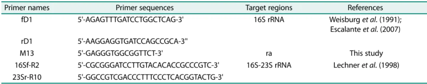
Table 1. List of PCR primers used in this study.
Primer names Primer sequences Target regions References
fD1 5'-AGAGTTTGATCCTGGCTCAG-3' 16S rRNA Weisburg et al. (1991);
Escalante et al. (2007)
rD1 5'-AAGGAGGTGATCCAGCCGCA-3''
M13 5'-GAGGGTGGCGGTTCT-3' ra This study
16Sf-R2 5'-CGCGGGATCCTTGTACACACCGCCCGTC-3' 16S-23S rRNA Lechner et al. (1998) 23Sr-R10 5'-GGCCGTCGACCCTTTCCCTCACGGTACTG-3'
ra
: Random amplification.
Antimicrobial activity of Bacillus strains against food- borne pathogens. Production of antimicrobial substances by Bacillus strains against closely related species of organisms or genera, including food-borne pathogens (L.
sakei DSM 20017
T, L. casei DSM 20011
T, S. aureus subsp. aureus ATCC 11632, E. coli ATCC 11229, L.
monocytogenes ATCC 19118, E. faecium ATCC 35667, B.
cereus MTCC 430, B. cereus MBU 1011, S. aureus MBU 1023, E. coli MBU 1035 and S. enterica serovar Typh- imurium MBU 1047) was determined using two meth- ods; the agar spot-on-lawn test as described by Schillinger and Lücke [32] and the colony overlay assay by Pugsley [33], as well as the agar well diffusion assay using cell-free supernatant as earlier described [34].
Safety assessment studies
Haemolysis on blood agar. Bacillus strains (B. subtilis U170B, B. subtilis U146A, B. clausii UBBC-07, B. cereus U175 and a reference haemolytic B. cereus MBU 1011) grown in nutrient broth (HiMedia) for 18 h at 37 ℃ were streaked on BHI agar, supplemented with 5% sheep blood, and incubated at 37 ℃ for 18−24 h. A β-haemo-
lytic activity is indicated by the presence of clear zone around the streaked area (positive), α-haemolysis is associated with partial clearance zone and greenish colouration around the streaked region, while ( γ-haemolysis) is without clearance zone, which is considered negative [35].
Antibiotic susceptibility testing. The susceptibility of Bacillus strains to antibiotics was determined by modifi- cation of the standard disk and agar overlay diffusion methods of Clinical and Laboratory Standards Institute [36]. Antibiotic disks analyzed include: penicillin G (P, 10 units/disk), ampicillin (AMP, 10 µg/disk) ( β-lactams);
erythromycin (E, 15 µg/disk), vancomycin (VA, 30 µg/
disk) (Gram+ve spectrum); nalidixic acid (NA, 30 µg/
disk) (Gram-ve spectrum); chloramphenicol (C, 30 µg/
disk), rifampicin (RIF, 5 µg/disk), tetracycline (TE, 30 µg/disk) (broad spectrum); kanamycin (K, 30 µg/disk), gentamicin (GEN, 10 µg/disk), streptomycin (S, 10 µg/
disk) (aminoglycosides); others were amoxyclav (AMC, 30 µg/disk), clindamycin (CD, 2 µg/disk), ciprofloxacin (CIP, 5 µg/disk), methicillin (MET, 5 µg/disk), trimetho-
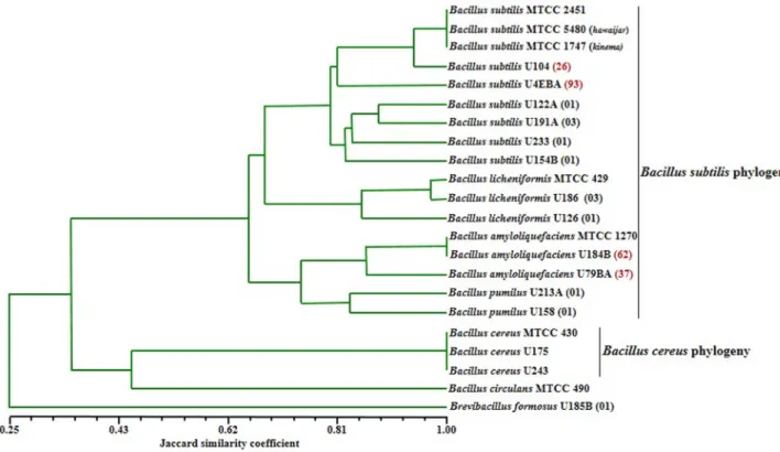
Fig. 1. Dendrogram based on UPGMA clustering of Jaccard similarity coefficient (Sj) of normalized combined ITS-PCR and
ITS-PCR-RFLP fingerprint patterns of Bacillus species isolated from iru and reference strains.
prim (TR, 5 µg/disk) and norfloxacin (NX, 10 µg/disk).
Already prepared BHI agar plates were overlaid with soft BHI agar (0.7%), containing 100 µl of bacterial iso- lates at log phase growth. After solidification, the antibi- otic disks (HiMedia) were aseptically placed onto the agar surface, and plates were incubated at 37 ℃ for 18−
24 h, to allow for bacterial-antibiotic interaction. Diame- ters of zones of inhibition in two replicate, including those of the disks (in mm) were measured and the results were expressed in terms of resistance (R) and susceptibility (S) in accordance to Performance Stan- dards for Antimicrobial Disk Susceptibility Tests, CLSI.
Results
Characterization and 16S rRNA gene phylogenetic of Bacillus species
Results of the combined dendrogram of ITS-PCR and ITS-PCR-RFLP characterized different species of bacilli as diverse. Fig. 1 shows two major clusters identified as B. subtilis and the B. cereus phylogeny. However, analysis of the ITS-PCR-RFLP further shows that B. subtilis
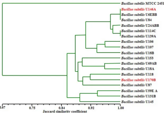
phylogeny is comprised of three sub-clusters that include strains of species identified as B. licheniformis, B. amyloliquefaciens and B. pumilus. Further analysis using RAPD-PCR indicated high strains genomic diver- gence and sub-types among the wild B. subtilis sensu stricto strains in iru (Fig. 2). These B. subtilis strains were grouped into three major sub-clusters, while the B.
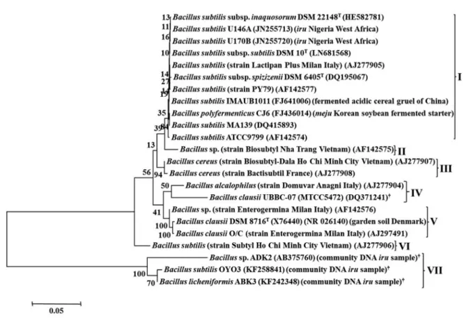
subtilis MTCC 2451 did not cluster with any of the groups (Fig. 2). Phylogenetic tree constructed using 16S rRNA gene sequences identified seven different clusters (Fig. 3). Cluster I indicated evolutionary relatedness and clonal relationship among B. subtilis strains from iru, B.
subtilis subsp. inaquosorum DSM 22148
T, B. subtilis subsp. spizizenii DSM 6405
T, B. subtilis subsp. subtilis DSM 10
Tand B. subtilis ATCC 9799 type strains, including one commercial Bacillus probiotic product (Lactipan Plus). Biosubtyl is the only member of cluster II, which is a sub-group of cluster I at bootstrap value
>80%. Cluster III consists of two B. cereus probiotic strains, marketed as Biosubtyl-Dala and Bactisubtil in Asia and Europe respectively. The fourth cluster com- prises of closely related strains of B. alcalophilus, pres- ent in registered probiotic product (Domuvar) and B.
Fig. 2. Dendrogram based on UPGMA clustering of Jaccard similarity coefficient (Sj) of normalized ITS-PCR, ITS-PCRRFLP
M13 RAPD-PCR finger-prints of divergent B. subtilis strains.
clausii probiotic reference strain. Cluster V has Bacillus sp. and B. clausii in Enterogermina probiotic products, in addition to B. clausii DSM 8716
T. B. subtilis Subtyl is also the only strain in cluster VI, while the last cluster comprises of Bacillus strains from bacterial community DNA analysis of iru samples.
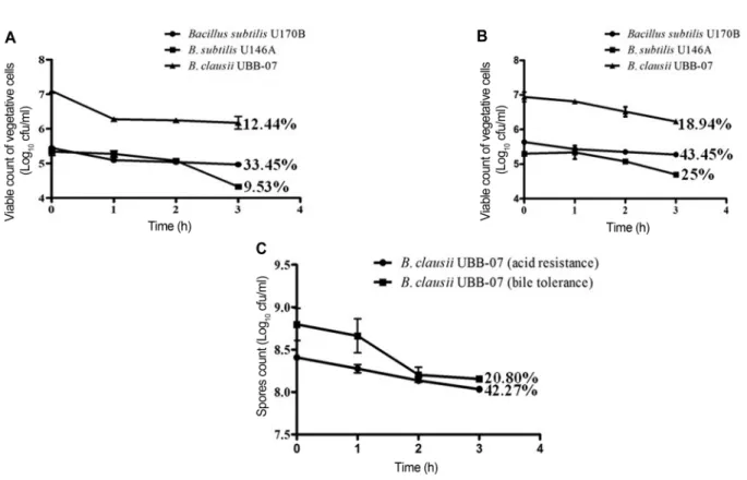
In vitro probiotic functional properties of Bacillus strains Resistance to acid and tolerance to bile salts. Vegetative cells of bacilli analyzed generally showed resistance to acid condition of pH 2.0 at 37 ℃. B. subtilis U170B and B.
clausii UBBC-07 (a probiotic reference strain obtained from India) had just < 1 Log unit reduction after 3 h incubation, while B. subtilis U146A had 1 Log unit lower (Fig. 4A). The survival rates (expressed in percentage) of the Bacillus strains based on initial viable colony counts and population counts after 3 h incubation at 37 ℃ were 33.45%, 12.44% and 9.53% for B. subtilis U170B, B.
clausii UBBC-07, and B. subtilis U146A, respectively.
Longer incubation up to 24 h did not show resistance but rather cell death. The three strains exhibited higher tol- erance to bile salts concentration of 0.3% (w/v), com- pared to their resistance to acidic pH. In terms of Log cycle loss, they all had < 1 Log unit reduction (Fig. 4B).
Cell viability of these strains after 3 h passage in bile salts was also higher compared to that of gastric juice. B.
subtilis U170B demonstrated the highest survival rate (43.45%), followed by B. subtilis U146A (25.00%) and B.
clausii UBBC-07 (18.94%). In general, vegetative cells of B. subtilis U170B were more acid-resistant and bile salts-tolerant than the other two strains tested. How- ever, only B. clausii UBBC-07 spores survived the acidic and bile salts conditions; others were found to be highly sensitive as no single spore count was recorded. The B.
clausii UBBC-07 spores thrived in both low pH and bile salts with an average marginal decrease of 0.5 Log cycle losses (Fig. 4C). Survival rates obtained were 42.27%
and 20.80% for acid and bile salts respectively.
Fig. 3. Phylogenetic tree of pairwise and multiple alignments of 16S rRNA gene sequences of Bacillus strains isolated from
iru, type, reference, and commercial probiotic Bacillus products. The tree was generated using the Molecular Evolutionary
Genetics Analysis (MEGA7) software.
†Accession no. of closest relative organisms of nucleotide sequences found in GenBank data-
base.
In vitro antagonistic potentials of Bacillus strains against food-borne pathogens. The production of antibacterial compounds by Bacillus strains against eleven indicator
bacterial strains was demonstrated. The bacilli tested could not inhibit most of the indicator organisms (Table 2). However, B. subtilis U170B and B. subtilis U146A Fig. 4. Growth and viability of Bacillus strains under simulated gastric juice and intestinal bile conditions. (A) Acid resistance and (B) bile resistance of vegetative cells of B. subtilis strains from iru and B. clausii UBB-07 reference probiotic strain; (C) acid and bile tolerance of B. clausii UBB-07 spores. Error bars represent the standard deviation of replicate determinations.
Table 2. Antagonistic activities of B. subtilis strains isolated from iru and reference probiotic B. clausii UBBC-07 against eleven indicator bacterial strains.
Indicator strains Test organisms
B. subtilis U170B B. subtilis U146A B. clausii UBBC-07
L. sakei DSM 20017
a- - nd
L. casei DSM 20011 - - nd
S. aureus subsp. aureus ATCC 11632 - - nd
E. coli ATCC 11229 - - nd
L. monocytogenes ATCC 19118 - - nd
E. faecium ATCC 35667 - - nd
B. cereus MTCC 430 + (2 mm)
b- nd
B. cereus MBU 1011 + (2 mm)
b+ (4 mm)
b+ (9 mm)
cS. aureus MBU 1023 - - -
E. coli MBU 1035 - - -
S. enterica serovar Typhimurium MBU 1047 - - -
a
DSM: Deutsche Sammlung von Mikroorganismen Gottingen, Germany; ATCC: American Type Culture Collection; MTCC: Microbial
Type Culture Collection, Chandigarh, India.
bWeak activity using agar spot-on-lawn method.
cStrong inhibition with agar well diffu-
sion assay. nd: not determined. Diameter of inhibition zone obtained after subtracting diameter of bored hole from the entire halo.
showed weak inhibition zone against B. cereus MTCC 430 and B. cereus MBU 1011 with the agar spot-on-lawn method. No zone of inhibition was detected using the cell-free supernatant of these strains. B. clausii UBBC- 07 displayed strong antimicrobial activity against B.
cereus MBU 1011 (Table 2), though wider inhibitory zone was recorded for agar spot-on-lawn.
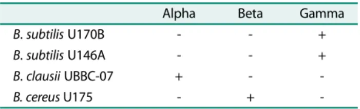
In vitro safety evaluation of Bacillus strains Haemolytic activity.
B. subtilis strains U170B and U146A isolated from iru did not lyse red blood cells when streaked on BHI agar containing sheep blood. This can be considered γ-haemo- lytic. B. clausii UBBC-07 had α-haemolysis with partial clearance zone and greenish colouration around the region streaked. B. cereus U175 also from iru and hae-
molytic B. cereus MBU 1011 used as positive controls produced complete clear zones, indicating β-haemolysis (Table 3).
Antibiotic susceptibility pattern. B. subtilis U170B and B. subtilis U146A were sensitive to most of the antibiotics;
however, the latter was resistant to methicillin (Table 4).
On the contrary, B. clausii UBBC-07 was resistant to majority of the antibiotics used, but only sensitive to chloramphenicol.
Discussion
There is dearth of information on the beneficial health potentials of Bacillus species consumed in large popula- tions with African fermented condiments. This study first provided information about the genetic diversity of these bacilli isolated from fermented condiments in Africa, and determined in vitro properties, to enable selection of multifunctional strains that can be used as starter cultures, as well as having ability to survive gut conditions. This endeavour can culminate in the use of safe strains with dual functions, with an overall objec- tive of improving gut health beneficial properties using a Table 3. Type of haemolysis on blood agar.
Alpha Beta Gamma
B. subtilis U170B - - +
B. subtilis U146A - - +
B. clausii UBBC-07 + - -
B. cereus U175 - + -
Table 4. Antibiotic sensitivity profiles of B. subtilis strains isolated from iru and reference probiotic B. clausii UBBC-07.
Antibiotics (symbol, μg) Zone of inhibition (mm)
‡B. subtilis U170B B. subtilis U146A B. clausii UBB-07
Kanamycin (K, 30) 25.10 ± 0.28
†(S) 27.40 ± 0.71 (S) 16.35 ± 0.07 (I)
Amoxyclav (AMC, 30) 37.15 ± 0.92 (S) 40.00 ± 0.85 (S) 03.20 ± 0.99 (R)
Ampicillin (AMP, 10) 24.10 ± 0.14 (S) 26.20 ± 0.14 (S) 01.40 ± 0.57 (R)
Chloramphenicol (C, 30) 25.15 ± 0.07 (S) 29.30 ± 0.28 (S) 25.00 ± 0.99 (S)
Clindamycin (CD, 2) 19.10 ± 0.14 (S) 34.25 ± 0.35 (S) 11.25 ± 0.78 (R)
Ciprofloxacin (CIP, 5) 33.15 ± 0.35 (S) 38.15 ± 0.78 (S) 14.25 ± 0.50 (R)
Erythromycin (E, 15) 23.10 ± 0.42 (S) 42.25 ± 0.07 (S) 10.05 ± 0.21 (R)
Gentamicin (GEN, 10) 27.20 ± 0.28 (S) 24.15 ± 0.35 (S) 13.15 ± 0.07 (I)
Methicillin (MET, 5) 10.00 ± 0.28 (I) 04.20 ± 0.99 (R) - (R)
Nalidixic Acid (NA, 30) 15.05 ± 0.35 (I) 28.35 ± 0.21 (S) 05.25 ± 0.35 (R)
Penicillin G (P, 10 units) 39.20 ± 0.57 (S) 34.35 ± 0.35 (S) - (R)
Rifampicin (RIF, 5) 24.20 ± 0.28 (S) 25.45 ± 0.35 (S) 05.25 ± 0.50 (R)
Streptomycin (S, 10) 23.10 ± 0.57 (S) 29.05 ± 1.20 (S) 12.20 ± 0.14 (I)
Tetracycline (TE, 30) 38.30 ± 0.71 (S) 34.30 ± 0.28 (S) 05.05 ± 1.06 (R)
Trimethoprim (TR, 5) 24.20 ± 0.14 (S) 24.15 ± 0.64 (S) - (R)
Vancomycin (VA, 30) 22.00 ± 1.13 (S) 28.60 ± 0.14 (S) 12.10 ± 1.13 (R)
Norfloxacin (NX, 10) 36.05 ± 0.50 (S) 32.35 ± 0.21 (S) 11.60 ± 0.42 (R)
‡