Probiotic Properties of Lactic Acid Bacteria Isolated Traditional Fermented Foods
Eun-Ji Kim, Seung-Wha Jo, Jin-Kyeong Kim and Do-Youn Jeong*
Microbial Institute for Fermentation Industry(MIFI), 61-27, Minsokmaeul-gil, Sunchang-eup, Jeonbuk 56048, Korea Received December 20, 2018 /Revised June 17, 2019 /Accepted June 24, 2019
This study performed to investigate the probiotic properties of lactic acid bacteria 200 strains isolated from traditional fermented foods. Based on being higher tolerance to bile salts and showing higher acid resistance, 4 LAB Strains were selected in the screening experiment; Lactobacillus plantarum SRCM 102224, Lb. plantarum SRCM102227, Lb. paracasei SRCM102329, Lb. paracasei SRCM102343. Antibacterial activity against various pathogens, acid and bile salt tolerance, hemolytic phenomenon, cell surface hydrophobicity, and antibiotic resistance were examined. Among the tested strains, SRCM 102343 (95.9%) was highly observed hydrophobicity compared to Lb. rhmanosus GG (13.4%) as control. In this study, the in vitro adhesion properties of 4 strains of LAB was investigated using human intestinal caco-2 cell cultures. SRCM102329 and SRCM102343showed higher adherence to caco-2 cells than Lb.
rhamnosus GG. The antibacterial activities of 4 strains LAB were investigated. the 3 strains showing strongly antimicrobial activity against Escherichia coli ATCC10798, Staphylococcus aureus KCCM11593, Listeria invanovii KCTC3444, Bacillus cereus ATCC11778 and S. enterica serovar. Typhi KCTC1926. These results suggest that selected strains have good probiotic potential for application in functional foods.
Key words : Adhesion, antibiotic resistance hydrophobicity, Lactobacillus, probiotic
*Corresponding author
*Tel : +82-63-650-2000, Fax : +82-63-653-9590
*E-mail : [email protected]
This is an Open-Access article distributed under the terms of the Creative Commons Attribution Non-Commercial License (http://creativecommons.org/licenses/by-nc/3.0) which permits unrestricted non-commercial use, distribution, and reproduction in any medium, provided the original work is properly cited.
Journal of Life Science 2019 Vol. 29. No. 6. 697~704 DOI : https://doi.org/10.5352/JLS.2019.29.6.697
서 론
유산균은 일반적으로 Lactobacillus 속, Pediococcus 속, Leuconostoc 속 등의 균종을 포함하고 있으며 유기산(젖산, 아 세트산 등), 저급지방산, hydrogen peroxide, 그리고 bacter- iocin과 같은 항균물질 등 다양한 대사물질을 생산하여 부패세 균의 생육을 억제한다고 알려져 있으며[32], 또한 정장 작용, 면역 조절, 항암 및 항돌연변이 효과, 콜레스테롤 저하, 항알레 르기 효과, 유당불내증 완화 등의 프로바이오틱 기능성이 알 려 있다[5, 19, 29].
프로바이오틱스는 숙주와 상호작용을 통해 숙주의 건강증 진에 영향을 주는 살아있는 미생물로 주로 인간의 장 건강에 유익한 영향을 미치며, 소화관의 안정성 유지, 설사 예방이나 증상의 완화, 콜레스테롤 수치의 감소, 고혈압 완화, 면역체계 의 개선 등의 효과가 있다고 알려져 있다[22]. 프로바이오틱스 생균제로 사용되고 있는 세균으로는 Lactobacillus 속, Enter- ococcus 속, Streptococcus 속, Bifidobacterium 속 등이 보고되어 있다. 대부분 산업적으로 이용되고 있는 프로바이오틱스는 인
간의 장관이나 유제품 등에서 분리한 동물 유래 유산균이었으 나, 김치, 장류 등과 같이 식물 유래 유산균에도 프로바이오틱 스 기능이 있다고 밝혀지고 있다[14]. 이러한 프로바이오틱스 유산균들은 각종 건강기능식품, 의약품 및 사료첨가제 등으로 광범위하게 이용되고 있다. 특히, 최근 서구화된 식습관으로 인해 현대인의 비만, 당뇨, 고혈압, 아토피 등과 같은 질병에 효과가 있다고 알려지면서 소비자의 관심이 급증하는 추세로 국내 시장 또한 크게 증가하고 있는 실정이다[3, 9]. 프로바이 오틱스가 인체에서 정장작용 및 여러 생리적 기능을 발휘하기 위하여 위산과 담즙이 존재하는 환경에서의 생존력, 대장상피 세포에 대한 높은 부착능 등의 조건을 갖춰야한다고 알려져 있으며, 항균력, 콜레스테롤 저하능 등의 특성이 확보되어야 한다. 그러나 이러한 특성들은 미생물의 종류에 따라 그 정도 가 다르게 나타나며, 같은 종에 속한 미생물의 경우에도 차이 가 있는 것으로 알려져 있다[33]. 따라서 본 연구에서는 안전성 이 확보 및 기능성을 가진 잠재적인 프로바이오틱스 특성을 지닌 균주를 분리하기 위하여 전통발효식품로부터 유산균을 선발하였으며, 용혈성, 내산성 및 내담즙성, 항균 활성, 항생제 에 대한 내성 그리고 장내 상피세포 부착능 등의 특성을 분석 하여 프로바이오틱스 소재로서의 적용 가능성을 검증하고자 하였다.
재료 및 방법
유산균의 분리, 동정 및 계통도
국내에 있는 가정에서 담근 전통식 된장, 간장, 고추장, 누 룩, 갓김치, 총각김치, 배추김치, 갓김치, 알타리 김치 등의 시 료 100여 종을 사용하여 각 시료 10 g을 멸균생리식염수 90 ml에 단계희석하여 lactobacilli MRS (Difco, MI, USA) 배지에 도말하여 37℃, 48시간 배양하였다. 각각의 유산균 200여종을 순수 분리하였고, 그 중 내산성, 내담즙성에서 활성이 가장 뛰어난 유산균 4종에 대하여 macrogen (Seoul, Korea)에 16S rDNA sequence 분석을 의뢰하였고, 분석된 염기서열은 NCBI의 data base와 비교하여 동정하였다. 16S rRNA 염기서 열 분석을 위해 universal 프라이머인 27F (5'-AGAGTTTGAT CCTGGCTCAG-3')와 1492R (5'-GGTTAC CTTGTTACGAC TT-3')을 사용하여 유전자를 증폭 후 염기서열을 해독하였다.
염기서열의 chromatogram을 이용하여 gap을 최소화한 후 NCBI에서 서열 일치도가 높은 표준 균주들의 16S rRNA 유전 자 염기서열을 확보하여 계통도를 작성하였다. 계통도 분석은 tamura-Nei 모델에 기초한 maximum likelihood 방법을 사용 하여 분석하였고[31], 산출한 각각의 계통수에서 각 분지에 대 한 통계학적 신뢰도를 산출하기 위해 bootstrap 분석을 1,000 회 실행하였으며, 분석은 MEGA 7.0.26 program을 사용하였 다[17].
내산성, 내담즙성 및 용혈성 측정
선별된 유산균의 내산성 평가는 pH 2.0, 3.0, 4.0, 5.0 (in 5 N HCl)로 조정한 MRS broth에 위산에 대한 저항성을 확인 하기 위하여 각각의 균주를 접종하여 30분 정치 후, 시료를 회수하여 생균수를 측정하였다. 대조구와 비교하여 생균수가 증가한 실험구를 내산성을 가진 것으로 평가하였다. 내담즙성 평가를 하기 위하여 oxgall (Neogen, Lansing, MI, USA)을 0.1%, 0.3%, 0.5%, 1.0%의 농도로 첨가한 MRS broth와 oxgall 을 첨가하지 않은 MRS broth에 각각의 유산균 접종하여 37℃
에서 6시간 배양하여 대조구와 비교하여 내담즙성을 평가하 였다. 용혈형태를 확인하기 위해 선별된 유산균 4종을 MRS (Difco) 액체배지에 1%(V/V) 접종하여 37℃에서 배양한 다음, 5% 면양혈액 한천배지(MB cell, Seoul, Korea)에 획선 도말하 여 균주의 용혈타입을 확인하였다. 획선 도말된 배지는 37℃
에서 48시간 동안 배양하였고, 배양이 끝난 다음 콜로니 주변 에 용혈현상으로 인해 생성된 환의 형태를 확인하였다[6].
세포 표면 소수성
세포의 장내 부착성은 세포표면의 조성 및 구조에 따라 많 은 영향을 받아 세포표면의 소수성이 주요한 요인으로 알려져 있다[18]. 용매에 대한 세포 표면 소수성(cell-surface hydro- phobicity) 검사는 Doyle 등의 방법으로 실시하였다[6]. 각각 의 균주를 MRS broth에 접종한 뒤 37℃에서 18시간 배양하였 다. 배양액의 균체 회수한 후, PBS로 두 번 세척한 다음 같은 용액으로 현탁하였다. 현탁액과 chloroform (Sigmaaldrich)을
각각 1:1의 비율로 첨가한 다음 2분 동안 vortex 후, 30분 동안 상온에 방치하였다. 현탁액(OD 580 nm reading 1)과 분리된 층에서 수용액 층(OD 580 nm reading 2)에 대한 흡광도를 580 nm에서 측정하였으며, 소수성은 다음과 같이 계산하여 측정하였다[16].
% hydrophobicity = [OD 580 nm reading 1–OD 580 nm reading 2]/ OD 580 nm reading 1 ×100
세포주 Caco-2에 대한 장내 부착능
실험에 사용한 인체 결장암 세포주 Caco-2 세포는 Korean Cell Line Bank (KCLB, Korea)로부터 분양 받아 사용하였다.
Caco-2 세포는 10% fetal bovine serum (FBS, Hyclone), 1%
streptomycin/penicillin (10,000 IU/ml, Hyclone), 1% non-es- sential amino acid (Hyclone), 10 mM HEPES, 1 mM L-gluta- mate, 1 mM sodium pyruvate (Hyclone)이 첨가된 Minimum essential medium (MEM, Gibco)배지를 사용하여 37℃, 5%
CO
2/ 95% air 조건으로 배양하였다. 5×10
5cells/well로 seed- ing하여 cell이 monolayer를 형성될 때까지 분화시켜 사용하 였다. 각각의 균주는 MRS 배지에서 18시간 배양한 후, 원심분 리(10,000 rpm, 3 min, 4℃)로 균체를 회수하여 PBS (pH 7.2)로 2회 세척한 후 serum free MEM에 10
7CFU/ml로 현탁하여 monolayer를 형성한 caco-2 세포가 있는 24 well tissue cul- ture plates에 각각 well에 0.5 ml씩 첨가하였다. 37℃, 5%
CO
2/ 95% air 조건하에 1시간 배양한 후, PBS로 3회 세척하고, 0.05% triton X-100 1 ml씩 넣고 10분간 고정시키고 이를 연속 희석을 통하여 생균수를 측정함으로써 대조구와 비교하여 Caco-2 cell에 대한 부착능을 측정하였다[7, 10, 27].
항균력 조사
항균 활성은 paper disc method를 이용하여 조사하였다 [30]. E. coli ATCC 10798, M. luteus KCTC1056, S. aureus KCCM 11593, L. invanovii KCTC34440, B. cereus ATCC11778 및 S. typhi KCTC1926에 대한 선별된 유산균의 항균력을 조사 하였다. 선별균주를 37℃에서 24시간 배양한 후 0.4 μm mem- brane 필터 (Millipore, Membrane filter, Ireland)를 이용하여 제균한 상등액을 시료로 사용하여 항균력을 측정하였다. 각각 의 지시균주가 10
5CFU/ml로 도말된 고체배지에 8 mm 직경 의 paper disc (Advantec, Tokyo, Japan)를 놓고 시료 100 μl씩 분주하여 생육저지환을 측정하였다.
항생제 감수성 조사
항생제 감수성 시험은 NCCLS (National Committee for
Clinical Laboratory Standards)의 disk diffusion법으로 실시
하였다. Clinical and Laboratory Standards Institute (CLSI)
guidelines을 따라 disc method에 의해 수행하였다[4]. 사용한
항생제디스크 (BBL, Sensi-disc, Becton Dickinson Co., NJ,
Fig. 1. Molecular Phylogenetic analysis by Maximum Likelihood method. The evolutionary history was inferred by using the Maximum Likelihood method based on the Tamura-Nei model [1]. The tree with the highest log likelihood (-3382.85) is shown. The percentage of trees in which the associated taxa clustered together is shown next to the branches. Initial tree(s) for the heuristic search were obtained automatically by applying Neighbor-Join and BioNJ algorithms to a matrix of pairwise distances estimated using the Maximum Composite Likelihood (MCL) approach, and then selecting the topology with superior log likelihood value. The tree is drawn to scale, with branch lengths measured in the number of substitutions per site.
The analysis involved 14 nucleotide sequences. All positions containing gaps and missing data were eliminated. There were a total of 1188 positions in the final dataset. Evolutionary analyses were conducted in MEGA7 [17].
USA)는 amikacin, amoxicillin-clavulanic acid, ampicillin, azithromycin, cefaclor, ceftazidime 등 총 30종의 항생제를 사 용하였다. 유산균 4종은 MRS 액체배지에서 배양한 후, 10
5CFU/ml가 되게 Mueller hinton agar (Merck, Darmstadf, Germany)에 도말한 다음, 30종의 항생제 디스크를 놓고 37℃
에서 24시간 방치한 다음, 각 항생제에 대한 저지환의 크기를 측정하고 감수성과 내성을 판정하였다.
결과 및 고찰
유산균의 분리, 동정 및 계통도
국내에 있는 가정에서 담근 전통식 된장, 간장, 고추장, 누 룩, 갓김치, 총각김치, 배추김치, 갓김치, 알타리 김치 등의 시 료 100여종을 수집하여 이를 대상으로 유산균 200여종을 순수 분리하여 -80℃에 보관하여 사용하였다. 분리된 균주에 대하 여 내산성, 내담즙성을 확인하였고, 저항성이 높은 유산균 4종 을 선발하여 SRCM102224, SRCM102227, SRCM102329, SRCM 102343로 명명하고, 분리된 균주는 16S rRNA sequencing을 통해 얻은 염기서열을 NCBI BLAST DB에서 검색한 결과,
99%의 상동성을 보이는 Lb. paracasei SRCM102329, Lb. para- casei SRCM102343, Lb. plantarum SRCM102224, Lb. plantarum SRCM102227로 명명하였다(Fig. 1). 그 중 SRCM102343은 한 국미생물보존센터(KCCM, Korean Culture Center of Microor- ganism)에 KFCC1197P 기탁하였다.
유산균의 내산성, 내담즙성 및 용혈성
유산균이 식품과 함께 섭취 시 프로바이오틱스로서의 효과
를 나타내기 위해서는 소화액 수준인 pH 3 이하의 낮은 pH
조건과, 장내 담즙산의 농도보다 고농도의 담즙산이 함유된
배지에서 생장 할 수 있는 조건이 요구되어야 한다[11]. 선별된
유산균 4종을 대상으로 pH에 대한 내성을 분석한 결과, 모든
균주가 pH 2에서 10
3CFU/ml 이상의 생균수를 나타내었고,
SRCM102224, SRCM102227 균주가 가장 높은 생균수를 보이
는 것을 확인하였다(Table 1). 유산균의 내산성은 당분해 과정
흐름의 변화, 세포내 pH 조절 능력과 세포막의 ATPase와 관
련이 있으며, 식품의 성분에 의해 저항성은 유의하게 증가되
는 것으로 보고되어 있다[24]. 또한 Mishra 등의 연구에 따르
면 프로바이오틱스로 사용되는 여러 균주들이 pH 2에서 10
4Table 1. pH resistance of selected 4 strains at various pH from 6.5 to 2.0 (Unit: Log CFU/ml)
Strains Survival of cells
pH 2.0 pH 3.0 pH 4.0 pH 5.0 pH 6.5
SRCM102224 SRCM102227 SRCM102329 SRCM102343
4.32±0.10ab 4.60±0.07a 3.70±0.21c 3.85±0.14bc
8.00±0.69a 8.00±0.03a 7.90±0.07a 8.00±0.10a
8.19±0.09a 8.28±0.21a 8.15±0.15a 8.29±0.07a
8.13±0.12a 8.19±0.21a 8.23±0.04a 8.23±0.12a
8.10±0.19a 8.21±0.00a 8.06±0.04a 8.05±0.17a Means in each column with different subscripts letters are significantly different at duncan's test (p<0.05).
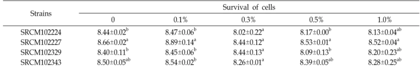
Table 2. Bile tolerance of selected 4 strains at various bile salt from 0 to 1.0% (Unit: Log CFU/ml)
Strains Survival of cells
0 0.1% 0.3% 0.5% 1.0%
SRCM102224 SRCM102227 SRCM102329 SRCM102343
8.44±0.02b 8.66±0.02a 8.40±0.11b 8.50±0.05ab
8.47±0.06b 8.89±0.14a 8.45±0.06b 8.54±0.02b
8.02±0.22a 8.44±0.12a 8.44±0.13a 8.26±0.01a
8.17±0.00b 8.53±0.01a 8.09±0.13b 8.39±0.05ab
8.13±0.04ab 8.52±0.04a 8.20±0.23ab 8.28±0.25ab Means in each column with different subscripts letters are significantly different at duncan's test (p<0.05).
Table 3. Adhesion ability to Caco-2 cells of selected 4 strains and LGG
Strains Initial cell count (Log CFU/ml) Adhered cell count (Log CFU/ml) Adhesion (%) SRCM102224
SRCM102227 SRCM102329 SRCM102343
LGG
7.01±0.51b 7.13±0.24b 7.51±0.18ab 7.69±0.11a 7.04±0.21b
4.89±0.16c 5.32±0.28b 5.73±0.12a 5.89±0.13a 5.16±0.19bc
69.92±1.97c 74.54±1.15ab
76.30±0.33a 76.58±0.84a 73.34±0.73b LGG; Lactobacillus rhamnosus GG, Values represent the mean ± SD (n=3).
Means in each column with different subscripts letters are significantly different at duncan's test (p<0.05).
Fig 2. Hydrophobicity of selected 4 strains and LGG. LGG;
Lactobacillus rhamnosus GG, Values represent the mean±
SD (n=3). Means in each column with different sub- scripts letters are significantly different at duncan's test (p<0.05).
CFU/ml 이하의 생균수를 보였고[20], 본 연구 결과와 유사한 결과를 나타내었다. 따라서 본 연구에서 선별된 유산균 4종의 경우 산성에 대한 우수한 내성 능력을 보유하는 것을 확인할 수 있었다. 담즙산은 지질로 구성된 미생물의 세포막에 영향 을 주어 미생물의 생장을 억제하는 것으로 알려져 있는데, Lactobacillus 속을 포함한 많은 종의 유산균에서는 담즙산염 가수분해효소를 생성하여 담즙산을 가수분해하고 이 같은 억 제작용을 감소시키는 것으로 보고되어 있다[28]. 선별된 균주 의 내담즙성을 확인한 결과 모든 균주가 1% 담즙엽 농도에서 10
8CFU/ml 이상의 매우 높은 생균수를 보여, 담즙염에 대해 우수한 내성을 보이는 것을 확인할 수 있었다(Table 2). 이와 같은 결과는 선별된 유산균 4종 모두 내산성 및 내담즙성이 프로바이오틱 균주로 적합하다고 판단되어진다. 용혈작용은 적혈구가 파괴되는 정상적인 작용과 적혈구의 유전적인 결함 이나 화학물질, 뱀 등의 독액, 미생물이 생성하는 독성물질에 의해서 형성되는 비정상적인 작용이다[15]. SRCM102224, SRCM102227, SRCM102329, SRCM102343 4종에 대하여 α형, β 형, γ형 용혈 타입을 확인한 결과, 선별된 유산균 4종 모두에
서 sheep blood가 포함된 한천배지에서 용혈 현상이 없는 γ형
으로 확인되었다. 이에 프로바이오틱으로 섭취하였을 시 인체
에 유해성을 나타낼 수 있는 적혈구 용혈에 대한 안전성이
Table 4. Antimicrobial activities of selected 4 strains against food pathogens
Strains E. coli
ATCC 10798
M. luteus KCTC 1056
Staphy. aureus KCCM 11593
L. invanovii KCTC 3444
S. enterica serovar.
Typhi KCTC 1926
B. cereus ATCC 11778 SRCM102224
SRCM102227 SRCM102329 SRCM102343
+ + + +
- + - -
+ + + +
+ + + +
+ + - -
+ + + + +, Positive activity; -, negative activity.
Values represent the mean ± SD (n=3).
Table 5. Antibiotic susceptibility of selected 4 strains
Antibiotics Code/Disc potency
(μg)
Result SRCM
102224
SRCM 102227
SRCM 102329
SRCM 102343 Amikacin
Amoxicillin Amoxicillin/clavulanic acid
Ampicillin Azithromycin
Cefaclor Ceftazidime
Cephalexin Ciprofloxacin
Clindamycin Erythromycin Fusidic acid
Gentamicin Imipenem Kanamycin Lincomycin Metronidazole Nalidixic acid Neomycin Netilmicin Ofloxacin Oxacillin Penicillin G Pipemidic acd
Piperacillin Rifampicin Roxithromycin
Teicoplanin Tetracycline
Trimethoprim-sulfamethoxazde
AN/30 AML/25 AMC/20 AM/10 AZM/15
CEC/30 CAZ/30 CL/30 CIP/5 CD/10 E/15 FC/30 GM/10 IMI/10 K/30 MY/15 MTZ/5 NA/30 N/30 NET/30
OFX/5 OX/5 P1 IU PI/20 PRL/100
RD/5 RXT/15 TEC/30 TE/30 SXT/25
S SS SS SS S S S SS
S S SS
S S SS
S S R R S S R S S R SS SS S R S S
S SS
S SS
S S S S R SS SS S S SS
R S R R S S R SS
S R SS
S S R S S
S SS
S SS SS S S S S SS SS R S S R SS
R R S S S S S R SS SS SS R SS
R
R SS
S SS
S S S S S SS SS R S S R SS
R R R S S S S R SS SS SS R S R Diameter, cm; R, resistance, S, 1.2∼3.0, SS, 3.1∼4.0. Values represent the mean ± SD (n=3).
확인되었다[27].
세포표면 소수성
세포표면의 소수성은 병원성 세균과의 부착 및 응집현상,
숙주세포의 방어 등 다양한 현상들과 연관성을 가진다고 알려
져 있다[2, 13]. 미생물의 탄화수소에 대한 부착능을 chloro-
form에 부착하는 정도를 측정한 결과, 선별된 유산균 4종은
10~95.9% 소수성을 나타내었는데, 그 중 SRCM102343가
chloroform에 대하여 가장 높은 소수성을 보였다. 또한 프로바 이오틱스 특성을 갖는 표준균주인 LGG (13.41%)에 비해 SRCM102227, SRCM102329도 높은 결과를 나타내었다(Fig.
2). 반면 SRCM102224는 ~로 다소 낮은 소수성을 나타내었다.
Perez 등의 연구에 따르면 세포 표면 소수성은 85% 이상이 되어야 장 상피 세포에 대한 부착성에 도움이 된다고 알려져 있어 본 연구결과와 비교했을 때, SRCM102343은 장 상피세포 의 부착성이 높을 것으로 사료된다[24].
세포주 Caco-2에 대한 장내 부착능
유산균이 프로바이오틱스의 특징을 지니려면 위와 십이지 장을 통과하여 최종 목적 부위인 장에 도달하여 장내 상피세 포에 정상적으로 부착되어야 그 기능을 할 수 있다[8]. LGG는 다른 유산균에 비해 장내부착능이 뛰어난 것으로 알려져 있는 데, 선별된 유산균의 Caco-2 세포에 대한 장내부착능을 확인 한 결과(Table 3), SRCM102224를 제외하고 LGG에 비해 높은 부착능을 보여주었다. 상기 소수성 결과와 유사하게 선별된 유산균 4종 중 SRCM102343 균주가 76.53%의 부착능이 가장 높게 나타나 프로바이오틱스의 소재로서의 가능성이 확인되 었다.
항균력 조사
유산균의 항균활성은 유산균에 의해 생성된 유기산, 저급지 방산, hydrogen peroxide, diacetyl, bacteriocin등을 생성하여 병원성 세균과 부패세균의 생육을 억제한다고 알려져 있다 [30]. 선별된 유산균을 식중독 및 유해균주에 대한 항균력을 측정한 결과(Table 4), 선별된 균주 4종 모두 E. coli, S. aureus, B. cereus, S. typhi에 대하여 항균활성을 가지는 것으로 확인되 었는데 Lb. plantarum SRCM102227만 M. luteus에 대하여 억제 환이 형성되는 것을 확인하였다. 기존의 여러 연구결과에서도 다양한 유산 균주가 S. aureus, E. coli, S. typhi, B. cereus 등에 항균효과가 있다고 보고되고 있다[1, 22, 25, 35]. 따라서 선별 된 균주는 부패균의 증식을 억제하여 장내 유해 세균의 억제 에 효과를 보일 것으로 판단된다.
선별된 균주의 항생제 내성
Lactobacillus 속은 본질적으로 광범위한 항생제에 대하여 저 항성을 나타내는 것으로 알려져 있다. 항생제에 대한 내성이 강할수록 생존 가능성이 높아 장관 내 증식하여 유용한 기능 성을 발휘할 수 있다고 알려져 있다[12, 16]. 선별된 유산균 4종에 대해 항생제 내성 특성은 Table 5와 같다. 항생제 감수성 을 조사한 결과, penicillin계 항생제인 penicillin G, amox- icillin, amoxicillin/clavulanic acid, ampicillin에 대하여 선별 된 유산균 4종 모두 생육이 억제됨을 확인하였고, tetracycline 계 tetracycline, cephem계 cephalexin, macrolide계 eryth- romycin, roxithromycin, lincomycin계 clindamycin, linco-
mycin에 대하여 선별된 유산균 4종 모두 감수성을 가지는 것 을 확인하였다. Aminoglycoside계 amikacin과 neomycin에 대하여 SRCM102343은 내성을 지니고 있으며, kanamycin에 대하여 SRCM102224를 제외하고 내성을 지니고 있음을 확인 하였다. 또한, quinolones계 항생제인 nalidixic acid와 glyco- peptides계 항생제인 teicoplanin에 대하여 선별된 유산균 4종 모두 내성을 가지는 것을 확인하였다.
References
1. Adesokan, I. A., Odetoyinbo, B. B. and Olubamiwa, A. O.
2008. Biopreservative activity of lactic acid bacteria on suya produced from poultry meat. Afr. J. Biotechnol. 7, 3799-3803.
2. Bar-Ness, R., Avrabamy, N., Matsuyama, T. and Rosenberg, M. 1988. Increased cell surface hydrophobicity of a Serraia marcescens NS 38 mutant lacking wetting activity. J. Bacter- iol. 170, 4361-436.
3. Chon, J. W., Kim, D. H., Kim, H. S., Kim, H. S., Hwang, D. G., Song, K. Y., Yim, J. H., Choi, D. S., Lim, J. S. and Seo, K. H. 2014. Effect of probiotics on risk factors for hu- man disease: A review. Kor. J. Dairy Sci. Technol. 32, 17-29.
4. Clinical and Laboratory Standards Institute. 2006. Methods for Antimicrobial Dilution and Disk Susceptibility Testing of Infrequently Isolated or Fastidious Bacteria: Approved Guideline M45-A. Clinical and Laboratory Standards Institute, 26.
5. Diep, D. B., Godager, L., Brede, D. and Nes, I. F. 2006. Data minig and characterization of a novel pediocin-like bacter- iocin system from the genome of Pediococcus pentosaceus ATCC 25745. Microbiology 152, 1649-1659.
6. Donohue, D. C. and Salminen, S. 1996. Safety of probiotic bacteria. Asia Pac. J. Clin. Nutr. 5, 25-28.
7. Doyle, R. J. and Rosenberg, M. 1995. Measurement of micro- bial adhesion to hydrophobic substrata. Methods Enzymol.
253, 532-550.
8. Dunne, C. L., O’Mahoney, L., Murphy, L., Thornton, D., Mor- rissey, D., O’Halloran, S., Feeney, M., Flynn, S., Fitzgerald, G., Daly, C., Kiely, B., O’Sullivan, G. C., Shanahan, F. and Collins, J. K. 2001. In vitro selection criteria for probiotic bac- teria of human origin: correlation with in vivo findings. Am.
J. Clin. Nutr. 73, 386S-392S.
9. Eor, J, Y,, Park, M. H. and Kim, S. H. 2015. Prevention of obesity and type 2 diabetes by using probiotics. J. Milk Sci.
Biotechnol. 33, 231-235.
10. Fernádez, M. F., Boris, S. and Barbés, C. 2003. Probiotic properties of human Lactobacilli strains to be used in the gastrointestinal tract. J. Appl. Microbiol. 94, 449-455.
11. Gilliland, S. E. and Speck, M. L. 1977. Deconjugation of bile acids by intestinal Lactobacilli. Appl. Environ. Microbiol. 33, 15-18.
12. Gueimonde, M. and Salminen, S. 2006. New methods for selecting and evaluating probiotics. Dig. Liver Dis. 38, S242- S247.
13. Hazen, K. C., Lay, J. G., Hazen, B. W., Fu, R. C. and Murthy,
S. 1990. Partial biochemical charaterization of cell surface hydrophobicity and hydrophilicity of Candida albicans.
Infect. Immun. 58, 3469-3476.
14. Jonganurakkun, B., Wang, Q., Xu, S. H., Tada, Y., Minami- da, K., Yasokawa, D., Sugi, M., Hara, H. and Asano, K. 2008.
Pediococcus pentosaceus NB-17 for probiotic use. J. Biosci.
Bioeng. 106, 69-73.
15. Kim, Y. M., Park, U. K., Mok, J. S. and Chang, D. S. 1995.
Physiological characteristics of Listeria monocytogenes YM-7.
Kor. Fish Aquatic Sci. 28, 443-450.
16. Kos, B., Šuškoviæ, J., Vukoviæ, S., Šimpraga, M., Frece, J.
and Matošiæ, S. 2003. Adhesion and aggregation ability of probiotic strain Lactobacillus acidophilus M92. J. Appl.
Microbiol. 94, 981-987.
17. Lim, K. S. and Huh, C. S. 2006. Adhesion of bifidobacteria to Caco-2 cells and in relation to cell surface hydrophobic- ity. Kor. J. Food Sci. Anim. Resour. 26, 497-502.
18. Maria, G. V. P., Franz, C . M., Schillinger, U. and Holzapfel, W. H. 2006. Lactobacillus spp. with i n vitro probiotic proper- ties from human faeces and traditional fermented products.
Int. J. Food Microbiol. 109, 205-214.
19. Mathara, J. M., Schillinger, U., Guigas, C., Franz, C., Kutima, P. M. and Mbugua, S. K., et al. 2008. Functional character- istics of Lactobacillus spp. from traditional Maasai fer- mented milk products in Kenya. Int. J. Food Microbiol. 126, 57-64.
20. Mishra, V. and Prasad, D. N. 2005. Application of in vitro methods for selection of Lactobacillus casei strains as poten- tial probiotics. Int. J. Food Microbiol. 103, 109-115.
21. Obadina, A. O., Oyewole, O. B., Sanni, L. O. and Tomlins, K. I. 2006. Bio-preservative activities of Lactobacillus planta- rum strains in fermenting Casssava ‘fufu’. Afr. J. Biotechnol.
5, 620-623.
22. Ouwehand, A. C., Salminen, S. and Isolauri, E. 2002. Probi- otics: an overview of beneficial effects. Antonie van Leeuwen- hoek 82, 279-289.
23. Puttalingamma, V., Begum, K. and Bawa, A. S. 2006. Anti microbial peptides-new weapons against enteric pathogens.
Pak. J. Nutr. 5, 432-435.
24. Radulović, Z., Miočinović, J., Mirković, N., Mirković, M., Paunović, D., Ivanović, M. and Seratlić, S. 2017. Survival of spray dried and free‐cells of potential probiotic Lacto bacillus plantarum 564 in soft goat cheese. Anim. Sci. J. 88, 1849-1854.
25. Ramos, C. L., Thorsen, L., Schwan, R. F. and Jespersen, L.
2013. Strain-specific probiotics properties of Lactobacillus fer- mentum, Lactobacillus plantarum and Lactobacillus brevis iso- lates from Brazilian food products. Food Microbiol. 36, 22-29.
26. Rim, E. J., Monia, E. B., Pilar, C. M., Karola, B., Inmaculada, C. F. N., Jorge, B. V. and Balkiss, B. Z. 2015. In vitro probiotic profiling of novel Enterococcus faecium and Leuconostoc mes- enteroides from Tunisian freshwater fishes. Can. J. Microbiol.
62, 60-71.
27. Sahadeva, R. P. K., Leong, S. F., Chua, K. H., Tan, C. H., Chan, H. Y., Tong, E. V., Wong, S. Y. W. and Chan, H.
K. 2011. Survival of commercial probiotic strains to pH and bile. Int. Food Res. J. 18, 1515-1522.
28. Sybesma, W., Hugenholtz, J., de Vos, W. M. and Smid, E.
J. 2006 Safe use of genetically modified lactic acid bacteria in food, bridging the gap between consumers, green groups, and industry. Electron. J. Biotechnol. 9, 424-448.
29. Tagg, J. R., Dajani, A. S. and Wannamake, L. W. 1976.
Bacteriocins of gram-positive bacteria. Bacteriol. Rev. 40, 722- 756.
30. Tulumoglu, S., Yuksekdag, Z. N., Beyatli, Y., Simek, O., Cinar, B. and Yasar, E. 2013. Probiotic properties of Lactobacilli species isolated from children's feces. Anaerobe 24, 36-42.
31. Yang, H. J., Jung, S. J., Jung, S. Y., Ryu, M. S. and Jung, D.
Y. 2018. Isolation of biogenic amine non-producing Lactoba- cillus brevis SBB07 and its potential probiotic properties. J.
Life Sci. 28, 68-77.
32. Wasilewski, A., Zielinska, M., Storr, M. and Fichna, J. 2015.
Beneficial effects of probiotics, prebiotics, synbiotics, and psychobiotics in inflammatory bowel disease. Inflamm. Bowel Dis. 21, 1674-1682.
초록:전통발효식품 유래 유산균의 프로바이오틱스 특성 연구 김은지․조승화․김진경․정도연*
(재단법인 발효미생물산업진흥원)
![Fig. 1. Molecular Phylogenetic analysis by Maximum Likelihood method. The evolutionary history was inferred by using the Maximum Likelihood method based on the Tamura-Nei model [1]](https://thumb-ap.123doks.com/thumbv2/123dokinfo/4996772.547599/3.892.187.686.596.991/molecular-phylogenetic-analysis-maximum-likelihood-evolutionary-inferred-likelihood.webp)