ISSN: 2233-601X (Print) ISSN: 2093-6516 (Online)
Received: August 31, 2015, Revised: October 19, 2015, Accepted: October 20, 2015, Published online: August 5, 2016
Corresponding author: Sung-Ho Jung, Department of Thoracic and Cardiovascular Surgery, Asan Medical Center, University of Ulsan College of Medicine, 88 Olympic-ro 43-gil, Songpa-gu, Seoul 05505, Korea
(Tel) 82-2-3010-5990 (Fax) 82-2-3010-6966 (E-mail) [email protected]
© The Korean Society for Thoracic and Cardiovascular Surgery. 2016. All right reserved.
This is an open access article distributed under the terms of the Creative Commons Attribution Non-Commercial License (http://creativecommons.org/
licenses/by-nc/4.0) which permits unrestricted non-commercial use, distribution, and reproduction in any medium, provided the original work is properly cited.
Long-Term Outcomes of Homografts in the Aortic Valve and Root Position: A 20-Year Experience
Joo Yeon Kim, M.D., Joon Bum Kim, M.D., Ph.D., Sung-Ho Jung, M.D., Ph.D., Suk Jung Choo, M.D., Ph.D., Cheol Hyun Chung, M.D., Ph.D., Jae Won Lee, M.D., Ph.D.
Department of Thoracic and Cardiovascular Surgery, Asan Medical Center, University of Ulsan College of Medicine
Background: The advantages of using a homograft in valve replacement surgery are the excellent hemody- namic profile, low risk of thromboembolism, and low risk of prosthetic valve infection. The aim of this study was to evaluate the long-term outcomes of homograft implantation in the aortic valve position. Methods: This is a retrospective study of 33 patients (>20 years old) who underwent aortic valve replacement or root re- placement with homografts between April 1995 and May 2015. Valves were collected within 24 hours from explanted hearts of heart transplant recipients (<60 years) and organ donors who were not suitable for heart transplantation. The median follow-up duration was 35.6 months (range, 0 to 168 months). Results:
Aortic homografts were used in all patients. The 30-day mortality rate was 9.1%. The 1- and 5-year survival rates were 80.0%±7.3% and 60.8%±10.1%, respectively. The 1-, 5-, and 10-year freedom from reoperation rates were 92.3%±5.2%, 68.9%±10.2%, and 50.3%±13.6%, respectively. The 1-, 5-, and 10-year freedom from significant aortic dysfunction rates were 91.7%±8.0%, 41.7%±14.2%, and 25.0%±12.5%, respectively.
Conclusion: Homografts had the advantages of a good hemodynamic profile and low risk of thromboembolic events, and with good outcomes in cases of aortitis.
Key words: 1. Aortic root 2. Aortic valve 3. Homograft 4. Allograft
Introduction
Homograft implantation has been used for a varie- ty of aortic root and aortic valve diseases. The ad- vantages of using homografts in valve replacement surgery include the excellent hemodynamic profile, good hemostasis, low risk of thromboembolism, and low risk of prosthetic valve infection [1-4]. In partic- ular, the aortic homograft has proven its value in complex aortic root pathology such as endocarditis with abscess formation [5].
However, the disadvantages are homograft tissue degeneration and destruction—durability is the pri- mary problem. Reoperation on the aortic root is also difficult because reimplantation of the coronary ar- teries can be complicated. Due to a shortage of the homograft bank reserves, access to the proper size of homograft can be limited.
Since the 1990s, only a few centers have used ho- mografts and only a few studies have been published in Korea, in part due to the scarcity of homografts.
The main concern has been durability and the qual-
http://dx.doi.org/10.5090/kjtcs.2016.49.4.258
Table 1. Baseline characteristics (N=33)
Variable Value
Mean age (yr) 47.2±2.6
Female 8 (24.2)
Hypertension 5 (15.2)
Diabetes mellitus 3 (10.1)
Cerebrovascular accident history 2 (6.1)
Behcet’s disease 5
Infective endocarditis 21
Complicated infection 18
Values are presented as mean±standard deviation or number (%).
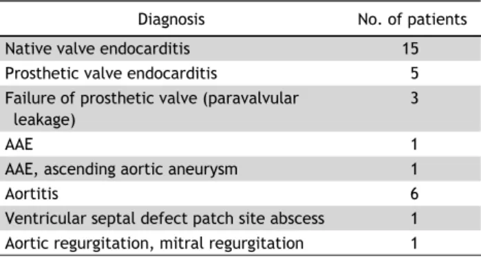
Table 2. Preoperative diagnosis (N=33)
Diagnosis No. of patients
Native valve endocarditis 15
Prosthetic valve endocarditis 5
Failure of prosthetic valve (paravalvular leakage)
3
AAE 1
AAE, ascending aortic aneurysm 1
Aortitis 6
Ventricular septal defect patch site abscess 1 Aortic regurgitation, mitral regurgitation 1 AAE, aortoannular ectasia.
ity of the homograft after thawing. The aims of this study were to evaluate long-term outcomes using ho- mografts in the aortic valve position, and to deter- mine the durability of homografts and the mode of failure.
Methods
1) Patient characteristics
This is a retrospective study of 33 patients (>20 years old) w ho underw ent aortic valve replacement or root replacement w ith a homograft in the Asan Medical Center between April 1995 and May 2015. The mean age at operation was 47.2±2.6 years. Twenty-five pa- tients were male (75.8%). Five patients had hyper- tension (15.2%) (Table 1). The underlying reasons for aortic valve surgery were native valve endocardi- tis (n=15, 45.5%), prosthetic valve endocarditis (n=5, 15.2%), and aortitis (n=6, 18.2%) (Table 2). The in- dication for use of a homograft w as not confirmed. We considered the use of a homograft based on the sur- geon’s preference, available homograft size, and de- gree of infection or aortitis. The primary endpoint was the day of death or explantation of the homograft.
The median follow-up duration was 35.6 months (range, 0 to 168 months).
2) Homograft preservation technique
Valves were collected within 24 hours from explanted hearts of heart transplant recipients (<60 years old).
Another source was from organ donors who were not suitable for heart transplantation. Valves that had a congenital anomaly, large fenestrations in the cusps, or calcification in the cusps or aortic wall were ex- cluded. Valves were extracted and placed in Roswell Park Memorial Institute (RPMI) medium with addi-
tion of low-dose antibiotics (50 IU/mL penicillin and 50 μg/mL streptomycin). The valves were held for 6 hours at 37
oC. After culture, freezing solutions w ere prepared w ith 90 mL RPMI medium plus 10 mL di- methyl sulfoxide (DMSO) solution (i.e., 90 mL RPMI medium plus 10 mL DMSO solution). The valves w ere frozen using a controlled-rate freezing machine, with the tissue temperature decreased at a rate of 1
oC per minute to −40
oC. The life-span of the homograft was 10 years. The homograft was used after confirming that cultures of the solutions and valves were negative.
3) Homograft thawing and dilution technique For implantation, the homograft should be trans- ported at a temperature below −100
oC. Just before being used, the homograft was thawed rapidly at 37
oC to 42
oC to prevent crystallization. The bag con- taining the homograft was soaked in 40
oC saline for 6 to 7 minutes before the ice had melted completely.
The homograft was taken out of the bag, and se- quentially placed for 5 minutes in each of four dilute solutions of RPMI plus DMSO at 5%, 2.5%, 0%, and 0%, respectively.
4) Implantation technique
Twenty-nine patients underwent the root replace-
ment technique, 2 underwent an intact noncoronary
sinus scalloped technique, and 1 underwent an in-
clusion technique. Every procedure was performed
under bicaval cannulation via median sternotomy. The
mean cardiopulmonary bypass time was 234.4±15.6
minutes; aortic cross-clamp time was 175.88±9.32
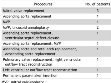
minutes. Mitral valve replacement was performed in
3 patients (10.1%), and other concomitant proce-
dures are summarized in Table 3.
Table 3. Concomitant procedures (N=33)
Procedures No. of patients
Mitral valve replacement 3
Ascending aorta replacement 1
MVP 1
MVP, tricuspid annuloplasty 2
Ascending aorta replacement, ventricular septal defect closure
1
Ascending aorta replacement, MVP 1
Ascending aorta and total arch replacement, descending aorta replacement
1
Pulmonary valve replacement, right ventricular outflow tract reconstruction
1
Left ventricular outflow tract reconstruction 1
Permanent pace-maker insertion 1
MVP, mitral valvuloplasty.
Fig. 2. Freedom from AV dysfunction: moderate to severe aortic regurgitation or aortic stenosis. AV, aortic valve.
Fig. 1. Survival rate after homograft implantation in aortic valve and root position.
5) Statistical analysis
Continuous variable data are expressed as mean±
standard deviation, and categorical variable data are expressed as percentages (%). Survival and freedom from reoperation rates were estimated using the Kaplan-Meier method. To determine differences in echocardiography results before and after surgery, the Wilcoxon signed rank test w as used. A p-value of
<0.05 was considered statistically significant. Statistical analysis was performed using PASW SPSS ver. 18.0 (SPSS Inc., Chicago, IL, USA).
Results
1) Echocardiography
Postoperative echocardiography was performed in all patients, excluding immediate postoperative deaths.
All patients had aortic regurgitation (AR) before sur- gery. Immediate postoperative echocardiography was repeated 6.6±2.1 days later. The mean preoperative AR grade w as 3.5±1.1; the immediate postoperative AR grade was 0.9±0.5, which was downgraded sig- nificantly (p<0.001). The aortic valve systolic pres- sure gradient was downgraded significantly from 59.1±28.8 to 15.8±7.9 (p=0.021); the aortic valve mean pressure gradient was also downgraded significantly from 35.1±17.7 to 8.4±4.7 (p=0.021).
2) Early outcomes
The 30-day mortality rate was 9.1%. Three patients died within 30 days postoperatively; the cause in each case w as sepsis w ith multi-organ failure. Tw o pa- tients had postoperative bleeding and underwent sur- gery to control the bleeding (6.1%). One patient re- quired an intra-aortic balloon pump, and another re- quired a biventricular assist device due to low car- diac output. Another patient was diagnosed with right ventricular failure due to right coronary artery ostium stenosis; coronary artery bypass grafting was performed in this patient.
3) Late outcomes
The 1- and 10-year survival rates were 80.3%±7.2%
and 63.4%±9.5%, respectively (Fig. 1). The 1-, 5-, and
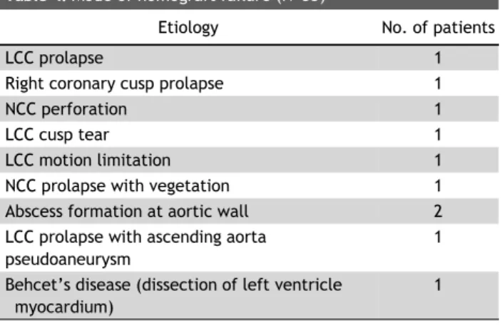
Table 4. Mode of homograft failure (N=33)