http://dx.doi.org/10.7180/kmj.2014.29.2.107
Original Article
Increased carotid atherosclerosis and carotid artery stiffness according to the disease duration and activity in patients with systemic lupus erythematosus
전신 홍반 루푸스 환자의 질환의 활성화 및 기간에 따른 경동맥 경직도와 경동맥 동맥경화 증가
Gi Sup Byun,
1Ji Hyun Lee,
1Kyoung Im Cho, 2 Hong Jik Lee, 1 Hyun Jung Yeo, 1 Min Jeong , Kim, 1 Ho Joon Im 1
변기섭, 1 이지현, 1 조경임, 2 이홍직, 1 여현정, 1 김민정, 1 임호준 1
1
Department of Internal Medicine, Maryknoll Medical Center, Busan, Korea,
2
Department of Internal Medicine, College of Medicine, Kosin University, Gospel Hospital, Busan, Korea
1 메리놀 병원 내과
2 고신대학교 복음병원 내과
Objective: The link among carotid intima-media thickness (IMT), vascular elastic property and the disease activity of systemic lupus erythematosus (SLE) is not well defined. We investigated the association between carotid ath- erosclerosis, elastic properties of the carotid arterial wall and clinical parameters of SLE.
Methods: Fifty-one SLE patients and fifty healthy controls were included. Peak systolic global circumferential and posterior radial strains of carotid artery were measured to assess the elastic properties. Beta stiffness index was used as conventional method for the distensibility of the carotid artery. Information concerning SLE duration, cumulative dose of steroids and/or immunosuppressive drug intake was recorded, and SLE activity was assessed by SLE disease activity index (SLEDAI) score.
Results: Carotid plaques were more common in SLE patients. SLE patients with plaques were older and showed the increased mean IMT, high sensitivity C-reactive protein (hs CRP), IgG anti-cardiolipin antibody (aCL), and longer disease duration compared with those without plaques. Peak systolic global circumferential and posterior radial strain as well β stiffness index were significantly lower in SLE group. Age, disease duration, hsCRP, IgG aCL showed significant correlations with mean IMT and parameters of carotid elastic property (all P’s<0.05).
Conclusions: Carotid atherosclerosis was more common in SLE patients, and carotid arterial stiffness had signif- icant correlation with disease duration, hsCRP and IgG aCL level. Speckle tracking strain imaging is a comparative method for the assessment of elastic properties of carotid artery of SLE patients.
Key Words: Carotid atherosclerosis, Strain, Systemic lupus erythematosus, Ultrasonography Corresponding Author : Kyoung Im Cho, Department of Internal Medicine, College of Medicine,
Kosin University, 262, Kamcheon-ro, Seo-gu, Busan, 602-702, Korea
TEL: +82-51-990-6990 FAX: +82-51-990-3049 E-mail: [email protected]
Received : April 9, 2013
Revised : July 19, 2013
Accepted : September 11, 2013
전신 홍반 루푸스(이하 루푸스)는 여러 기관을 침범하는 전 신적인 자가면역 질환으로 정상인에 비해 일과성 허혈 발 작, 뇌경색, 심근 경색 등의 심혈관 질환의 이환률이 증가되 어 있다. 1 부검을 통한 연구에서 대조군은 심한 동맥경화증 이 2 %인 반면에 루푸스 환자에서는 40 %로 매우 높았고, 루 푸스 환자는 이른 시기부터 동맥경화증이 항진된다는 보고 가 있다. 2,3 또한, 루푸스 환자에서 관상동맥 질환이나 뇌경 색으로 인한 사망 위험도는 약 3배 정도, 20~40대 사이의 루 푸스 환자에서는 약 16배까지 증가하는 것으로 보고되고 있 다. 4 루푸스에서 관찰되는 심혈관 질환의 이환률의 증가는 항인지질 항체로 인한 과응고 상태나 만성적으로 항진된 동 맥경화증이 원인으로 제시되고 있으며 루푸스에서 동맥경 화증의 발생은 전통적인 심혈관계 위험인자 외에 진단 당시 고령, 질병 활성도, 전염증성 고밀도 지질단백질, 부신 피질 호르몬의 사용량, 호모시스테인 등과 관련이 있다. 5
심혈관계 발생 고위험군에서 동맥경화증발생을 측정하기 위한 다양한 검사법들이 사용되고 있으며 대표적인 검사로 비침습적인 방법인 혈관의 내피세포의존성 확장반응(flow mediated dilatation), 맥파 혈류 속도측정(pulse wave veloc- ity), 관상동맥 석회화측정(calcium scoring CT), 그리고 경동 맥 내중막 두께(intima-media thickness, IMT)와 죽상경화 판 검사, 망막 혈관 촬영 등이 있다. 6 이 중 경동맥 내중막 두 께 측정법은 임상적 증상이 나타나기 전에 혈관의 상태를 알 수 있는 선행지표로 사용될 수 있어 무증상 환자에서도 일차 선별검사로 심혈관 질환의 위험성을 예측하는데 사용된다. 7 또한 경동맥 초음파 촬영시 심박동 주기에 포함된 수축기와 이완기에 혈관의 직경을 측정하여 변형(strain)을 측정함으 로써 경동맥 경직도를 평가할 수 있다. 특히 최근 이면성 (2- dimensional) 경동맥 초음파 검사의 speckle tracking 방법을 이용한 변형 측정 방법은 조직 도플러 영상을 이용한 방법에 비하여 다양한 방향의 변형 (longitudinal, circumferential, radial strain)을 측정할 수 있는 장점이 있다. 8,9
최근 루푸스에서 경동맥 내중막 두께를 이용하여 심혈관계 위험성을 평가한 몇몇 연구가 있었으나, 10-13 경동맥의 신전 성(distensibility) 혹은 경직성(stiffness)에 대한 연구는 부족 한 상태이다. 이에 본 연구에서는 speckle tracking 방법을 이 용한 이면성 경동맥 초음파 검사를 이용하여 루푸스 환자에 서 경동맥의 변형 및 경동맥 내중막 두께를 측정하였으며 이 지표 및 질병 활성도와 자가 항체와의 연관성을 알아보고자 하였다.
연구대상 및 방법
1. 연구 대상
본 연구는 단일기관에서 시행된 전향적 코호트 연구이며, 병원 임상 윤리 위원회의 승인을 받았다 (MMC/19-2[148]).
대상 환자는 메리놀 병원 류마티스 내과에서 치료 중인 루푸 스 환자 중 연구 참여에 동의한 51명과 성별, 나이가 유사하 면서 ANA가 음성인 건강인 50명을 대상으로 하였다. 루푸 스 환자는 모두 미국 류마티스 학회(American College of Rheumatology)의 진단 기준을 만족하였다. 환자군과 대조 군 모두 최근 5년 내의 흡연력, 고혈압, 당뇨병, 심부전, 신부 전, 말초혈관질환, 갑상선 이상 및 임신, 최근 3개월 내 지질 강하제 복용력 등이 있는 사람은 제외하였다.
2. 경동맥 초음파
경동맥 초음파 측정은 Vivid 7 (GE Medical System, Mil-
waukee, WI)를 사용하였으며 측정 시 오차를 줄이기 위하여
동일한 검사자(KI Cho)에 의해 측정하였다. 경동맥 내중막
두께는 처음 반사면의 선연으로부터 두번째 반사면의 선연
까지를 측정하였다. 경동맥 내중막 두께를 측정하기 위하여
환자를 앙와위로 하고 전사위, 측위에서 초음파를 투시하여
좌측과 우측 경동맥의 종단면을 따라 경동맥이 내경동맥과
외경동맥으로 분리되는 분지점의 근위부에 위치한 총경동
맥의 원벽에서 내중막 두께가 최고인 지점을 정하고 이를 중
심으로 근위 10 mm, 원위 10 mm 위치에서 내중막 두께를 측 정하였고 3부위의 산술 평균값(평균 내중막 두께)과 3부위 중 최대값(최대 내중막 두께)을 구하였다. 이때 경동맥 내 중 막 두께의 측정이 예정된 부위가 석회화되었거나 내강 내로 돌출된 병변이면서 초음파상 이질성을 보이는 죽전(plaque) 이 있는 경우에는 죽전이 포함되지 않은 근위부에서 측정을 시행하였다. 관찰자 내 변동성(intraobserver variability)은 0.037 mm (5.1%)였다.
3. 경동맥 경직도 지표
경동맥의 경직도를 전형적인 방법으로 측정하기 위해서 심 장의 주기 중 수축기 혈압과 이완기 혈압에 의한 혈관 변형 을 측정하였다. 수축기 혈압과 이완기 혈압의 차이가 기계 적인 스트레스가 되고 이에 대한 경동맥의 경직을 반영하는 지표는 다음과 같은 공식으로 구할 수 있다. 14 -16
1) 변형 (strain, %) = (SD-DD)/DD x100,
SD - 수축기 총경동맥 내경, DD - 이완기 총경동맥 내경 2) 경직도 (β stiffness index) = ln (수축기 혈압/이완기 혈압)/
변형x100
3) 신전성(distensibility )= = 1/ [ln (SBP/DBP)/strain × IMT]
4. Speckle tracking 방법을 이용한 변형 및 변형률 측정
경동맥 변형 및 변형률은 speckle tracking 방법을 이면성 초음파에 적용하여 전용 프로그램인 EchoPAC Dimension system (General Electric, Horten, Norway)으로 측정하였다.
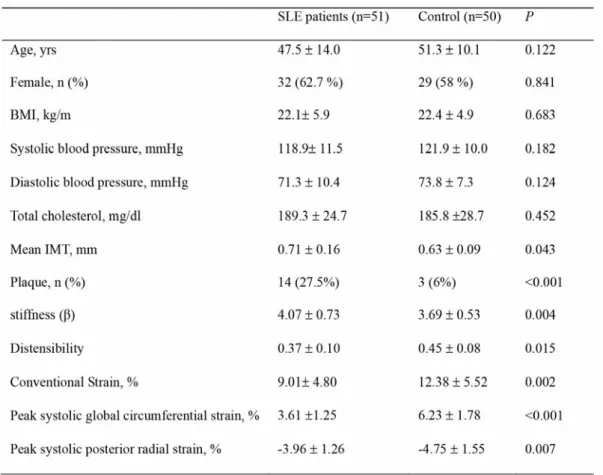
총경동맥의 단축 (short axis view)에서 보이는 경동맥 내중 막을 따라 1x1 mm 면적의 측정 부위(region of interest, ROI) 를 정하고 ROI의 폭을 조절하면 연속적인 화면에서 혈관의 움직임 및 두께를 추적할 수 있으며, 목표로 하는 두 지점 사 이의 상대적 거리 변화는 조직의 수축과 이완을 반영하게 된 다(그림 1). Circumferential peak systolic strain (%)은 단축면 의 전반적인 변형을 측정할 수 있지만 기계의 한계로 radial peak systolic strain은 총경동맥의 후벽부 특정부위 (20 × 20
pixels)에서 측정한다. 수축기시 혈관벽이 늘어나면서 얇아 지기 때문에 circumferential strain은 양의 값이 되고 radial strain은 음의 값이 된다. 화면값(frame rate)은 60 frames/sec 로 측정하였고, 관찰자 내 변동성(intraobserver variability) 은 10~12% 였다.
5. 검사실 검사 및 질병 활성도 측정
모든 환자와 대조군에서 최소 8시간 이상 금식을 시킨 후 채혈하여 백혈구, 혈색소, 혈소판, 적혈구 침강속도, 혈청 C-반응 단백질, 혈청 크레아티닌, 간기능 검사, 총 콜레스 테롤을 측정하였다. 환자군에서는 항핵항체, C3, C4, 항 double stranded DNA항체, 항 Sm항체, 항 Ro항체, 항 La항 체 및 IgM/IgG 항 cardiolipin항체를 측정하였으며 질병활성 도의 측정을 위해 Systemic Lupus Erythematosus Disease Activity Index (SLEDAI) score를 측정하였으며 SLEDAI score 는 경동맥 초음파를 시행하기 직전에 측정하였다.
6. 통계적 분석
통계 분석은 PASW Statistics version 18.0 for Windows
Figure 1. An automated tracking algorithm outlined the intima-
media complex of common carotid artery (a). Systolic and di-
astolic diameters of common carotid artery were obtained from
short axis view (b). Regions of interest with computation area
of 1x1 mm were placed in the intima-media complex from the
short axis view of the common carotid artery (c and d). Peak
systolic global circumferential strain (c) and posterior radial
strain (d) and during systole were used for the analysis.
(SPSS Inc.,Chicago, IL, USA)를 이용하였고, 모든 자료는 평 균값 ± 표준편차 및 퍼센트로 표시하였다. 각 군 간의 비교 는 Student’s t-test를 이용하였고, 그룹 간 빈도의 비교는 카 이 검정법을 이용하였다. 경동맥 내중막 두께에 영향을 미 치는 인자를 확인하기 위하여 Pearson 및 Spearman 상관분 석을 시행하였고, 통계학적으로 유의한 결과를 보인 변수들 의 상관성을 판단하기 위하여 다중회귀분석을 이용하였으 며 P 값이 0.05 미만인 경우를 통계학적으로 유의한 것으로 판정하였다.
결과
1. 환자의 임상적 특징
총 51명의 환자를 대상으로 하였으며 남녀 수는 각각 19 명 (37.3 %), 32 명(62.7 %)이었고 평균연령은 47.06 ± 14.7 세, 유병기간은 평균 5.9± 4.8년 이었다. 연구 시점에서 46명 (92 %)의 환자가 스테로이드를 복용 중이었으며 사용한 스 테로이드 평균 누적량은 4513 ± 5404 mg (prednisolone 기준) 이었다. 그 외 30명의 환자 (60%)가 비스테로이드성 소염제 를, 30명의 환자 (60%)가 항말라리아제를 복용중이었으며 8 명 (16%)가 cyclophosphamide 및 azathioprine과 같은 면역 억제제를 복용중이었다. 37명 (78%)은 단백뇨가 관찰되지 않았으며 4명 (8%)의 환자는 신증후군 정도의 단백뇨가 관 찰되었다.
2. 경동맥 초음파 소견
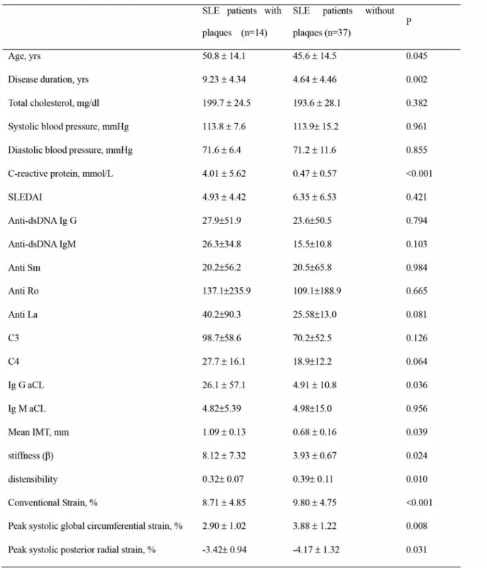
루푸스 환자에서 대조군에 비해 경동맥 중막 두께가 두꺼웠 으며(0.71±0.16 vs. 0.63±0.09 mm, p=0.043) 죽전의 빈도 (27.5 vs. 6 %, P=0.002) 및 β stiffness index (4.07±0.73 vs.
3.69±0.53 %, P=0.004) 역시 현저히 높았다. Speckle tracking method로 측정한 변형 지표인 circumferential strain (9.01±4.80 vs. 12.38±5.52 %, P=0.002) 과 radial strain (- 3.96±1.26 vs. -4.75±1.55 %, P=0.007) 은 환자군에서 감소
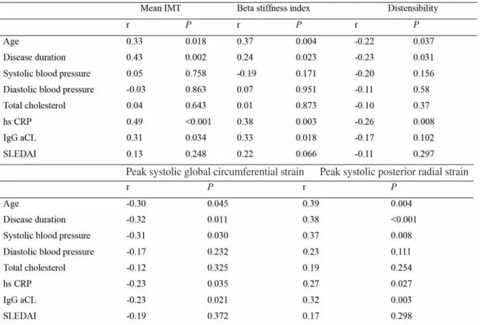
되어 있었고 통계적 유의성을 보였다 (표 1). 전체 환자군을 죽전의 유무에 따라 나누어 임상적인 특징을 비교하였다 (표 2). 죽전이 관찰된 군이 그렇지 않은 군에 비하여 평균 연령 이 더 높았고 (50.8±14.1 vs. 45.6±14.5 years, P=0.045), 유병 기간도 더 길었으며 (9.23±4.34 vs. 4.64±4.46 years, P=0.002), 평균 경동맥 중막 두께(1.09±0.13 vs. 0.68±0.16 mm, P=0.039) 및 β stiffness index(8.12±7.32 vs. 3.93±0.67 years, P=0.024) 가 증가되어 있었고 circumferential strain(2.90±1.02 vs. 3.88±1.22 %, P=0.008) 과 radial strain(- 3.42±0.94 vs. -4.17±1.32 %, P=0.031) 이 유의하게 감소되 어 있었다.
3. 경동맥 초음파와 임상 소견과의 상관관계
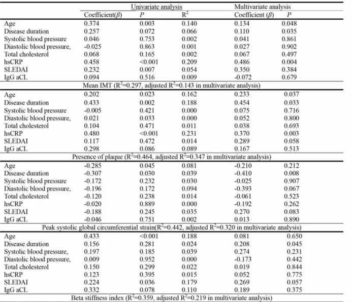
경동맥 중막 두께와 동맥 경직도에 영향을 미치는 여러 인자 들을 알아보기 위하여 상관분석을 시행하였는데 경동맥 중 막 두께와 동맥 경직도는 연령, 유병기간, 혈청 C-반응 단백 질 및 IgG 항 cardiolipin항체와 유의한 양의 상관관계를 보 였다 (all Ps<0.05) (표 3). 상관분석을 토대로 연령, 유병기 간, SLEDAI score, 총콜레스테롤, 혈청 C-반응 단백질 및 IgG 항 cardiolipin항체를 인자로 하여 다중회귀분석을 시행 하였는데, 연령, 유병기간, 혈청 C-반응 단백질이 경동맥의 동맥경화에 영향을 주는 독립적인 위험인자로 나타난 반면 경동맥 변형도에는 유병기간만이 의미 있는 독립인자로 분 석되었다 (표 4).
고찰
본 연구는 심혈관계 위험 인자가 없는 루푸스 환자에서 경동
맥 초음파를 이용하여 동맥 경화 정도 및 경동맥 경직을 측
정한 연구로 루푸스 환자에서 위험인자를 가지지 않은 정상
인에 비해 통계적으로 유의하게 경동맥 중막 두께가 증가되
어 있었으며 여러 임상 변수와의 상관 관계를 조사한 결과로
는 나이 및 질병 이환 기간, 혈청 C-반응 단백질 및 IgG 항
cardiolipin항체와는 양의 상관 관계를 보였으나 총 스테로 이드 투여량, 항핵항체, C3, C4, 항 double stranded DNA항 체, 항 Sm항체, 항 Ro항체, 항 La항체 및 IgM 항 cardiolipin 항체 등 다른 변수와는 유의한 상관 관계가 없었다. 또한 동 맥 경직도를 보기 위해 시행한 경동맥 변형 역시 루푸스 환 자군에서는 감소되어 있었고 이 또한 나이 및 질병 이환 기 간, 혈청 C-반응 단백질 및 IgG 항 cardiolipin항체와는 양의 상관 관계를 나타내었다.
이제까지 루푸스 환자에서 심혈관계 질환 증가의 위험인자 로는 흡연, 고혈압, 당뇨, 고지혈증, 비만 및 고호모시스테 인증 등이 알려져 있으며, 그 외 스테로이드 사용으로 인한 직접적인 동맥경화증의 악화나 면역복합체, 자가항체에 의
한 내피 세포 손상 등이 루푸스에서 동맥경화증을 악화시킨 다고 알려져 있다. 17 또한 루푸스에서 관찰되는 심혈관 질환 의 이환률의 증가는 항인지질 항체로 인한 과응고 상태나 만 성적으로 항진된 동맥경화증이 원인으로 제시되고 있
다. 11,18 루푸스 환자에서 심혈관계 합병증을 조기에 발견하
고 이를 치료하기 위해서는 심혈관계 합병증과 관련이 있는 혈관의 변화를 조기에 측정하는 것이 매우 중요하다.
심혈관계 위험성을 평가하는데 사용되는 방법 중 경동맥 초 음파는 경동맥의 내강-내막 접촉면과 중막-외막 접촉면인 두개의 선 사이의 두께를 측정하며 일반적으로 1.0mm 이상 이면 비정상으로 간주된다. 19 경동맥 중막 두께의 증가는 심 혈관 질환이 없는 군에서 심근경색증과 뇌졸중의 위험을 증
Table 1 Clinical characteristics and parameters of the carotid artery of the study population
All values are presented as the mean ± SD
BMI body mass index, SLE systemic lupus erythematous, IMT intima-media thickness
Table 2 Characteristics of SLE patients with and without carotid arterial plaque
All values are presented as the mean ± SD
SLE systemic lupus erythematous, SLEDAI Systemic Lupus Erythematosus Disease Activity Index, anti-dsDNA
Ab anti-double stranded DNA antibodies aCL anti-cardiolipin antibody, IMT intima-media thickness
가시키며, 경동맥 중막 두께 자체가 전통적인 위험인자와는 별도로 독립적인 예측인자로 이용할 수 있다는 보고도 있 다. 20 루푸스에서 경동맥 중막 두께를 측정한 연구 결과는 다수가 보고되어 있으나 10-13,21-23 그 결과는 일관적이지 못하고 동맥 경직도에 대한 연구는 소수이다. 12,13
변형이란 가해진 힘에 비례한 조직의 상대적인 변화를 뜻하 며 이러한 변형은 공간적으로 세 가지 축을 기준으로 longi- tudinal, circumferential, radial strain으로 구분된다. 24 변형 은 혈관의 경직도 및 신축성을 반영하는 것으로 내피세포의 손상 및 회복 정도와 관련이 있으며 이는 동맥경화의 조기 발현과 관련이 깊다. 25 1980년대 초음파를 이용하여 심장의 변형을 측정하기 시작한 이래로 현재는 조직 도플러 영상을
이용하거나 이면성 초음파 영상에 speckle tracking 방법을 이용하여 변형의 정도를 측정하고 있다. 그러나 기존의 조 직 도플러 영상을 이용한 방법의 경우, 조직에 의한 끌림 현 상에 영향을 받고 초음파 방향과 조직의 움직임이 평행하지 않을 때에는 정확한 정도를 얻지 못하는 제한점 등이 보고되 었다. 26 이에 비해 이면성 경동맥 초음파 검사의 speckle tracking 방법을 이용한 변형 및 변형률 측정 방법은 조직 도 플러 영상을 이용한 방법에 비하여 다양한 방향의 변형을 측 정할 수 있는 장점이 있고, 또한 혈압의 변동에 영향을 받지 않는 다는 장점이 있다. 본 연구에서 루푸스 환자의 동맥경 직도를 측정하는데 이용한 speckle tracking 방법을 이용하 여 측정한 circumferential strain과 radial strain정도는 저자 들이 이전에 시행한 연구에서 전통적으로 측정하는 수축기
Table 3 Correlation coefficients between the parameters of carotid artery and clinical parameters of SLE group (n=51)
IMT intima-media thickness, hs CRP high sensitivity C-reactive protein, aCL anti-cardiolipin antibody, SLEDAI Systemic Lupus Erythematosus Disease Activity Index.
Peak systolic global circumferential strain Peak systolic posterior radial strain
와 이완기시 혈관의 내경의 차와 맥압차로 구하는 beta stiffness index와 유의한 상관 관계를 보인다. 27 본 연구에서 는 혈압에 의존되는 전통적인 방법인 방법이 아닌, 혈관 자 체의 변형을 보여주는 strain parameter를 이용하여 동맥 경 직도를 측정하였고 더욱 예민한 지표를 사용했다는 점이 기 존의 다른 연구와 차이점이 있다.
본 연구의 의의는 심혈관계 위험 인자가 없는 루푸스 환자 에서 동맥 경화도와 동맥 경직도가 증가되어 있음을 확인 하
였고, 전형적인 심혈관계 위험인자인 연령 외에 루푸스에 이환된 기간과 염증 정도, 항인지질 항체와의 연관성을 보 여 줌으로서 과응고 상태나 만성적으로 항진된 동맥경화증 이 루푸스 환자의 심혈관계 위험의 원인이 됨을 보여 주고 있다. 본 연구가 연구 대상을 보편적인 심혈관 질환의 위험 이 없는 집단으로 제한하였음에도 환자군에서 경동맥 중막 두께 증가가 관찰된 것은 루푸스라는 질환이 심혈관계의 위 험 인자임을 추측하게 한다. 본 연구에서 제시된 환자의 나
Table 4 Multiple linear regression analysis of mean IMT and parameters of arterial stiffness of common carotid artery in SLE groups.
hs CRP high sensitivity C-reactive protein, SLEDAI Systemic Lupus Erythematosus Disease Activity Index,
aCL anti-cardiolipin antibody.
이는 현재 나이이며 발병 연령은 명시하지 않았으나 질환 유 병 기간을 변수로 분석하였다. 본 연구에서 관찰되는 경동 맥 초음파의 변화가 SLEDAI와는 관련이 없으나 항체혈청 C-반응 단백질과 유의한 관계가 있다는 결과를 보여준 것 은 루푸스에서 관찰되는 질병 활성도보다 루푸스에서 동반 된 염증이 경동맥 초음파의 변화와 더 관련이 있음을 제시한 다. 또한 본 연구에서는 심혈관 질환의 위험도를 최소화하 기 위하여 연구 대상을 적어도 5년 이내에 흡연력, 당뇨, 고 혈압, 갑상선 기능 저하증이 없는 집단으로 제한하였으며 이로 인해 고령이나 유병 기간이 오래된 환자들이 제외되었 고 그로 인해 포함된 환자군의 질병 활성도가 그리 높지 않 았다. 향후 질병 활성도가 높은 환자를 더 많이 포함한 추가 연구가 필요하리라 생각된다. 또한 더 많은 증례 분석을 통 해 경동맥 초음파의 변화가 루푸스에서 관찰되는 심혈관 질 환의 증가를 조기에 반영할 수 있는지를 확인하고 경동맥의 변화의 단계를 정량적으로 치료에 적용하는 방법이 적용되 어야 하리라 생각된다.
결론적으로, 본 연구에서 루푸스 환자는 정상군에 비해 경 동맥 중막 두께와 죽전의 수가 증가되어 있었고 speckle tracking으로 측정한 변형의 정도의 감소 및 beta stiffness가 증가되어 있었다. 이러한 동맥경화증의 발생은 전통적인 심 혈관계 위험인자 외에 진단 당시 연령, 질병 이환 기간, 항체 혈청 C-반응 단백질 및 IgG 항 cardiolipin항체와 유의한 관 련이 있었다.
참고문헌
1. J.Wajed, Y.Ahmad, P.N. Durrington, Bruce IN. Prevention of cardiovascular disease in systemic lupus erythematosus?
proposed guidelines for risk management. Rheumatology 2004;43:7-12.
2. Manzi S, Meilahn EN, Rairie JE, Conte CG, Medsger TA Jr.
Jansen-McWilliams L, et al. Age-specific incidence rates of myocardial infarction and angina in women with systemic lupus erythematosus:comparison with the Framingham Study.
Am J Epidemiol 1997;145:408-15.
3. Roman MJ, Shanker BA, Davis A, Lockshin MD, Sammari- tano L, Simantov R, et al. Prevalence and correlates of accel- erated atherosclerosis in systemic lupus erythematosus. N Engl J Med 2003;349:2399-406.
4. Bjornadal L, Yin L, Granath F, Klareskog L, Ekborn A. Car- diovascular disease a hazard despite improved prognosis in patients with systemic lupus erythematosus: results from a Swedish population based study. J Rheumatol 2004;31:713- 5. Tassiulas IO, Boumpas DT. Textbook of Rheumatology. 8th9.
ed. P. 1429-51, Philadelphia, Saunders, 2009.
6. Alam TA, Seifalian AM, Baker D. A review of methods cur- rently used for assessment of in vivo endothelial function. Eur J Vasc Endovasc Surg 2005; 29:269-76.
7. Sohn IS, Lee JB, Cho BH, Park JH, Jin ES, Cho JM, et al.
Carotid intima-media thickness and arterial stiffness in hy- pertensive patients with first attack of ischemic stroke. J Ko- rean Soc Hypertens 2010;16:14-21.
8. Hoeks AP, Brands PJ, Smeets FA, Reneman RS. Assessment of the distensibility of superficial arteries Ultrasound Med Biol 1990;16:121-8.
9. Selzer RH, Mack WJ, Lee PL, Kwong-Fu H, Hodis HN. Im- proved common carotid elasticity and intima-media thickness measurements from computer analysis of sequential ultra- sound frames. Atherosclerosis 2001;154:185-93.
10. El-Magdmi M, Bodill H, Ahmad Y, Durrington PN, Mack- ness M, Walker M, et al. Systemic lupus erythematosus. An independent risk factor for endothelial dysfunction in women.
Circulation 2004;110:399-404.
11. Svenuungsson E, Jensen-Urstad K, Heimburger M, Silveira A, Hamsten A, de Faire U, et al. Risk factors for cardiovas- cular disease in systemic lupus erythematosus. Circulation 2001;104:1887-93.
12. Greene ER, Lanphere KR, Sharrar J, Roldan CA. Arterial dis- tensibility in systemic lupus erythematosus. Conf Proc IEEE Eng Med Biol Soc 2009;2009:1109-12.
13. Cacciapaglia F, Zardi EM, Coppolino G, Buzzelini F, Mar- giotta D, Arcarese L, et al. Stiffness parameters, intima-media thickness and early atherosclerosis in systemic lupus erythe- matosus patients. Lupus 2009;18:249-56.
14. Barenbrock M, Kosch M, Joster E, Kisters K, Rahn KH, Hausberg M. Reduced arterial distensibility is a predictor of cardiovascular disease in patients after renal transplantation.
J Hypertens 2002;20:79-84.
15. Van Popele NM, Grobbee DE, Bots ML, Asmar R, Topouch- ian J, Reneman RS, et al. Association between arterial stiff- ness and atherosclerosis: the Rotterdam Study. Stroke 2001;
32:454-60.
16. Harloff A, Strecker C, Reinhard M, Kollum M, Handke M,
Olschewski M, et al. Combined measurement of carotid stiff- ness and intima-media thickness improves prediction of com- plex aortic plaques in patients with ischemic stroke. Stroke 2006;37:2708-12.
17. Manzi S. Systemic lupus erythematosus; a model for athero- genesis? Rheumatology 2000;39:353-9.
18. Ames PR, Margarita A, Alves JD. Antiphospholipid antibod- ies and atherosclerosis: insights from systemic lupus erythe- matosus and primary antiphospholipid syndrome. Clin Rev Allergy Immunol 2009;37:29-35.
19. Bots ML, Dijk JM, Oren A, Grobbee DE. Carotid intima- media thickness, arterial stiffness and risk of cardiovascular disease: current evidence. J Hypertens 2002;20:2317-25.
20. Greenland P, Abrams J, Aurigemma GP, Bond MG, Clark LT, Criqui MH, et al. Prevention Conference V: Beyond sec- ondary prevention: identifying the high-risk patient for pri- mary prevention: noninvasive tests of atherosclerotic burden:
Writing Group III. Circulation 2000;101:E16-22.
21. de Leeuw K, Smit AJ, de Groot E, van Roon AM, Kallenberg CG, Bijl M. Longitudinal study on premature atherosclerosis in patients with systemic lupus erythematosus. Atherosclero- sis 2009;206:546-50.
22. Manzi S, Selzer F, Sutton-Tyrrell K, Fitzgerald SG, Rairie JE, Tracy RP, et al. Prevalence and risk factors of carotid plaque in women with systemic lupus erythematosus. Arthri- tis Rheum 1999;42:51-60.
23. Roman MJ, Shanker BA, Davis A, Lockshin MD, Sammari- tano L, Simantov R, et al. Prevalence and correlates of accel- erated atherosclerosis in systemic lupus erythematosus. N Engl J Med 2003;349:2399-406.
24. Gilman G, Khandheria BK, Hagen ME, Abraham TP, Seward JB, Belohlavek M. Strain rate and strain: a step-by-step ap- proach to image and data acquisition. J Am Soc Echocardiogr 2004;17:1011-20.
25. Laurent S, Boutouyrie P, Lacolley P. Structural and genetic bases of arterial stiffness. Hypertension 2005;45:1050-5.
26. Golemati S, Sassano A, Lever MJ, Bharath AA, Dhanjil S, Nicolaides AN. Carotid artery wall motion estimated from B- mode ultrasound using region tracking and block matching.
Ultrasound Med Biol 2003;29:387-99.
27. Lee JH, Cho KI, Kim SM. Carotid arterial stiffness in patients with rheumatoid arthritis assessed by speckle tracking strain imaging: its association with carotid atherosclerosis Clin Exp Rheumatol 2012;30:720-8.