• 검색 결과가 없습니다.

Reduction of Chronic Temporomandibular Joint Dislocation by Surgical Traction: Two Cases Report

N/A
N/A
Protected

Academic year: 2021

Share "Reduction of Chronic Temporomandibular Joint Dislocation by Surgical Traction: Two Cases Report"

Copied!
5
0
0

로드 중.... (전체 텍스트 보기)

전체 글

(1)

pISSN 2288-9272 eISSN 2383-8493 J Oral Med Pain 2017;42(2):44-48 https://doi.org/10.14476/jomp.2017.42.2.44

Reduction of Chronic Temporomandibular Joint Dislocation by Surgical Traction: Two Cases Report

Hye-Youn Lim, Sang-Jun Park, Tae-Young Jung

Department of Oral and Maxillofacial Surgery, Inje University Busan Paik Hospital, Busan, Korea

Received April 26, 2017 Revised June 9, 2017 Accepted June 13, 2017

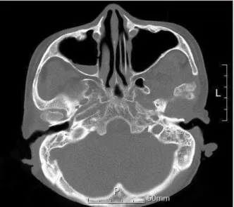
Chronic temporomandibular joint dislocation is defined as an acute dislocation that cannot be reduced or that recurs for more than one month. The management of dislocation depends on patient status and the duration of dislocation and ranges from conservative reduction to a surgical approach. In the present cases, a 64-year-old male was referred to our department for treatment of chronic dislocation for 6 weeks. The dislocation might be occurred by endotra- cheal intubation. A 70-year-old female was referred to our department with repeat right con- dyle dislocation after reduction of dislocation at a local clinic. When she visited for later treat- ment of new dentures, her condyle had been dislocated again for several weeks. In both cases, we tried to treat the dislocation with several manipulations, which were unsuccessful. Finally, chronic dislocation was successfully treated by surgical traction under general anesthesia with- out relapse. Surgical traction is a simple, effective option with the lowest morbidity of surgical procedures for chronic dislocation when conservative reduction is unsuccessful.

Key Words: Condyle; Joint dislocations; Temporomandibular joint; Traction

Correspondence to:

Tae-Young Jung

Department of Oral and Maxillofacial Surgery, Inje University Busan Paik Hospital, 75 Bokji-ro, Busanjin-gu, Busan 47392, Korea

Tel: +82-51-890-6369 Fax: +82-51-896-6675 E-mail: [email protected]

JOMP

Journal of Oral Medicine and Pain

Copyright Ⓒ 2017 Korean Academy of Orofacial Pain and Oral Medicine. All rights reserved.

CC This is an open-access article distributed under the terms of the Creative Commons Attribution Non-Commercial License (http://creativecommons.org/licenses/by-nc/4.0/), which permits unrestricted non-commercial use, distribution, and reproduction in any medium, provided the original work is properly cited.

INTRODUCTION

Dislocation of the temporomandibular joint (TMJ) typi- cally occurs when the mandibular condyle is located in an abnormal position in the glenoid fossa.

1)

An anterior posi- tion to the articular eminence is the most common type and bilateral dislocation is more common than unilateral dis- location.

2)

Females have a higher incidence of dislocation than males.

3)

The causes of dislocation include excessive movement during yawning, dental treatment, airway intubation and endoscopic operation for digestive tract, joint hypermobil- ity associated with systemic diseases, such as Ehlers-Danlos and Marfan syndromes, congenital joint weakness, and psychogenic and neurological disorders.

1,4)

Dislocation may also be caused by internal derangement of the TMJ, such as loosening and weakening of the TMJ capsular ligaments, masticatory muscle dysfunction, occlusal disharmony, or

atrophy of the articular eminence.

5)

In addition, in edentu- lous patients without opposing dentition, the TMJ capsule and lateral ligament can be stretched due to overclosure for a long time, leading to disclocation.

5)

Dislocation of the TMJ is usually acute. Recurrent dislo- cation or habitual dislocation is defined as repeated occur- rence of acute dislocation.

2)

Chronic dislocation can also be defined as an acute dislocation lasting for more than 1 month due to lack of reduction or recurrence.

6)

Chronic dislocation can cause fibrous adhesions between the disc and condyle and fibrous changes in jaw muscles and ligaments.

3)

This can lead to TMJ disorders such TMJ myalgia or spasm, inability to close the mouth, occlusal and facial changes, speech disorder, and excessive salivation, and makes reduction difficult.

2)

The treatment depends on patient status and the dura-

tion of dislocation, and ranges from conservative reduction,

such as bimanual reduction or botulinum toxin injection to

(2)

surgical approaches, such as condylectomy, condylotomy, inverted L-shaped ramus osteotomy, vertical ramus oste- otomy, myotomy, or surgical traction to lower the border.

5,7)

The purpose of this study is to present the authors’ experi- ence of chronic TMJ dislocation and review literature relat- ed to its management.

CASE REPORT

1. Case 1

A 64-year-old male was referred to our department with deviation of the jaw toward the right side. He had been in- volved in a traffic accident and stayed in the intensive care unit (ICU) for about 9 weeks. When endotracheal intuba- tion was changed to tracheostomy on his sixth week in the ICU, the dislocation was found and he was referred to our department. The dislocation might have been caused by en- dotracheal intubation, because both condyles of the man- dible were in the normal position on facial bone comput- ed tomographic (CT) image taken in the emergency room whereas his left condyle was located anterior to the articu- lar eminence on facial bone CT taken when he was referred (Fig. 1). He had prior treatment experience of temporoman- dibular disorder with mouth opening limitation and pain.

Traditional manual reduction was performed but failed be- cause of muscle rigidity. Muscle relaxants were given and exercises for mouth opening continued. Manual reductions

were tried several times; however, the muscle relaxant was stopped due to nausea and vomiting and manual reduction was not successful because of muscle rigidity. Four weeks after the first examination, the patient was asked to un- dergo surgery because his general condition had improved.

Surgical traction was performed under general anesthesia.

Arch bars on the maxilla and mandible were applied. An incision was made from the retromolar area to the buccal vestibule at the first molar on the left side and detached the periosteum of the ramus, the posterior aspect of mandible, sigmoid notch, and condyle. In addition, a 1-cm extraoral incision was made under the left mandibular angle, a strip- per was inserted from the posterior mandibular border into the sigmoid notch to draw the mandible downward, and the left condyle was reduced by surgical traction with the stripper. The maxillomandibular fixation (MMF) was used to correct the occlusion and continued for 3 weeks. After the occlusion stabilized, the MMF was removed. The pa- tient was required to do mouth opening exercises. Mouth opening was 30-mm 1-month after surgery (Fig. 2). After another 2 months of follow-up, there was no recurrence or change in mouth opening and occlusion.

2. Case 2

A 70-year-old female was referred to our department with the chief complaint of repeat right condyle disloca- tion after reduction of left condyle dislocation at the local clinic. Traditional manual reduction was performed suc- cessfully. She had a previous history of TMJ dislocation

Fig. 2. Postoperative clinical photograph for case 1 shows a 30-mm range of mouth opening.

Fig. 1. Preoperative computed tomographic image for case 1.

(3)

during denture treatment 2 years previously. At that time, she underwent reduction of the dislocation at the local clinic. She visited our clinic for extraction of the root rest and new denture treatment. After 5 months from the on- set of treatment, our colleague dentist found her TMJ dis- location during denture treatment. She tried to wear her old dentures, but failed because of anterior deviation of the jaw. Radiologic examinations were undertaken with a pan- oramic radiograph and TMJ four-cuts view (Fig. 3). Both radiographs showed that her mandibular condyles had been displaced out of the glenoid fossas and anterior to the ar- ticular eminences, but she was unaware of this. As a result, the authors were unable to determine the exact duration of dislocation and assumed it to be several weeks. Manual

reduction was attempted to reposition the condyles to the original position but this attempt failed. Muscle relaxants were given and several manual reductions were tried, but these attempts also failed. At this point, surgical interven- tion was indicated and discussed with the patient. The au- thor performed surgical traction under general anesthesia as for the first case. Extraoral incisions were made under both mandibular angles and the strippers were inserted from the posterior mandibular borders into the sigmoid notches to draw the mandible downward. Both condyles were reduced by surgical traction with the strippers (Fig. 4). The authors placed MMF by excavating holes in her denture and used wire traction to correct the occlusion (Fig. 5). The MMF wire was removed after 2 weeks and the occlusion was stable.

Fig. 3. Preoperative temporomandibular panoramic image for case 2 shows both temporomandibular joint dislocations.

Fig. 5. Maxillomandibular fixation using existing dentures to correct the occlusion in case 2.

Fig. 4. Image of surgical traction to draw the mandible downwards

in case 2.

(4)

The patient was required to do mouth opening exercises.

Mouth opening was 40-mm 1-month after surgery. After another 1-week of follow-up, we continued treatment for new dentures because there was no recurrence or change in mouth opening.

DISCUSSION

Chronic TMJ dislocation is relatively rare and its reduc- tion is difficult because of fibrotic changes of soft tissue and soft tissue in-growth into the glenoid fossa with dislo- cation.

8)

Huang et al.

8)

suggested that acute inflammation slowed down and repairing granulation was formed when an acute dislocation was left untreated. They reported no successful cases of conventional reduction when the dis- location lasted for more than 30 days, even under general anesthesia.

Conservative treatment is usually tried before more ag- gressive therapies are used. Conservative treatment includes bimanual reduction, local injection of fibrosing solutions to tighten the joint capsule, or botulinum toxin injection into the lateral pterygoid muscle.

5)

Bimanual reduction is the mainstay of treatment. This involves manipulation of the mandibular condyle downward and backward into the glenoid fossa with local or general anesthesia, sedation, or by oneself.

9)

The use of autologous blood reported by Brachmann

9)

in 1964 is based on the principle of restrict- ing excessive mandibular movements by inducing fibro- sis in the upper joint space and the pericapsular tissues.

10)

Botulinum toxin A temporarily blocks acetylcholine and weakens muscles; however, it may not be appropriate if the ligaments are loose or the muscles are weak.

11)

Huang et al.

8)

suggested that surgical procedures were re- quired to correct dislocations lasting more than 6 months.

There have been many reports of surgical treatments, such as condylectomy, condylotomy with or without coronoidot- omy, eminectomy, inverted L-shaped ramus osteotomy, ver- tical ramus osteotomy, myotomy, periosteal stripping, and surgical traction with wire to the lower border.

1)

For surgi- cal procedures like condylectomy, condylotomy or eminec- tomy, exposure of the TMJ is achieved by the preauricular approach under general anesthesia.

4)

Condylectomy is the method used to expose the zygomatic arch and condyle

head. The condyle head is resected and, when the mouth is closed, the condyle head is placed on the condyle fossa.

4)

Eminectomy reduces the eminence to permit free movement of the condyle. Augmentation of the eminence limits an- terior movement of the mandibular condyle by increasing the height of the eminence with grafting.

12)

According to Adekeye et al.,

13)

in more resistant cases with limited man- dibular movement, condylectomy or condylotomy may be necessary and possible together with coronoidotomy be- cause of the impingement of the coronoid on the condylar process. Also, inverted L-shaped ramus osteotomy or modi- fied vertical ramus osteotomy allows repositioning of the mandible and achieves normal occlusion without modifi- cation of the joint.

13)

Myotomy limits mandibular transla- tion and allows only rotational movement of the condyle through resection of the insertion of the external pterygoid muscle.

2)

Many researchers have used surgical traction for reduc- tion of chronic TMJ dislocation. In 1968, Rowe and William used a bone hook, which was passed through the subperi- osteal tunnel from a small incision below the mandibular angle, reached the sigmoid notch, and then pulled down- ward.

3,14)

In 1981, Stakesby Lewis

15)

used a bristows eleva- tor through the temporal fascia, in the same manner as the Gillies technique of elevation of the depressed zygomatic bone or arch. In 1986, El-Attar and Ord

16)

used a traction hook inserted into bur holes in the angle of the mandible.

In the present cases, the authors thought that traction would be a simple, less invasive, and effective surgical op- tion. When a conservative approach is not successful, sur- gical traction under general anesthesia is the least morbid surgical intervention for reduction of chronic TMJ disloca- tion. Our two patients did not show relapse or functional mobility disorder 3 months after surgical traction. Further recurrence of dislocation may require more invasive open surgery, such as condylectomy or eminectomy to prevent recurrence of chronic dislocation.

CONFLICT OF INTEREST

No potential conflict of interest relevant to this article

was reported.

(5)

REFERENCES

1. Baur DA, Jannuzzi JR, Mercan U, Quereshy FA. Treatment of long term anterior dislocation of the TMJ. Int J Oral Maxillofac Surg 2013;42:1030-1033.

2. Shakya S, Ongole R, Sumanth KN, Denny CE. Chronic bilateral dislocation of temporomandibular joint. Kathmandu Univ Med J (KUMJ) 2010;8:251-256.

3. Sidalingapa MN, Singh M, Dwarakanath S, Mittal N. Chronic bi- lateral condylar dislocation. Int J Contemp Dent 2011;2:12-15.

4. Kim CH, Kim DH. Chronic dislocation of temporomandibular joint persisting for 6 months: a case report. J Korean Assoc Oral Maxillofac Surg 2012;38:305-309.

5. Schwartz AJ. Dislocation of the mandible: a case report. AANA J 2000;68:507-513.

6. Saikia D. Long standing temporomandibular joint dislocation: a case report. IOSR-JDMS 2000;13:3-8.

7. Ardehali MM, Kouhi A, Meighani A, Rad FM, Emami H. Tem- poromandibular joint dislocation reduction technique. Ann Plast Surg 2009;63:176-178.

8. Huang IY, Chen CM, Kao YH, Chen CM, Wu CW. Management of long-standing mandibular dislocation. Int J Oral Maxillofac Surg 2011;40:810-814.

9. Brachmann F. Eigenblutinjektionen bei rezidivierendren, nicht- fixierten Kiefergelenkluxationen. Zahnarztl 1964;15:97.

10. Machon V, Abramowicz S, Paska J, Dolwick MF. Autologous blood injection for the treatment of chronic recurrent temporo- mandibular joint dislocation. J Oral Maxillofac Surg 2009;67:114- 119.

11. Moore AP, Wood GD. Medical treatment of recurrent temporo- mandibular joint dislocation using botulinum toxin A. Br Dent J 1997;183:415-417.

12. Vasconcelos BC, Porto GG, Neto JP, Vasconcelos CF. Treatment of chronic mandibular dislocations by eminectomy: follow-up of 10 cases and literature review. Med Oral Patol Oral Cir Bucal 2009;14:e593-e596.

13. Adekeye EO, Shamia RI, Cove P. Inverted L-shaped ramus oste- otomy for prolonged bilateral dislocation of the temporoman- dibular joint. Oral Surg Oral Med Oral Pathol 1976;41:568-577.

14. Rowe NL, William JLI. Maxillofacial injuries. Vol. 1. 2nd ed.

Edinburgh London, Madrid Melbourne, New York and Tokyo:

Churchill Livingstone; 1994. pp. 421-422.

15. Stakesby Lewis JE. A simple technique for reduction of long- standing dislocation of the mandible. Br J Oral Surg 1981;19:52- 56.

16. El-Attar A, Ord RA. Long-standing mandibular dislocations: re-

port of a case, review of the literature. Br Dent J 1986;160:91-94.

수치

Fig. 2. Postoperative clinical photograph for case 1 shows a 30-mm  range of mouth opening.
Fig. 3. Preoperative temporomandibular  panoramic image for case 2 shows both  temporomandibular joint dislocations.

참조

관련 문서

Conclusion : The treatment using facet joint block with lumbar surgical operation seems to have short term clinical effects, however can't prevent

This report, therefore, analyses two important policy issues: (1) the appropriateness of the government allocated GHG reduction targets by sector and industry; (2) the

The reduction of power series ( , m→ ∞) to polynomials (m is finite) is a great advantage.. because then we have solutions for all x,

Purpose: The purpose of this study is to report the safety and effectiveness of arthroscopic-assisted implantation of Cartistem®, an articular cartilage

Every seconds complete mixing across the cross section of the river (but not longitudinally) occurs. An instantaneous injection of a conservative tracer results in a

ƒ Reduction (환원) : reduction of metal oxides using gases such as.. ƒ Reduction (환원) : reduction of metal oxides using gases such as hydrogen

In linear stability problems of the nongyroscopic conservative type all the critical loads are supplied not only by the kinetic approach but also by the

The Jacobi energy integral is one type of invariant of motion associated with