• 검색 결과가 없습니다.

Cardiometabolic Effects of Obstructive Sleep Apnea and Treatment Effects of Oral Appliance: An Updated Review for Dentists JOMP

N/A
N/A
Protected

Academic year: 2021

Share "Cardiometabolic Effects of Obstructive Sleep Apnea and Treatment Effects of Oral Appliance: An Updated Review for Dentists JOMP"

Copied!
9
0
0

로드 중.... (전체 텍스트 보기)

전체 글

(1)

pISSN 2288-9272 eISSN 2383-8493 J Oral Med Pain 2018;43(3):61-69 https://doi.org/10.14476/jomp.2018.43.3.61

Cardiometabolic Effects of Obstructive Sleep Apnea and Treatment Effects of Oral Appliance: An Updated Review for Dentists

Hye-Kyoung Kim, Mee-Eun Kim

Department of Oral Medicine, College of Dentistry, Dankook University, Cheonan, Korea

Received June 14, 2018 Revised August 24, 2018 Accepted August 29, 2018

Obstructive sleep apnea (OSA) is a relatively common, but greatly underdiagnosed sleep-related breathing disorder, characterized by recurrent collapse of the upper airway during sleep. OSA has been associated with a variety of cardiometabolic disease, such as hypertension, coronary artery disease, cardiac arrhythmia, cerebrovascular disease and metabolic dysfunction. Neuro- cognitive impairment, including excessive daytime sleepiness, increased risk of motor vehicle accidents, is also related to OSA. Sleep fragmentation and related arousals during sleep lead to intermittent hypoxia, sympathetic activation, oxidative stress, systemic inflammation and metabolic dysregulation which provide biological plausibility to this pathologic mechanism.

Extensive studies demonstrated that OSA is a modifiable risk factor for the above mentioned diseases and oral appliances (OAs), although continuous positive air pressure (CPAP) is a first- line therapy of OSA, are not inferior to CPAP at least in mild OSA, and may be an alternative to CPAP in CPAP-intolerant subjects with OSA. The goal of this article is to provide a current knowledge of pathologic link between OSA and cardiovascular disease, focusing on intermit- tent hypoxia, sympathetic activation, oxidative stress and metabolic dysregulation. Then, previous epidemiologic studies will be reviewed to understand the causal relationship between OSA and cardiovascular disease. Finally, the effects of OAs will be updated via recent meta- analyses compared to CPAP.

Key Words: Cardiovascular; Mandibular advancement device; Obstructive sleep apnea; Oral appliance

Correspondence to:

Mee-Eum Kim

Department of Oral Medicine, College of Dentistry, Dankook University, 119 Dandae-ro, Dongnam-gu, Cheonan 31116, Korea

Tel: +82-41-550-1915 Fax: +82-505-434-7951 E-mail: [email protected]

JOMP

Journal of Oral Medicine and Pain

Copyright Ⓒ 2018 Korean Academy of Orofacial Pain and Oral Medicine. All rights reserved.

CC This is an open-access article distributed under the terms of the Creative Commons Attribution Non-Commercial License (http://creativecommons.org/licenses/by-nc/4.0/),

INTRODUCTION

The third edition of the International Classification of Sleep Disorders-3 identified seven major categories consist- ing of insomnia disorders, sleep-related breathing disorders, central disorders of hypersomnolence, cardiac rhythm sleep- wake disorders, sleep-related movement disorders, para- somnias, and other sleep disorders (Table 1).

1)

Among them, obstructive sleep apnea (OSA) is one of the major pheno- types of sleep-related breathing disorders (Table 2). In terms of mechanical view, OSA is a disease associated with mani- festation of ever-increasing resistance to airflow in upper airway cause by intermittent collapse of the upper airway

during sleep.

As consequences of repetitive obstruction of airflow, the diagnosis of OSA requires either signs/symptoms (associated sleepiness, fatigue, insomnia, snoring, subjective nocturnal respiratory disturbance, or observed apnea) or associated medical (hypertension [HTN], coronary artery disease [CAD], arterial fibrillation, congestive heart failure, stroke, diabetes) or psychiatric disorder (cognitive dysfunction, or mood dis- order) coupled with five or more predominantly obstructive respiratory events per hour of sleep during polysomnogra- phy (PSG).

2)

Alternatively, a frequency of obstructive respi- ratory events ≥15/h satisfies the criteria.

OSA is a common health problem that affects 3% to 7%

(2)

of the adult population aged 30 to 70 years in western countries.

3)

In Korea, Kim’s epidemiologic study

4)

that used PSG found that the prevalence of sleep-disordered breath- ing (SDB, apnea-hypopnea index, AHI ≥5) was 27% and 16% in men and women aged 40 to 69 years.

4)

When OSA was defined by an AHI ≥5 and excessive daytime sleepi- ness, its prevalence was 4.5% in men and 3.2% in women.

4)

However, OSA still remains greatly underdiagnosed.

5)

This prevalence is likely to increase because of increasing obe- sity and the aging population.

6)

Undiagnosed and untreated OSA may lead to a variety of cardiometabolic disorders.

Long-term observation study confirmed a clear association between OSA and cardiovascular disease.

7)

The dentist should have a sound understanding of sys- temic effect of untreated OSA in order to effectively diag- nose and treat OSA.

In this review, intermediate mechanisms underlying the systemic effects of OSA on cardiovascular disease (CVD) will be described and epidemiological evidence supports such a relation also be discussed. Finally, the treatment ef- fects of oral appliance (OA) on CVD will be updated.

UNDERLYING MECHANISM OF OSA FOR CARDIOVASCULAR DISEASE

1. Intermittent Hypoxia

Unique characteristic pattern of OSA is a repetitive epi- sodic hypoxia followed by reoxygenation. Animal studies of Fletcher’s group

8,9)

have shown that intermittent hypoxia associated with recurrent upper-airway obstruction leads to a significant increase in blood pressure (BP) that that is dependent on peripheral carotid chemoreceptors. This was independent of hypercapnia. Exposure to intermittent

hypoxia for 2 weeks also elevated daytime BP and sympa- thetic activity of healthy subjects.

10)

The renin-angiotensin system might be a possible explanation for secondary surge in BP on exposure to intermittent hypoxia. Fletcher’s ani- mal

11)

experiment demonstrated that sustained alteration in the renin-angiotensin system following acute alteration in BP with intermittent hypoxia and suggested that upregula- tion of the tissue angiotensin II system appears to be nec- essary for the chronic BP changes that occur from episodic hypoxia.

11)

All of these evidence support the importance of intermittent hypoxia, as the critical abnormality in OSA, leading to the immediate-term and long-term cardiovascu- lar consequences of OSA including systemic HTN, left ven- tricular hypertrophy, and endothelial dysfunction.

6)

2. Sympathetic Activation

During hypoxia, activation of carotid chemoreceptors results in hyperventilation to enhance oxygen delivery to blood, which is followed by sympathetically mediated vaso- constriction to redistribute oxygenated blood flow to vital organs and simultaneously, parasympathetically activat- ed bradycardia to reduce myocardial oxygen demand.

12-14)

Unfortunately, this oxygen-conserving reflex becomes dys- functional and pathological with the imbalance between sympathetic and parasympathetic activity when the increase in sympathetic activity is sustained in OSA. Somers et al.

13)

has demonstrated that patients with OSA have high sym- pathetic activity even when awake, with further increases in BP and sympathetic activity during sleep. Kumar and Prabhakar

15)

have suggested the concept of the carotid body plasticity as a result of intermittent hypoxia. It is thought that the sympathetic hyperactivity persists through the day- time owing to the memory effect of plasticity in the sympa- thetic activation

6)

and this sensitization appears to be asso- ciated with reactive oxygen species (ROS) and hypoxia-in- ducible factor-1.

16)

Ultimately, sustained sympathoexcitation

Table 2. Sleep-related breathing disorders Disorder Obstructive sleep apnea disorders Central sleep apnea syndromes Sleep-related hypoventilation disorders Sleep-related hypoxemia disorder Table 1. Seven major sleep disorders categorized by ICSD-3

Diagnostic section Insomnia

Sleep-related breathing disorders Central disorders of hypersomnolence Circadian rhythm sleep-wake disorders Parasomnias

Sleep-related movement disorders Other sleep disorders

ICSD, International Classification of Sleep Disorders.

(3)

can be seen as a critical mediator between OSA and HTN and has a catastrophizing effect on cardiovascular system.

Particularly, in patients with HTN, impaired baroreceptor- chemoreceptor reflex may predispose to excessive auto- nomic responses to chemoreflex and drug-resistant HTN.

12)

3. Oxidative Stress

Repetitive intermittent hypoxia followed by reoxygen- ation, as mentioned above, promotes the carotid chemore- ceptors via an ROS-dependent pathway

17)

and may cause adenosine triphosphate depletion, xanthine oxidase acti- vation, activation of polymorphoneuclear neutrophils and increases the generation of oxygen-derived free radicals.

18)

This pathological phenomenon is analogous to a cardiac ischemia-reperfusion injury.

19)

An imbalance between pro- and antioxidant system with overproduction of ROS leads to oxidative stress which imparts an tremendous burden the cardiovascular disease. Excessive accumulation of ROS in various organs and systems promotes vascular diseases through direct and irreversible oxidative damage to mac- romolecules, as well as disruption of redox-dependent vas- cular wall signaling processes.

20)

Vascular damage via in- creased oxidative stress is involved in the pathogenesis of endothelial dysfunction with likely contribution to cogni- tive impairment as well as vascular lesion, atherosclerosis, HTN, myocardial injury and stroke.

6)

Recurrent intermittent hypoxia contributes vascular dysfunction via systemic in- flammation as well as oxidative stress. OSA patients present increased circulating leukocyte,

21)

C-reactive protein (CRP),

22)

serum amyloid A

23)

and cytokines,

24)

such as tumor necrosis factor- α and interleukin-6.

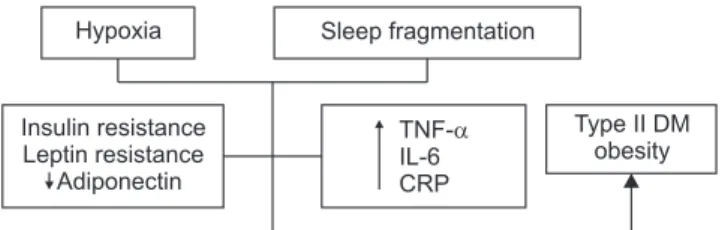
4. Metabolic Dysregulation

Many studies have shown that OSA is associated with the development of metabolic syndrome, independent of obesity.

25-30)

Coughlin et al.

31)

reported that metabolic syn- drome was 9.1 times more likely to be present in subjects with OSA. Metabolic syndrome, as a major risk factor for cardiovascular morbidity and mortality,

32)

is defined by the presence of at least three of the following signs: abdominal obesity, hypertriglyceridemia, low plasma high-density li- poprotein, hyperglycemia, and elevated BP.

27)

Among them, insulin resistance is a primary manifestation of metabolic

syndrome and typical feature of OSA.

31,33)

Human experi- ments have demonstrated that acute exposure to hypoxia worsened insulin sensitivity and glucose tolerance.

34,35)

It is thought that sympathetic hyperactivity caused by hypoxia in OSA stimulates glycogenolysis, gluconeogenesis and li- polysis and as a result, promotes hyperinsulinemia and in- sulin resistance.

28,33)

Sleep restriction or sleep loss could also affect the process, such as dysfunctional glucose me- tabolism and upregulation of appetite which can increase the risk of developing type II diabetes by 2 to 3 times.

36-38)

Adipose-derived hormones or cytokines should be consid- ered in the understanding of the mechanisms underlying this relation. Previous studies reported the dysfunctional role of leptin and ghrelin, an adipocyte-derived hormone that controls appetite, and increased leptin resistance in

OSA.

28,29,39)

Adiponectin is an adipocyte-derived cytokine

with regulatory function in glucose and lipid metabolism and this anti-inflammatory cytokine is decreased in OSA as well as obesity and metabolic syndrome.

28,40,41)

Inflammatory responses with elevated pro-inflammatory cytokines and CRP are also implicated.

28)

Fig. 1 illustrates the impact of sleep disturbance and restriction of OSA on metabolic regulation.

EPIDEMIOLOGIC STUDIES ON THE ASSOCIATION BETWEEN OSA AND

CARDIOVASCULAR DISEASE

1. Systemic HTN and OSA

Systemic HTN is the most common CVD leading to a sig- nificant portion of CVD-related mortality in developed soci- eties and is the best-established cardiovascular consequenc- es of OSA.

6)

Insulin resistance Leptin resistance Adiponectin

Type II DM obesity Hypoxia Sleep fragmentation

TNF- IL-6 CRP


Fig. 1. Impact of sleep disturbance and restriction of obstructive sleep

apnea on endocrine-metabolic regulation. TNF-α, tumor necrosis

factor-α; IL-6, interleukin-6; CRP, C-reactive protein; DM, diabetes

mellitus.

(4)

A landmark study which studied the association between OSA and HTN was the Wisconsin Sleep Cohort Study with 709 healthy subjects. In this study, 184 of these participants were followed for 8 years with the assessment of baseline PSG. The odds ratio for the presence of HTN was 1.42 (1.13<

confidence interval [CI] <1.78) for 0.1< AHI <4.9; 2.03 (1.29< CI <3.17) for 5< AHI <14.9; 2.89 (1.46< CI <5.64) for 15<AHI. This study demonstrated a dose-response re- lationship as well as a critical causative relationship be- tween OSA and HTN.

42)

Interestingly, this relationship was independent of well-known risk factor of HTN. The 2018 meta-analysis on the association of OSA and HTN also con- firmed this strong relationship and dose-response relation- ship.

43)

Particularly, OSA was an increased risk factor for resistant HTN (odds ratio [OR], 2.84; 1.70< CI <3.98). In the Wisconsin Sleep Cohort Study, 60% to 80% of resistant HTN which is defined as BP that still remains higher than a reference level despite more than 3 types of antihyperten- sive showed OSA with the characteristics of non-dipping of BP during sleep. This abnormal pattern of BP pattern dur- ing sleep suggests that patients with OSA present dysfunc- tional autonomic regulation of BP with chronic resetting of the baroreceptors to higher set point of BP.

44,45)

2. CAD and OSA

Although not as extensively studied as HTN, CAD also shares likely causative relationship with OSA.

A prospective cohort study of 1,651 men including OSA patients or simple snorers or healthy men with a follow-up period for a mean of 10 years revealed that untreated se- vere OSA significantly increased the risk of fatal cardiovas- cular events, such as death from myocardial infarction or stroke (OR, 2.87; 95% CI, 1.17-7.51). For non-fatal cardio- vascular events (non-fatal myocardial infarction, non-fatal stroke, coronary artery bypass surgery, and percutaneous transluminal coronary angiography) showed adjusted odds ratio of 3.17 (95% CI, 1.12-7.51) compared with healthy subjects.

7)

According to the Dong’s meta-analysis

46)

of pro- spective studies on the relation between OSA and cardio- vascular risk, individuals with moderate-severe OSA had an almost 2.5-fold risk of cardiovascular events and 1.37 for coronary heart disease. In addition, the severity of OSA is significantly correlated with the severity of CAD in the Jia’s

prospective study. The Cox regression model presented that the severity of OSA was an independent risk factor of long- term major adverse cardiovascular events (hazard ratio [HR], 1.61; 95% CI, 1.06-3.86).

47)

The important underlying pathophysiology linking the association of OSA and CAD is vascular endothelial dys- function. Impaired endothelium-dependent vascular relax- ation facilitates the atherosclerotic changes and is a prog- nostic marker of CVD.

48,49)

3. CA and OSA

The association of OSA and cardiac arrhythmia (CA) has been focused in recent years because CA is one of the im- portant cause of cardiovascular morbidity, such as stroke.

Previous epidemiologic studies have revealed an increased prevalence of atrial arrhythmia, the most common CA, in patients with OSA.

6)

A large, cross-sectional study of the Sleep Heart Health Study reported that the prevalence of CA was more com- mon in subjects with SDB compared with those without SDB: 4.8% versus 0.9% (p=0.003) for atrial fibrillation (AF);

5.3% versus 1.2% (p=0.004) for nonsustained ventricular tachycardia; 25.0% versus 14.5% (p=0.002) for complex ventricular ectopy.

50)

Particularly, subjects with severe SDB presented 2 to 4 times higher adjusted odds of complex ar- rhythmias than controls.

A meta-analysis of 6 observational studies concluded that OSA increases recurrence risk after cardiac surgery of AF by 25% (risk ratio [RR], 1.25; 95% CI, 0.91-1.27) and OSA diagnosed by PSG is a modifiable risk factor as well as a strong predictor of AF recurrence (RR, 1.40; 95% CI, 1.16-1.27).

51)

As detailed earlier, multifactorial process, such as inter- mittent hypoxia, hypercapnia, an increased sympathetic activity, increased vascular volume and inflammation in sleep disordered breathing including central sleep apnea as well as OSA leads to myocardial structural and functional changes prompt to CAs.

52)

4. Cerebrovascular Disease and OSA

OSA has been found to be an independent risk factor for

stroke in previous epidemiologic studies.

53-56)

A cross-sec-

tional results of the Sleep Heart Health Study reported that

(5)

sleep disordered breathing was associated with self-report- ed stroke (The relative odd, 1.58; 95% CI, 1.02-2.46).

56)

In a systemic review including 37 studies with 3,242 patients, the prevalence of OSA (apnea hypopnea index [AHI] >10) ranged from 34.5% to 92.3%, the random-effects pooled prevalence was 61.9%.

57)

Among them, AHI greater than 30, 20 and 5 was 30.1%, 39.5% and 70.4%, respectively.

Central type was only 8.3%. In the study on the prevalence of SDB amongst stroke survivors at 3 years compared to normal elderly patients, prevalence of SDB (AHI ≥5) was 81%.

58)

Particularly, hemorrhagic stroke and stroke sever- ity at 1 month were important predictors for the occurrence of AHI ≥15.60.

59)

Although two long-term studies showed inconsistent results on the association between the severity of OSA and poor functional outcome of stroke,

58,59)

meta- analysis of 17 prospective cohort studies revealed that the pooled relative risks for patients with moderate-severe OSA with the reference group were 2.02 (95% CI, 1.40-2.90).

46)

Many epidemiologic studies clearly show that OSA inde- pendently increases the risk of stroke. However, the mecha- nism of this relation is still unclear. In the experiment of Urbano et al.,

10)

patients with OSA have decreased cerebral artery blood flow velocity at baseline and delayed cerebro- vascular compensatory response to changes in BP but not to CO

2

. The results of this study suggests impaired cerebral autoregulation in OSA which may increase the risk of cere- bral ischemia during OSA.

60)

On the other hand, cerebrovas- cular reactivity (CVR) is associated with an increased risk of stroke did not differ between OSA and non-OSA subjects for isoxic tension despite the increased CVR in hypoxia.

61)

TREATMENT EFFECTS OF OA FOR OSA

In this section, treatment effects of OAs for OSA will be discussed in terms of respiratory effect as a direct outcome (e.g., AHI) and systemic effect as an indirect health outcome (e.g., BP), O2 desaturation) through reviewing updated sys- temic review and meta-analyses. Particularly, respiratory and systemic effects of OA will be compared with the CPAP, which is currently the gold standard of OSA treatment.

A systemic review and meta-analysis was conducted with a total of 7 studies (4 observational studies, 2 randomized clinical trials [RCT]) which were published until December

15, 2011 to evaluate the effect of OAs on BP.

62)

The pooled estimate of mean changes for AHI reduction was 12.07%

(95% CI, 9.7 to 14.3; p<0.001). The mean reduction in systolic and diastolic pressure was 2.7 mmHg (95% CI,

– 0.5 to 4.6; p=0.040) and 2.7 mmHg (95% CI, 0.9 to

– 4.6; p=0.004), respectively. The mean arterial pressure also showed significant change of 2.4 mmHg (95% CI, 4.01 to

– 0.80; p=0.003). The OAs revealed a favorable respiratory and systemic effect on the previous meta-analysis. The AHI reduction and magnitude of change in BP showed signifi- cantly moderate correlation (r=0.31, p=0.002). Although the number of 2.7 mmHg in reduction of BP seems small at a glance, this small reduction of BP has enormous clinical im- plication. A decrease in 2 mmHg diastolic BP would result in a decrease in the prevalence of HTN by 17%, in the risk of coronary heart disease by 6%, and in the risk of stroke and transient ischemic attacks by 15%.

63)

Furthermore, a re- duction of 5 mmHg in diastolic BP across a population of 420,000 individuals was associated with ≥34% less stroke and 21% less coronary heart disease.

64)

In 2015, a meta- analysis was conducted to compare the treatment effect of CPAP versus mandibular advancement devices (MADs) versus inactive control on BP.

65)

Unlike a meta-analysis in 2013,

62)

this study in 2015 included only RCTs. Among re- trieved 55 RCTs, there was a total of 4 RCTs comparing the effect of CPAP versus MADs. Comparing with an inactive control, CPAP and MADs presented a reduction in systolic BP of 2.5 mmHg (95% CI, 0.8 to 2.3 mmHg; p<0.001) and 2.1 mmHg (95% CI, 0.8 to 3.4 mmHg; p=0.002), respec- tively. In diastolic BP, CPAP and MADs comparing with an inactive control also showed superior effect of 2.0 mmHg (95% CI, 1.3 to 2.7 mmHg; p<0.001) and 1.9 mmHg (95%

Cl, 0.5 to 3.2 mmHg; p=0.008), respectively. There was no

significant difference between CPAP and MADs in the sys-

temic effect of BP reduction. This recent meta-analysis in-

cluding only RCT studies demonstrated that both of CPAP

and OAs are associated with reductions in BP and effect of

OAs in BP is not inferior to CPAP. In a meta-analysis on

the various cardiovascular effects (e.g., BP, heart rate, heart

rate variability, circulating cardiovascular biomarkers, en-

dothelial function and arterial stiffness) of OAs in OSA,

pooled data of the 11 RCTs showed that OAs and CPAP was

equally effective in reducing BP like the previous systemic

(6)

reviews.

66)

However, OAs did not show generally significant effect in reduction of heart rate. Unlike the significant ef- fect of CPAP from extensive literature on cardiovascular outcomes including improved inflammatory markers,

67)

de- crease in arterial stiffness

68)

and reduction of risk of cardio- vascular mortality,

69)

the treatment effects of OAs on heart rate variability, circulating cardiovascular biomarkers, en- dothelial function and arterial stiffness were inconclusive due to small numbers of included subjects and heteroge- neity of studies. Only one observation study observed the effect of CPAP versus OAs on cardiovascular mortality in 570 subjects with severe OSA (AHI ≥30/h) and 269 subjects as a control group (AHI <5/h) for a median of 79 months (Interquartile range, 76-88 months).

70)

Although residual AHI for OAs-treated patients was significantly higher than CPAP-treated patients, there was no difference in cardio- vascular death rate between two treatments (HR, 1.08; 95%

CI, 0.55-1.74; p=0.71).

On the other hand, the systemic review on RCTs of MADs and CPAP for OSA, published in 2016, particularly com- pared the treatment effect (AHI reduction, subjective sleepi- ness) of MADs with CPAP according to the severity of OSA.

71)

73 Both of MADs and CPAP significantly reduced AHI by 9.3/h for MADs (p<0.001) and 25.4/h for CPAP (p<0.001). For subgroup analysis, differences in AHI be- tween MADs and CPAP was significant for both moderate and severe OSA (p<0.001). There was no direct comparison between CPAP versus MADs in mild OSA, but in compari- son with a control of conservative management, the esti- mated reduction in AHI was 2.4 events/h for CPAP and

– 7.79 events/h for MADs. The results of direct and indirect comparisons suggest that CPAP is the most clinically effect treatment at reducing AHI in moderate to severe OSA, but MADs may be not inferior or superior to CPAP in mild cas- es. Particularly, for CPAP-intolerant patients, MADs, as an alternative treatment option, are better than no treatment for moderate and severe cases. In subjective sleepiness as- sessed by Epworth Sleepiness Scale, both CPAP and MAD reduced sleepiness to a similar extent.

71)

In one randomized crossover study, effects of CPAP ver- sus MADs on health outcomes consisting two dimension (cardiovascular and neurobehavioral outcomes) were com- pared after 1 month of treatment.

72)

Quality of life as well as

sleepiness overall improved on both treatments by similar amounts and this result was consistent with the outcomes of a recent meta-analysis on health-related quality of life related to effects of CPAP and MADs.

73)

As described ear- lier, CPAP was more efficacious than MAD in reducing the majority of respiratory index including AHI and MADs was not inferior to CPAP in reducing BP. However, the com- pliance of CPAP (5.2±2.0 h/night) was significantly low- er than MADs (6.5±1.3 h/night) (p<0.0001) and treatment preference of MADs (51.0%) was superior to that of CPAP (23.1%).

73)

This relative superiority of MADs in compliance suggests important clinical implication that greater efficacy of CPAP might be offset by inferior compliance compared to MAD, leading to similar effectiveness.

On the other hand, driving performance, as another im- portant aspect of health outcomes of OSA, should be con- sidered. Untreated OSA is a significant contributor to mo- tor vehicle crashes

74)

and it is well known that CPAP sig- nificantly reduces motor vehicle collisions.

75)

In one RCT study, OAs like CPAP showed substantial improvement in driving performance after treatment during 2-3 months.

76)

Lastly, can treatment of OSA, as an independent risk factor for type 2 diabetes, improve glucose metabolism? Although current data available is still limited and less clear, it is con- sistently suggested that CPAP could affect glucose metabo- lism via decreasing insulin resistance, increasing insulin sensitivity and possibly increasing glycemic control.

77,78)

The studies on the effect of OAs on glucose metabolism in OSA and diabetes are more limited than CPAP. Further studies on the metabolic effects of OAs are warranted.

CONCLUSION

OSA is not a simple problem of repetitive mechanical ob- struction of upper airway but a complex systemic disease that can adversely affect the cardiovascular, endocrine- metabolic and neurocognitive system and in vice versa.

Intermittent hypoxia, sympathetic activation, oxidative

stress, systemic inflammation and metabolic dysregulation

provide biological plausibility to this pathologic mecha-

nism. Although CPAP, a gold standard treatment of OSA,

presents the most efficacious outcomes in respiratory index

and most of pathologic systemic conditions, OAs are still

(7)

of importance with CPAP in the treatment of OSA in that compliance and adherence is better than CPAP.

OSA is an intriguing field expanding the horizon of den- tistry and treatment area and the review on the understand- ing of vicious cycles between OSA and systemic condition and treatment effects of OAs will contribute to the insights for dentists.

CONFLICT OF INTEREST

No potential conflict of interest relevant to this article was reported.

ACKNOWLEDGEMENTS

Part of this paper was presented at the 2018 spring con- ference of the Korean Academy of Dental Sleep Medicine at Kyung Hee University Dental Hospital in Seoul, Korea.

REFERENCES

1. American Academy of Sleep Medicine. International classifica- tion of sleep disorders. 3rd ed. Darien, III: American Academy of Sleep Medicine; 2014.

2. Sateia MJ. International classification of sleep disorders-third edi- tion: highlights and modifications. Chest 2014;146:1387-1394.

3. Durán J, Esnaola S, Rubio R, Iztueta A. Obstructive sleep apnea- hypopnea and related clinical features in a population-based sample of subjects aged 30 to 70 yr. Am J Respir Crit Care Med 2001;163:685-689.

4. Kim J, In K, Kim J, et al. Prevalence of sleep-disordered breathing in middle-aged Korean men and women. Am J Respir Crit Care Med 2004;170:1108-1113.

5. Netzer NC, Hoegel JJ, Loube D, et al. Prevalence of symptoms and risk of sleep apnea in primary care. Chest 2003;124:1406- 1414.

6. Khayat R, Pleister A. Consequences of obstructive sleep apnea:

cardiovascular risk of obstructive sleep apnea and whether con- tinuous positive airway pressure reduces that risk. Sleep Med Clin 2016;11:273-286.

7. Marin JM, Carrizo SJ, Vicente E, Agusti AG. Long-term cardio- vascular outcomes in men with obstructive sleep apnoea-hypop- noea with or without treatment with continuous positive airway pressure: an observational study. Lancet 2005;365:1046-1053.

8. Fletcher EC, Lesske J, Qian W, et al. Repetitive, episodic hypoxia causes diurnal elevation of blood pressure in rats. Hypertension 1992;19:555-561.

9. Fletcher EC, Lesske J, Behm R, et al. Carotid chemoreceptors, sys- temic blood pressure, and chronic episodic hypoxia mimicking

sleep apnea. J Appl Physiol 1992;72:1978-1984.

10. Tamisier R, Pépin JL, Rémy J, et al. 14 nights of intermittent hy- poxia elevate daytime blood pressure and sympathetic activity in healthy humans. Eur Respir J 2011;37:119-128.

11. Fletcher EC, Orolinova N, Bader M. Blood pressure response to chronic episodic hypoxia: the renin-angiotensin system. J Appl Physiol 2002;92:627-633.

12. Abboud F, Kumar R. Obstructive sleep apnea and insight into mechanisms of sympathetic overactivity. J Clin Invest 2014;124:1454-1457.

13. Somers VK, Dyken ME, Clary MP, Abboud FM. Sympathetic neural mechanisms in obstructive sleep apnea. J Clin Invest 1995;96:1897-1904.

14. Somers VK, Dyken ME, Mark AL, Abboud FM. Parasympathetic hyperresponsiveness and bradyarrhythmias during apnoea in hy- pertension. Clin Auton Res 1992;2:171-176.

15. Kumar P, Prabhakar NR. Peripheral chemoreceptors: function and plasticity of the carotid body. Compr Physiol 2012;2:141-219.

16. Peng YJ, Yuan G, Ramakrishnan D, et al. Heterozygous HIF-1al- pha deficiency impairs carotid body-mediated systemic responses and reactive oxygen species generation in mice exposed to inter- mittent hypoxia. J Physiol 2006;577:705-716.

17. Turnbull CD. Intermittent hypoxia, cardiovascular disease and obstructive sleep apnoea. J Thorac Dis 2018;10(Suppl 1):33-39.

18. Zamarrón C, Valdés Cuadrado L, Alvarez-Sala R. Pathophysi- ologic mechanisms of cardiovascular disease in obstructive sleep apnea syndrome. Pulm Med 2013;2013:521087.

19. Neri M, Riezzo I, Pascale N, Pomara C, Turillazzi E. Ischemia/re- perfusion injury following acute myocardial infarction: a critical issue for clinicians and forensic pathologists. Mediators Inflamm 2017;2017:7018393.

20. Münzel T, Camici GG, Maack C, Bonetti NR, Fuster V, Kovacic JC.

Impact of oxidative stress on the heart and vasculature: part 2 of a 3-part series. J Am Coll Cardiol 2017;70:212-229.

21. Dyugovskaya L, Lavie P, Lavie L. Increased adhesion molecules expression and production of reactive oxygen species in leu- kocytes of sleep apnea patients. Am J Respir Crit Care Med 2002;165:934-939.

22. Rutter MK, Meigs JB, Sullivan LM, D’Agostino RB Sr, Wilson PW.

C-reactive protein, the metabolic syndrome, and prediction of cardiovascular events in the Framingham offspring study. Circu- lation 2004;110:380-385.

23. Svatikova A, Wolk R, Shamsuzzaman AS, Kara T, Olson EJ, Somers VK. Serum amyloid a in obstructive sleep apnea. Circula- tion 2003;108:1451-1454.

24. Imagawa S, Yamaguchi Y, Ogawa K, et al. Interleukin-6 and tu- mor necrosis factor-α in patients with obstructive sleep apnea- hypopnea syndrome. Respiration 2004;71:24-29.

25. Zamarron C, García Paz V, Riveiro A. Obstructive sleep apnea syndrome is a systemic disease. Current evidence. Eur J Intern Med 2008;19:390-398.

26. Javaheri S, Barbe F, Campos-Rodriguez F, et al. Sleep apnea:

types, mechanisms, and clinical cardiovascular consequences. J Am Coll Cardiol 2017;69:841-858.

27. Schmid SM, Hallschmid M, Schultes B. The metabolic burden of

(8)

sleep loss. Lancet Diabetes Endocrinol 2015;3:52-62.

28. Mesarwi OA, Sharma EV, Jun JC, Polotsky VY. Metabolic dys- function in obstructive sleep apnea: a critical examination of underlying mechanisms. Sleep Biol Rhythms 2015;13:2-17.

29. Vgontzas AN, Bixler EO, Chrousos GP. Sleep apnea is a manifes- tation of the metabolic syndrome. Sleep Med Rev 2005;9:211- 224.

30. Knutson KL. Impact of sleep and sleep loss on glucose homeosta- sis and appetite regulation. Sleep Med Clin 2007;2:187-197.

31. Coughlin SR, Mawdsley L, Mugarza JA, Calverley PM, Wilding JP. Obstructive sleep apnoea is independently associated with an increased prevalence of metabolic syndrome. Eur Heart J 2004;

25:735-741.

32. Expert Panel on Detection, Evaluation, Treatment of High Blood Cholesterol in Adults. Executive summary of the third report of the national cholesterol education program (NCEP) expert panel on detection, evaluation, and treatment of high blood cholesterol in adults (adult treatment panel III). JAMA 2001;285:2486-2497.

33. Harsch IA, Wallaschofski H, Koebnick C, et al. Adiponectin in pa- tients with obstructive sleep apnea syndrome: course and physi- ologic relevance. Respiration 2004;71:580-586.

34. Larsen JJ, Hansen JM, Olsen NV, Galbo H, Dela F. The effect of altitude hypoxia on glucose homeostasis in men. J Physiol 1997;

504:241-249.

35. Braun B, Rock PB, Zamudio S, et al. Women at altitude: short- term exposure to hypoxia and/or alpha (1)-adrenergic blockade reduces insulin sensitivity. J Appl Physiol 2001;91:623-631.

36. Kawakami N, Takatsuka N, Shimizu H. Sleep disturbance and on- set of type 2 diabetes. Diabetes Care 2004;27:282-283.

37. Nilsson PM, Rööst M, Engström G, Hedblad B, Berglund G. Inci- dence of diabetes in middle-aged men is related to sleep distur- bances. Diabetes Care 2004;27:2464-2469.

38. Mallon L, Broman JE, Hetta J. High incidence of diabetes in men with sleep complaints or short sleep duration: a 12-year follow- up study of a middle-aged population. Diabetes Care 2005;28:

2762-2767.

39. Ip MS, Lam KS, Ho C, Tsang KW, Lam W. Serum leptin and vas- cular risk factors in obstructive sleep apnea. Chest 2000;118:580- 586.

40. Zhang XL, Yin KS, Wang H, Su S. Serum adiponectin levels in adult male patients with obstructive sleep apnea hypopnea syn- drome. Respiration 2006;73:73-77.

41. Masserini B, Morpurgo PS, Donadio F, et al. Reduced levels of ad- iponectin in sleep apnea syndrome. J Endocrinol Invest 2006;29:

700-705.

42. Peppard PE, Young T, Palta M, Skatrud J. Prospective study of the association between sleep-disordered breathing and hypertension.

N Engl J Med 2000;342:1378-1384.

43. Hou H, Zhao Y, Yu W, et al. Association of obstructive sleep ap- nea with hypertension: a systematic review and meta-analysis. J Glob Health 2018;8:010405.

44. Somers VK, Mark AL, Abboud FM. Interaction of baroreceptor and chemoreceptor reflex control of sympathetic nerve activity in normal humans. J Clin Invest 1991;87:1953-1957.

45. Chapleau MW, Hajduczok G, Abboud FM. Mechanisms of reset-

ting of arterial baroreceptors: an overview. Am J Med Sci 1988;

295:327-334.

46. Dong JY, Zhang YH, Qin LQ. Obstructive sleep apnea and cardio- vascular risk: meta-analysis of prospective cohort studies. Ath- erosclerosis 2013;229:489-495.

47. Jia S, Zhou YJ, Yu Y, et al. Obstructive sleep apnea is associated with severity and long term prognosis of acute coronary syn- drome. J Geriatr Cardiol 2018;15:146-152.

48. Ross R. The pathogenesis of atherosclerosis: a perspective for the 1990s. Nature 1993;362:801-809.

49. Ip MS, Tse HF, Lam B, Tsang KW, Lam WK. Endothelial func- tion in obstructive sleep apnea and response to treatment. Am J Respir Crit Care Med 2004;169:348-353.

50. Mehra R, Benjamin EJ, Shahar E, et al. Association of nocturnal arrhythmias with sleep-disordered breathing: the sleep heart health study. Am J Respir Crit Care Med 2006;173:910-916.

51. Ng CY, Liu T, Shehata M, Stevens S, Chugh SS, Wang X. Meta- analysis of obstructive sleep apnea as predictor of atrial fibrilla- tion recurrence after catheter ablation. Am J Cardiol 2011;108:47- 51.

52. Bitter T, Horstkotte D, Oldenburg O. [Sleep disordered breathing and cardiac arrhythmias: mechanisms, interactions, and clinical relevance]. Dtsch Med Wochenschr 2011;136:431-435.

53. Barone DA, Krieger AC. Stroke and obstructive sleep apnea: a re- view. Curr Atheroscler Rep 2013;15:334.

54. Godoy J, Mellado P, Tapia J, Santin J. Obstructive sleep apnea as an independent stroke risk factor: possible mechanisms. Curr Mol Med 2009;9:203-209.

55. Yaggi HK, Concato J, Kernan WN, Lichtman JH, Brass LM, Mohs- enin V. Obstructive sleep apnea as a risk factor for stroke and death. N Engl J Med 2005;353:2034-2041.

56. Shahar E, Whitney CW, Redline S, et al. Sleep-disordered breath- ing and cardiovascular disease: cross-sectional results of the sleep heart health study. Am J Respir Crit Care Med 2001;163:19-25.

57. Dong R, Dong Z, Liu H, Shi F, Du J. Prevalence, risk Factors, out- comes, and treatment of obstructive sleep apnea in patients with cerebrovascular disease: a systematic review. J Stroke Cerebro- vasc Dis 2018;27:1471-1480.

58. Cadilhac DA, Thorpe RD, Pearce DC, et al. Sleep disordered breathing in chronic stroke survivors. A study of the long term follow-up of the SCOPES cohort using home based polysomnog- raphy. J Clin Neurosci 2005;12:632-637.

59. Camilo MR, Schnitman SV, Sander HH, et al. Sleep-disordered breathing among acute ischemic stroke patients in Brazil. Sleep Med 2016;19:8-12.

60. Urbano F, Roux F, Schindler J, Mohsenin V. Impaired cerebral autoregulation in obstructive sleep apnea. J Appl Physiol (1985) 2008;105:1852-1857.

61. Ryan CM, Battisti-Charbonney A, Sobczyk O, Duffin J, Fisher J.

Normal hypercapnic cerebrovascular conductance in obstructive sleep apnea. Respir Physiol Neurobiol 2014;190:47-53.

62. Iftikhar IH, Hays ER, Iverson MA, Magalang UJ, Maas AK. Effect of oral appliances on blood pressure in obstructive sleep apnea:

a systematic review and meta-analysis. J Clin Sleep Med 2013;9:

165-174.

(9)

63. Cook NR, Cohen J, Hebert PR, Taylor JO, Hennekens CH. Implica- tions of small reductions in diastolic blood pressure for primary prevention. Arch Intern Med 1995;155:701-709.

64. MacMahon S, Peto R, Cutler J, et al. Blood pressure, stroke, and coronary heart disease. Part 1, Prolonged differences in blood pressure: prospective observational studies corrected for the re- gression dilution bias. Lancet 1990;335:765-774.

65. Bratton DJ, Gaisl T, Wons AM, Kohler M. CPAP vs Mandibular advancement devices and blood pressure in patients with ob- structive sleep apnea: a systematic review and meta-analysis.

JAMA 2015;314:2280-2293.

66. de Vries GE, Wijkstra PJ, Houwerzijl EJ, Kerstjens HAM, Hoekema A. Cardiovascular effects of oral appliance therapy in obstructive sleep apnea: a systematic review and meta-analysis. Sleep Med Rev 2018;40:50-68.

67. Baessler A, Nadeem R, Harvey M, et al. Treatment for sleep apnea by continuous positive airway pressure improves levels of inflam- matory markers - a meta-analysis. J Inflamm (Lond) 2013;10:13.

68. Lin X, Chen G, Qi J, Chen X, Zhao J, Lin Q. Effect of continu- ous positive airway pressure on arterial stiffness in patients with obstructive sleep apnea and hypertension: a meta-analysis. Eur Arch Otorhinolaryngol 2016;273:4081-4088.

69. Fu Y, Xia Y, Yi H, Xu H, Guan J, Yin S. Meta-analysis of all- cause and cardiovascular mortality in obstructive sleep apnea with or without continuous positive airway pressure treatment.

Sleep Breath 2017;21:181-189.

70. Anandam A, Patil M, Akinnusi M, Jaoude P, El-Solh AA. Car- diovascular mortality in obstructive sleep apnoea treated with continuous positive airway pressure or oral appliance: an obser- vational study. Respirology 2013;18:1184-1190.

71. Sharples LD, Clutterbuck-James AL, Glover MJ, et al. Meta-anal- ysis of randomised controlled trials of oral mandibular advance- ment devices and continuous positive airway pressure for ob- structive sleep apnoea-hypopnoea. Sleep Med Rev 2016;27:108- 124.

72. Phillips CL, Grunstein RR, Darendeliler MA, et al. Health out- comes of continuous positive airway pressure versus oral appli- ance treatment for obstructive sleep apnea: a randomized con- trolled trial. Am J Respir Crit Care Med 2013;187:879-887.

73. Kuhn E, Schwarz EI, Bratton DJ, Rossi VA, Kohler M. Effects of CPAP and mandibular advancement devices on health-related quality of life in OSA. Chest 2017;151:786-794.

74. Tregear S, Reston J, Schoelles K, Phillips B. Obstructive sleep ap- nea and risk of motor vehicle crash: systemic review and meta- analysis. J Clin Sleep Med 2009;5:573-581.

75. George CF. Reduction in motor vehicle collisions following treat- ment of sleep apnoea with nasal CPAP. Thorax 2001;56:508-512.

76. Hoekema A, Stegenga B, Bakker M, Brouwer WH, de Bont LGM, Wijkstra PJ. Stimulated driving in obstructive sleep apnoea- hypopnoea; effects of oral appliances and continuous positive airway pressure. Sleep Breath 2007;11:129-138.

77. Zhu B, Ma C, Chaiard J, Shi C. Effect of continuous positive airway pressure on glucose metabolism in adults with type 2 diabetes: a systematic review and meta-analysis of randomized controlled trials. Sleep Breath 2018;22:287-295.

78. Martínez-Ceron E, Fernández-Navarro I, Garcia-Rio F. Effects of

continuous positive airway pressure treatment on glucose me-

tabolism in patients with obstructive sleep apnea. Sleep Med Rev

2016;25:121-130.

수치

Table 2. Sleep-related breathing disorders Disorder Obstructive sleep apnea disorders Central sleep apnea syndromes Sleep-related hypoventilation disorders Sleep-related hypoxemia disorderTable 1
Fig. 1. Impact of sleep disturbance and restriction of obstructive sleep  apnea on endocrine-metabolic regulation

참조

관련 문서

Case Report of Three Cases of Obstructive Sleep Apnea Improved with Oriental Medicine Treatment.. Hwan-Suck Chung 1 , Ho-Sun

Objective: The over-arching purpose of this paper is to describe the phenomenon of adherence to positive airway pressure (PAP) in adults with obstructive sleep apnea (OSA) from

An alternative treatment of snoring and obstructive sleep apnea (OSA) with 2 orthodontic microimplants anchored to the mandible providing skeletal anchorage for mandibular

We also observed increased IL-10, an anti-inflammatory cytokine, in OSA patients, which is an identical same result reported by Sahlman et al. 20) This finding might represent

Oral appli- ance therapy versus nasal continuous positive airway pres- sure in obstructive sleep apnea: a randomized, placebo-con- trolled trial.. Cephalometric

A Case of Manic and Hypomanic Episodes After the Use of Continuous Positive Airway Pressure (CPAP) for the Treatment of Obstructive Sleep Apnea (OSA).. Haeran Na, MD, Eun-Ho Kang,

Riley RW, Powell NB, Guilleminault C: Inferior mandibular osteoto- my and hyoid myotomy suspension for obstructive sleep apnea: A Review of 55 Patients. Riley RW,