Background and Purpose Responses to oral appliances (OAs) in obstructive sleep apnea (OSA) vary, and have not been fully evaluated in Korean patients. In this study we aimed to determine the efficacy of OAs for the first-line treatment of Korean patients with moderate or severe OSA.
Methods This multicenter prospective observational study included 45 patients with mod- erate or severe OSA that had been newly diagnosed between March 2017 and May 2018 and who underwent OA treatment for 1 month. Questionnaires were completed and polysomnog- raphy (PSG) was performed before and after OA treatment. The primary outcome measures were improvement in the absolute apnea-hypopnea index (AHI) and the percentage reduction in the AHI. The secondary outcomes were improvements in the questionnaire scores related to sleep-associated symptoms and PSG parameters.
Results The patients were aged 47.4±12.1 years (mean±SD), only two of them were female, and their AHI at baseline was 29.7±10.9/h. After OA treatment the AHI had reduced by 63.9±
25.8%, with the reduction was similar between the patients with moderate OSA and those with severe OSA. Overall 31.1% of the patients achieved a normal AHI (<5/h), and 64.4% had an AHI of ≤10/h after the treatment. The body mass index (BMI) was the most reliable factor for predicting the percentage reduction in the AHI. The OAs also improved the sleep architec- ture and subjective sleep-related symptoms.
Conclusions The OAs were effective in patients with moderate or severe OSA. The OAs re- duced the mean AHI to 63.9% of the baseline value, and this reduction was influenced by the BMI.
Key Words oral appliance, obstructive sleep apnea, treatment, apnea, hypopnea.
Efficacy of Oral Appliance Therapy as a First-Line Treatment for Moderate or Severe Obstructive Sleep Apnea:
A Korean Prospective Multicenter Observational Study
INTRODUCTION
Patients with moderate or severe obstructive sleep apnea (OSA) are prone to cardiovascu- lar and metabolic comorbidities, and so they should receive treatment.1 Although continu- ous positive airway pressure (CPAP) is a treatment mainstay for moderate and severe OSA, oral appliance (OA) therapy can be a useful alternative option. An OA is a device that ad- vances the mandible in order to reduce the collapsibility of the upper airway, which will im- prove OSA in most patients.2 OAs are generally recommended for patients with mild or moderate OSA and those who are intolerant of or refuse CPAP therapy.3 Some studies have found that OAs may even be useful for treating severe-OSA patients.4,5
There is only weak evidence for the use of OA treatment as a first-line therapy in Korean patients with moderate or severe OSA. Asian patients with OSA generally have a lower Jung-Ick Byuna,b
Dongha Kima Su-Jin Ahnc Kwang Ik Yangd Yong Won Choe Peter A. Cistullif,g Won Chul Shina,b
a Department of Neurology, Kyung Hee University Hospital at Gangdong, Seoul, Korea
b Department of Neurology,
School of Medicine, Kyunghee University, Seoul, Korea
c Department of Biomaterials &
Prosthodontics, Kyung Hee University Hospital at Gangdong,
School of Dentistry,
Kyung Hee University, Seoul, Korea
d Sleep Disorders Center, Department of Neurology, Soonchunhyang University College of Medicine,
Cheonan Hospital, Cheonan, Korea
e Department of Neurology, Keimyung University
Dongsan Medical Center, Daegu, Korea
f Charles Perkins Centre, School of Medicine,
University of Sydney, Sydney, Australia
g Centre for Sleep,
Department of Respiratory &
Sleep Medicine,
Royal North Shore Hospital, Sydney, Australia
pISSN 1738-6586 / eISSN 2005-5013 / J Clin Neurol 2020;16(2):215-221 / https://doi.org/10.3988/jcn.2020.16.2.215
Received August 30 2019 Revised October 16, 2019 Accepted October 18, 2019 Correspondence Won Chul Shin, MD, PhD Department of Neurology, Kyung Hee University Hospital at Gangdong,
892 Dongnam-ro, Gangdong-gu, Seoul 05278, Korea
Tel +82-2-440-6166 Fax +82-2-440-7262 E-mail [email protected]
cc This is an Open Access article distributed under the terms of the Creative Commons Attribution Non-Com- mercial License (https://creativecommons.org/licenses/by-nc/4.0) which permits unrestricted non-commercial use, distribution, and reproduction in any medium, provided the original work is properly cited.
JCN
Open AccessORIGINAL ARTICLE
Efficacy of OA for Moderate-to-Severe OSA
JCN
body mass index (BMI), smaller mandibles, and more-col- lapsible airways.6 Nonanatomical pathophysiological factors such as the arousal threshold, muscle responsiveness, and ventilatory control instability have also been suggested to be less important in Asians.7 Therefore, the influence of race on OA treatment outcomes needs to be clarified. A few retro- spective Korean studies found that the rates of successful OA treatment in patients with moderate OSA and those with severe OSA were 71–82% and 70–75%, respectively.8,9 One prospective study performed in Thailand included a subset of these patients, and found success rates of 55% and 75% in individuals with moderate OSA and those with severe OSA, respectively.10
Since the responses to OA treatment vary, careful patient selection is crucial for achieving treatment success in clini- cal practice. Previous studies have applied various cutoff cri- teria to indicate successful treatment, such as a follow-up ap- nea-hypopnea index (AHI) of <5/h or <10/h. Researchers have reported that being younger and having a lower BMI, smaller neck circumference, lower baseline AHI, and posi- tional OSA are predictors of a good outcome, but these asser- tions have not been consistent across studies.11,12 These vari- able outcomes might be due to OSA being a heterogeneous disorder. Many factors contribute to OSA, including upper airway anatomical abnormalities, airway dilator muscle dys- function, an increased loop gain, and a decreased arousal threshold.13 OAs improve the anatomy of the upper airway by decreasing its collapsibility, but they appear to have no effect on muscle function, loop gain, or the arousal threshold.14 The responses to OAs may be greatly affected by nonanatomical characteristics of OSA. We hypothesized that OAs can ame- liorate the portion of the AHI that is affected by anatomical factors. Therefore, we evaluated factors that are associated with the percentage reduction in the AHI rather than the cut- off criteria of the AHI.
In this multicenter prospective observational study we evaluated the efficacy of OA treatment in Korean patients with moderate or severe OSA. We evaluated the objective and sub- jective efficacies at 1 month after treatment initiation, and de- termined the clinical predictors of the percentage reductions in the AHI.
METHODS
Patients
This multicenter prospective observational study was per- formed in three sleep centers in South Korea (Kyung Hee University Hospital at Gangdong, Keimyung University Dong- san Medical Center, and Soonchunhyang University Hospital Cheonan). Patients who underwent overnight polysomnog-
raphy (PSG) and were newly diagnosed with moderate or se- vere OSA between March 2017 and May 2018 were consid- ered for inclusion. Moderate OSA was defined as an AHI of 15–29.9/h, and severe OSA was defined as an AHI ≥30/h.
The exclusion criteria were 1) OA contraindications (peri- odontal disease, insufficient teeth, or temporomandibular joint dysfunction), 2) predominantly central sleep apnea, and 3) significant pulmonary or cardiac disease. This study was approved by the Institutional Review Board of each center (IRB No. Kyung Hee University Hospital at Gangdong: 2017- 02-013-010, Keimyung University Dongsan Medical Center:
2017-02-046, Soonchunhyang University Hospital Cheonan:
2017-03-027), and written informed consent to participate was obtained from all of the enrolled patients.
Procedures
At baseline we obtained medical histories, performed physi- cal examinations, collected self-reported questionnaires, and gathered overnight PSG data. PSG was performed using a digital polygraph system (Grass-Telefactor twin version 2.6, West Warwick, RI, USA) according to standard protocols. Air- flow was measured using both an oronasal thermal sensor and a nasal pressure sensor. The data were manually scored according to version 2.2 of the Manual for the Scoring of Sleep and Associated Events published by American Academy of Sleep Medicine.15 The patients who met the inclusion crite- ria received a customized two-piece OA (SomnoDent, Som- noMed, Sydney, Australia). The treatment protocol for the OA was identical in all centers: the OA was incrementally titrated to the maximum comfortable limit over an acclimatization period lasting 4–6 weeks, and the results were confirmed by a qualified dentist. The degree of mandibular advancement was set by the dentist according to the maximum comfort- able (or tolerable) limit for each patient. The patients were followed up 1 month after the completion of OA titration, and the questionnaires and PSG were repeated. Compliance dur- ing OA treatment was determined objectively after 1 month using a temperature-sensitive microsensor (Dentitrac, Brae- bon, Ontario, Canada) embedded in the upper right side of the OA device.
Questionnaires
Questionnaires were completed by the participants prior to each PSG session. Sleep-related symptoms were evaluated us- ing the Korean version of the Pittsburgh Sleep Quality Index (PSQI), the Epworth Sleepiness Scale (ESS), and the Insomnia Severity Index (ISI). The PSQI measures the quality and pat- terns of sleep over a 4-week period, the ESS is an eight-item self-reported questionnaire that evaluates the level of daytime sleepiness, and the ISI is a seven-item questionnaire that mea-
Byun JI et al.
JCN
sures insomnia as perceived by the patient. Depression was evaluated using Beck Depression Inventory-II (BDI-II), which includes 21 multiple-choice questions, each of which is scored from 0 to 3 points.
Outcomes
The primary outcomes were the improvement in the AHI af- ter the treatment, as quantified by the absolute change and the percentage reduction. The improvement was classified into the following three groups according to the 1-month follow- up AHI: 1) <5/h, 2) <10/h, and 3) >50% reduction. The sec- ondary outcomes were the improvement in sleep architec- ture and the improvements in questionnaire scores evaluating sleep, depression, and OA compliance.
Statistical analysis
All analyses were performed on the intention-to-treat prin- ciple, with dropouts and missing values excluded from the analysis. Continuous data were tested for conformity to a normal distribution using the Kolmogorov-Smirnov test, and are presented as mean±SD values. Continuous data were compared using the t-test or Mann-Whitney U-test as appro- priate, and the chi-square test was used to analyze categori- cal data. We used the paired t-test or Wilcoxon signed-rank test to evaluate changes from baseline to 1 month after treat- ment titration. An initial linear regression was applied to iden- tify factors associated with the percentage change in AHI, and the results are expressed as Pearson correlation coefficients.
A multivariate regression model was then developed using stepwise selection, with age and the p-value criterion for in- clusion in the model set at <0.10. The criterion for statistical significance was set at p<0.05. All statistical comparisons were performed with SPSS (version 22.0, Armonk, NY, USA).
RESULTS
Clinical features and demographics
Fifty patients (25 with moderate OSA and 25 with severe OSA) were considered for enrollment, of which three patients were excluded for the following reasons: 2 had dental prob- lems and 1 withdrew consent. Two further patients (1 with moderate OSA and 1 with severe OSA) were lost during the 1-month follow-up, and so the remaining 45 patients (AHI=
15.0–49.0/h; 21 with moderate OSA and 24 with severe OSA) were included in the analysis (Supplementary Fig. 1 in the online-only Data Supplement). They were aged 47.4±
12.1 years, and only two of them were female. Their BMI was 26.8±3.3 kg/m2, and 15.6% of the patients were obese (BMI ≥30 kg/m2), with one patient being morbidly obese (BMI ≥35 kg/m2). Their neck circumference was 39.1±2.5 cm,
which was similar between the patients with moderate OSA and those with severe OSA. The mean OA compliance rate (the percentage of days on which the OA was worn for more than 4 h) was 63.4±28.4%, which was higher in the severe- OSA patients than in the moderate-OSA patients (72.8±
24.6% vs. 52.7±29.2%, p=0.018); however, the average daily usage time was similar between the two groups (Table 1).
Primary outcomes (changes in respiratory indices)
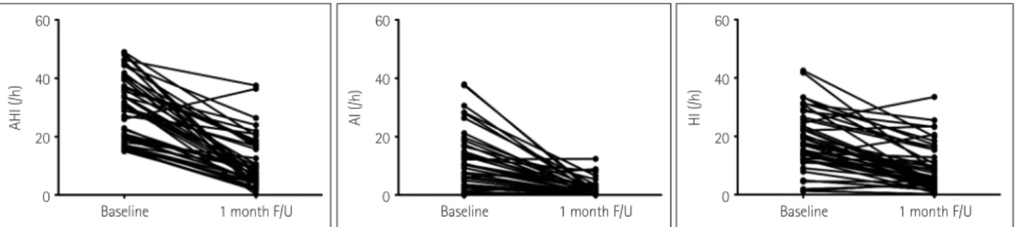
The overall mean percentage reduction in the AHI was 63.9±25.8% (range=40.0–99.4%), which was similar between the patients with moderate OSA and those with severe OSA. The AHI decreased from 29.8±11.0/h to 11.6±10.7/h, the apnea index decreased from 10.5±10.4/h to 1.6±2.7/h, and the hypopnea index decreased from 18.6±9.9/h to 9.0±7.7/h 1 month after OA therapy compared with the same param- eters at baseline (all p<0.001) (Fig. 1, Table 2).
The patients with moderate or severe OSA included 31.1%
with a normal AHI and 64.4% with an AHI of <10/h at the 1-month follow-up. The proportion of patients who had an AHI of <10/h at the follow-up was higher among those with moderate OSA (81.0% vs. 50.0%, p=0.03). However, the pro- portion of patients with a normal AHI at the follow-up was similar between the two groups. Three-quarters of the pa- tients exhibited a >50% reduction in the AHI (Table 2).
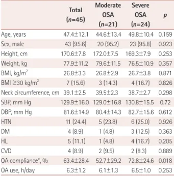
Table 1. Baseline characteristics of moderate and severe OSA patients Total
(n=45)
Moderate OSA (n=21)
Severe OSA (n=24)
p
Age, years 47.4±12.1 44.6±13.4 49.8±10.4 0.159 Sex, male 43 (95.6) 20 (95.2) 23 (95.8) 0.923 Height, cm 170.6±7.8 172.0±7.5 169.3±7.9 0.253 Weight, kg 77.9±11.2 79.6±11.5 76.5±10.9 0.357 BMI, kg/m2 26.8±3.3 26.8±2.9 26.7±3.8 0.871 BMI ≥30 kg/m2 7 (15.6) 3 (14.3) 4 (16.7) 0.826 Neck circumference, cm 39.1±2.5 39.5±2.3 38.7±2.7 0.298 SBP, mm Hg 129.9±16.0 129.0±16.8 130.8±15.5 0.72 DBP, mm Hg 81.6±14.9 80.4±14.3 82.7±15.6 0.612
HTN 11 (24.4) 5 (23.8) 6 (25.0) 0.926
DM 4 (8.9) 1 (4.8) 3 (12.5) 0.363
HL 5 (11.1) 1 (4.8) 4 (16.7) 0.205
CVD 4 (8.9) 2 (9.5) 2 (8.3) 0.889
OA compliance*, % 63.4±28.4 52.7±29.2 72.8±24.6 0.018 OA use, h/day 6.3±1.2 6.1±1.3 6.5±1.0 0.253 Data are mean±SD or n (%) values.
*Percentage of days that OA worn for >4 h.
BMI: body mass index, CVD: cardiovascular disease, DBP: diastolic blood pressure, DM: diabetes mellitus, HL: hyperlipidemia, HTN: hyper- tension, OA: oral appliance, OSA: obstructive sleep apnea, SBP: systolic blood pressure.
Efficacy of OA for Moderate-to-Severe OSA
JCN
Secondary outcomes (questionnaire scores and PSG results)
Some of the PSQI, ESS, ISI, and BDI-II scores improved sig- nificantly after 1 month of OA treatment. The ESS and BDI- II scores improved only in the patients with moderate OSA.
The treatment increased the proportions of time spent in sleep stages N1 and N3, and also the minimum oxygen satu- ration, and decreased the incidence of wake after sleep on- set (WASO) and the arousal index. The changes in WASO and the minimum oxygen saturation were significant only in the severe-OSA patients. All of the respiratory indices de- creased, but the proportion of hypopnea in the AHI increased after the treatment (Table 3 and Supplementary Table 1 in the online-only Data Supplement).
Clinical factors associated with the AHI percentage reduction
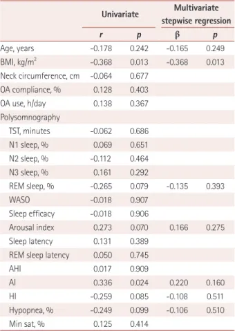
In the initial linear regression analysis, the percentage re- duction in the AHI was negatively correlated with the BMI (r=-0.368, p=0.013) and positively correlated with the ap- nea index (r=0.336, p=0.024). The percentage reduction in the AHI tended to be negatively correlated with the propor- tion of time spent in REM sleep (r=-0.265, p=0.079) and the hypopnea index (r=-0.259, p=0.085), and tended to be pos- itively correlated with the arousal index (r=0.273, p=0.070).
Stepwise multivariate linear regression analysis revealed an independent negative correlation between the BMI and the percentage reduction in the AHI (β=-0.368, p=0.013) (Table 4, Fig. 2).
DISCUSSION
This study found OA treatment to be an effective first-line therapy in Korean patients with moderate or severe OSA. The Table 2. Respiratory outcome after 1 month of OA therapy
Total (n=45)
Moderate OSA (n=21)
Severe OSA (n=24)
p
1-month F/U AHI <5/h 14 (31.1) 9 (42.9) 5 (20.8) 0.111 1-month F/U AHI <10/h 29 (64.4) 17 (81.0) 12 (50.0) 0.030 Reduction in AHI, /h 19.0±11.1 11.1±6.0 25.9±10.0 <0.001 Reduction in AI, /h 8.9±10.3 3.1±4.6 13.9±11.4 <0.001 Reduction in HI, /h 9.6±8.5 8.0±6.8 11.0±9.7 0.245 Reduction, % 63.9±25.8 59.8±29.5 67.4±22.0 0.340
>50% reduction in AHI 34 (75.6) 16 (76.2) 18 (75.0) 0.685 Data are mean±SD or n (%) values.
AHI: apnea-hypopnea index, AI: apnea index, F/U: follow-up, HI: hy- popnea index, OSA: obstructive sleep apnea.
Table 3. Questionnaire scores and PSG results at baseline and after 1 month of OA therapy
Baseline 1-month F/U p Questionnaires
PSQI score 8.1±3.3 6.1±2.9 <0.001
ESS score 8.0±4.0 7.1±4.7 0.029
ISI score 11.1±5.8 7.4±4.7 <0.001
BDI-II score 9.5±7.2 6.9±6.0 0.001
PSG
TST, minutes 303.4±72.2 308.3±65.4 0.404 N1 sleep, % 23.2±12.8 15.6±8.6 <0.001
N2 sleep, % 46.7±11.8 48.6±12.1 0.096
N3 sleep, % 14.5±13.4 19.1±15.7 0.001
REM sleep, % 15.6±6.4 16.5±7.3 0.410
WASO, minutes 59.9±52.3 38.8±31.8 0.009 Sleep efficacy, % 82.7±12.2 86.7±10.1 0.040 Arousal index, /h 39.7±14.5 24.2±10.9 <0.001 Sleep latency, minutes 5.5±5.6 8.1±13.8 0.909 REM sleep latency, minutes 115.2±65.1 98.2±58.4 0.053
AHI, /h 29.7±10.9 10.7±8.8 <0.001
AI, /h 10.5±10.4 1.6±2.7 <0.001
HI, /h 18.6±9.9 9.0±7.7 <0.001
Hypopnea, % 65.6±29.1 85.3±20.1 <0.001
Min sat, % 81.0±8.2 84.5±6.3 0.001
Data are mean±SD or n (%) values.
AHI: apnea-hypopnea index, AI: apnea index, BDI-II: Beck Depression Inventory-II, ESS: Epworth Sleepiness Scale, F/U: follow-up, HI: hypop- nea index, ISI: Insomnia Severity Index, Min sat: minimum oxygen sat- uration, OA: oral appliance, PSG: polysomnography, PSQI: Pittsburgh Sleep Quality Index, REM: rapid eye movement, TST: total sleep time, WASO: wake after sleep onset.
60 40 20 0
60 40 20 0
60 40 20 0
AHI (/h) AI (/h) HI (/h)
Baseline 1 month F/U Baseline 1 month F/U Baseline 1 month F/U Fig. 1. Changes in the AHI, AI, and HI. AHI: apnea-hypopnea index, AI: apnea index, F/U: follow-up, HI: hypopnea index.
Byun JI et al.
JCN
overall percentage reduction in the AHI was 63.9±25.8%, and was similar between patients with moderate OSA and those with severe OSA. The baseline BMI was the best predic- tive factor for the reduction. Approximately one-third (31.1%) of the patients had a normal AHI of <5/h at 1 month after the initiation of OA therapy, while around two-thirds (64.4%) had an AHI of <10/h. The treatment not only ameliorated respiratory symptoms during sleep but also improved the sleep quality and depression level.
This study evaluated the effect of OAs as a first-line treat- ment in Korean patients with moderate or severe OSA. The cohorts in previous retrospective9,16,17 and prospective10 stud- ies only included some patients with moderate OSA (30–
60%) or severe OSA (6–57%). One study evaluated the effect of OA treatment in 34 Chinese patients with severe OSA who refused CPAP therapy.18 Most of the patients enrolled in the present study were males, who are known to respond less to OSA treatment,19 and most of them were not obese (BMI <30 kg/m2), which is known to be associated with a better re- sponse.4 However, the proportions of patients with AHIs of
<5/h or <10/h at the follow-up were similar to those in pre- vious studies.9,10,16,17 The objectively measured percentage of days with good adherence (i.e., the percentage of days on which the OA was worn for >4 h) was 63.4±28.4%, and the duration of OA use was 6.3±1.2 h/day, which is similar to the results of previous studies.9,20,21
The present patients with severe OSA benefited from the OA treatment. Half of the patients had an AHI of <10/h, and 20% had a normal AHI after the treatment. The OA improved not only the respiratory indices of the participants but also Table 4. Results of univariate and multivariate linear regression anal-
yses
Univariate Multivariate stepwise regression
r p β p
Age, years -0.178 0.242 -0.165 0.249
BMI, kg/m2 -0.368 0.013 -0.368 0.013
Neck circumference, cm -0.064 0.677 OA compliance, % 0.128 0.403
OA use, h/day 0.138 0.367
Polysomnography
TST, minutes -0.062 0.686
N1 sleep, % 0.069 0.651
N2 sleep, % -0.112 0.464
N3 sleep, % 0.161 0.292
REM sleep, % -0.265 0.079 -0.135 0.393
WASO -0.018 0.907
Sleep efficacy -0.018 0.906
Arousal index 0.273 0.070 0.166 0.275
Sleep latency 0.131 0.389 REM sleep latency 0.050 0.745
AHI 0.017 0.909
AI 0.336 0.024 0.220 0.160
HI -0.259 0.085 -0.108 0.511
Hypopnea, % -0.249 0.099 -0.106 0.510
Min sat, % 0.125 0.414
AHI: apnea-hypopnea index, AI: apnea index, BMI: body mass index, HI: hypopnea index, Min sat: minimum oxygen saturation, OA: oral appliance, TST: total sleep time, WASO: wake after sleep onset.
100
50
0
-50
100
50
0
-50
Percent reduction in AHI (%) Percent reduction in AHI (%)
10 20 30 40 50 60 20 25 30 35 40
Baseline AHI BMI
Fig. 2. Correlations of the percentage reduction in the AHI with the baseline AHI (A) and BMI (B). AHI: apnea-hypopnea index, BMI: body mass index.
A B
Efficacy of OA for Moderate-to-Severe OSA
JCN
their sleep architecture and subjective sleep-related symp- toms. OAs are known to improve subjective daytime sleepi- ness,22 but contradictory results have also been reported.9 The subjective sleep quality and insomnia improved in both the patients with moderate OSA and those with severe OSA, but daytime sleepiness and the depression level improved only in those with moderate OSA. Patients with severe OSA had more frequent arousal and a lower minimum oxygen satura- tion during sleep, which could have led to this discrepancy.
We hypothesized that OAs can improve the part of the AHI associated with anatomical factors. The overall reduction in the AHI after OA treatment in our study was 63.9±25.8%, which was similar between patients with different severities of OSA. The percentage reduction in the AHI after OA treat- ment was reported previously in small studies or in specific OSA types. One study found that the reduction after OA therapy in seven patients ranged from 98.1% to 100%.23 A reduction of 62.6±7.5% was found during non-REM sleep, with a median reduction of 13.4% during REM sleep.14 Re- ductions of 74.69±16.92% and 46.03±36.44% were found in patients with positional and nonpositional OSA, respective- ly.24 OAs can improve airway collapsibility, but they do not affect muscle function, loop gain, or the arousal threshold.14 The heterogeneity in the results obtained might therefore be attributable to the various pathophysiological characteris- tics underlying OSA, such as increased loop gain and a de- creased arousal threshold.
The BMI and apnea index were associated with the per- centage reduction in the AHI in this study. Most previous studies have used different criteria to assess treatment suc- cess, including various cutoff values,11 and naturally a lower baseline AHI will be predictive of a good response. The base- line apnea index—but not the AHI—was associated with the AHI percentage reduction in this study. Apnea and hypop- nea episodes have different mechanisms, with the former representing the absence of flow due to a static obstruction and the latter representing flow limitations due to a dynamic obstruction.25 Anatomical changes produced by OAs might affect static obstructions more than dynamic obstructions, thus explaining the increased proportion of hypopnea epi- sodes after OA treatment.
The strongest predictive factor for the AHI percentage re- duction was the BMI. Most studies have found the BMI to be a predictor of a poor response to OA therapy.20,26 A previ- ous review showed that the BMI has large negative predictive value along with CPAP pressure and cephalometry measures.11 However, some studies performed in Western countries have found that age, the baseline AHI, and cephalometric mea- sures were more significant predictors than the BMI.27 The BMI might be more important for predicting OA treatment
responses in Koreans. Higher BMIs are associated with fat deposition, which can narrow pharyngeal wall diameters and increase upper airway collapsibility. Higher BMIs can also increase the loop gain,28 which is an independent predictor of a poor response to OA therapy, and the arousal threshold,29 which is not changed when using an OA. Moreover, a high BMI can cause pharyngeal dilator muscle dysfunction and reduce the respiratory functional residual volume.30 Our re- sults support the idea that a higher BMI can contribute to OSA in more ways than only via affecting the anatomy of the upper airway.
This was the first prospective study of Korean patients with moderate or severe OSA, but the treatment period was only one month, which might have been too short to provide a full evaluation of the compliance and treatment effects. More- over, the sample was of modest size and predominantly con- sisted of males, which limits the generalizability of our find- ings. Moreover, we did not consider cephalometric measures, sleeping position, or REM predominance, which can be im- portant predictors of OA treatment responses.
In conclusion, This study found that OA therapy was ef- fective for patients with either moderate or severe OSA. OA therapy improved not only respiratory indices but also sub- jective and objective sleep indices. The OAs reduced the mean AHI to 63.9% of the baseline value, and the percentage reduc- tion was lower in patients with higher BMIs. It can therefore be predicted that the follow-up AHI after OA therapy in pa- tients with moderate or severe OSA will be one-third of their baseline AHI, with some variation according to the BMI. Fu- ture larger studies are required to confirm the efficacy of OA therapy, treatment predictors for patients with moderate or severe OSA, and the influence of race on outcomes.
Supplementary Materials
The online-only Data Supplement is available with this arti- cle at https://doi.org/10.3988/jcn.2020.16.2.215.
Author Contributions
Conceptualization: Jung-Ick Byun, Su-Jin Ahn, Peter A. Cistulli, Won Chul Shin. Data curation: Jung-Ick Byun, Dong-Ha Kim, Su-Jin Ahn. Formal analysis: Jung-Ick Byun, Dong-Ha Kim. Funding acquisition: Won Chul Shin. Investigation: Jung-Ick Byun, Dong-Ha Kim, Su-Jin Ahn, Kwang Ik Yang, Yong Won Cho. Methodology: Jung-Ick Byun, Su-Jin Ahn, Won Chul Shin. Supervision: Won Chul Shin. Writing—original draft: Jung-Ick Byun. Writing—review & editing: Kwang Ik Yang, Yong Won Cho, Peter A.
Cistulli, Won Chul Shin.
ORCID iDs
Jung-Ick Byun https://orcid.org/0000-0002-6224-4575 Dongha Kim https://orcid.org/0000-0003-1879-9976 Su-Jin Ahn https://orcid.org/0000-0003-2128-1561 Kwang Ik Yang https://orcid.org/0000-0001-6343-6520 Yong Won Cho https://orcid.org/0000-0002-6127-1045 Peter A. Cistulli https://orcid.org/0000-0002-7920-4924
Byun JI et al.
JCN
Won Chul Shin https://orcid.org/0000-0003-3044-9397 Conflicts of Interest
The authors have no potential conflicts of interest to disclose.
Acknowledgements
This study was supported by grants from SomnoMed, but this entity played no role in the study design or in data acquisition, analysis, or in- terpretation.
REFERENCES
1. Flemons WW. Clinical practice. Obstructive sleep apnea. N Engl J Med 2002;347:498-504.
2. Sutherland K, Vanderveken OM, Tsuda H, Marklund M, Gagnadoux F, Kushida CA, et al. Oral appliance treatment for obstructive sleep apnea: an update. J Clin Sleep Med 2014;10:215-227.
3. Ramar K, Dort LC, Katz SG, Lettieri CJ, Harrod CG, Thomas SM, et al. Clinical practice guideline for the treatment of obstructive sleep apnea and snoring with oral appliance therapy: an update for 2015. J Clin Sleep Med 2015;11:773-827.
4. Sutherland K, Takaya H, Qian J, Petocz P, Ng AT, Cistulli PA. Oral appliance treatment response and polysomnographic phenotypes of obstructive sleep apnea. J Clin Sleep Med 2015;11:861-868.
5. Doff MHJ, Hoekema A, Wijkstra PJ, van der Hoeven JH, Huddleston Slater JJ, de Bont LGM, et al. Oral appliance versus continuous posi- tive airway pressure in obstructive sleep apnea syndrome: a 2-year follow-up. Sleep 2013;36:1289-1296.
6. Li KK, Kushida C, Powell NB, Riley RW, Guilleminault C. Obstruc- tive sleep apnea syndrome: a comparison between Far-East Asian and white men. Laryngoscope 2000;110:1689-1693.
7. Lee RWW, Sutherland K, Sands SA, Edwards BA, Chan TO, SS Ng S, et al. Differences in respiratory arousal threshold in Caucasian and Chinese patients with obstructive sleep apnoea. Respirology 2017;22:
1015-1021.
8. Park P, Jeon HW, Han DH, Won TB, Kim DY, Rhee CS, et al. Thera- peutic outcomes of mandibular advancement devices as an initial treatment modality for obstructive sleep apnea. Medicine (Baltimore) 2016;95:e5265.
9. Lee CH, Mo JH, Choi IJ, Lee HJ, Seo BS, Kim DY, et al. The mandib- ular advancement device and patient selection in the treatment of obstructive sleep apnea. Arch Otolaryngol Head Neck Surg 2009;135:
439-444.
10. Banhiran W, Kittiphumwong P, Assanasen P, Chongkolwatana C, Metheetrairut C. Adjustable thermoplastic mandibular advancement device for obstructive sleep apnea: outcomes and practicability. La- ryngoscope 2014;124:2427-2432.
11. Okuno K, Pliska BT, Hamoda M, Lowe AA, Almeida FR. Prediction of oral appliance treatment outcomes in obstructive sleep apnea: a systematic review. Sleep Med Rev 2016;30:25-33.
12. Ng JH, Yow M. Oral appliances in the management of obstructive sleep apnea. Sleep Med Clin 2019;14:109-118.
13. Shin W, Jen R, Li Y, Malhotra A. Tailored treatment strategies for ob- structive sleep apnea. Respir Investig 2016;54:2-7.
14. Edwards BA, Andara C, Landry S, Sands SA, Joosten SA, Owens RL, et al. Upper-airway collapsibility and loop gain predict the response to oral appliance therapy in patients with obstructive sleep apnea. Am J Respir Crit Care Med 2016;194:1413-1422.
15. Berry RB, Brooks R, Gamaldo CE, Harding SM, Lloyd RM, Marcus CL, Vaughn BV; the American Academy of Sleep Medicine. The AASM manual for the scoring of sleep and associated events: rules, ter- minology and technical specifications, version 2.2. Darien, IL: Ameri- can Academy of Sleep Medicine, 2015.
16. Park P, Jeon HW, Han DH, Won TB, Kim DY, Rhee CS, et al. Thera- peutic outcomes of mandibular advancement devices as an initial treatment modality for obstructive sleep apnea. Medicine (Baltimore) 2016;95:e5265.
17. Fukuda T, Tsuiki S, Kobayashi M, Nakayama H, Inoue Y. Selection of response criteria affects the success rate of oral appliance treatment for obstructive sleep apnea. Sleep Med 2014;15:367-370.
18. Lam B, Sam K, Lam JCM, Lai AYK, Lam CL, Ip MSM. The efficacy of oral appliances in the treatment of severe obstructive sleep apnea.
Sleep Breath 2011;15:195-201.
19. Ng ATM, Darendeliler MA, Petocz P, Cistulli PA. Cephalometry and prediction of oral appliance treatment outcome. Sleep Breath 2012;
16:47-58.
20. Dieltjens M, Verbruggen AE, Braem MJ, Wouters K, Verbraecken JA, De Backer WA, et al. Determinants of objective compliance during oral appliance therapy in patients with sleep-disordered breathing: a prospective clinical trial. JAMA Otolaryngol Head Neck Surg 2015;
141:894-900.
21. Ingman T, Arte S, Bachour A, Bäck L, Mäkitie A. Predicting compli- ance for mandible advancement splint therapy in 96 obstructive sleep apnea patients. Eur J Orthod 2013;35:752-757.
22. Ahrens A, McGrath C, Hägg U. Subjective efficacy of oral appliance design features in the management of obstructive sleep apnea: a sys- tematic review. Am J Orthod Dentofacial Orthop 2010;138:559-576.
23. Zhao M, Barber T, Cistulli P, Sutherland K, Rosengarten G. Compu- tational fluid dynamics for the assessment of upper airway response to oral appliance treatment in obstructive sleep apnea. J Biomech 2013;
46:142-150.
24. Chung JW, Enciso R, Levendowski DJ, Morgan TD, Westbrook PR, Clark GT. Treatment outcomes of mandibular advancement devices in positional and nonpositional OSA patients. Oral Surg Oral Med Oral Pathol Oral Radiol Endod 2010;109:724-731.
25. Farré R, Rigau J, Montserrat JM, Buscemi L, Ballester E, Navajas D.
Static and dynamic upper airway obstruction in sleep apnea: role of the breathing gas properties. Am J Respir Crit Care Med 2003;168:
659-663.
26. Zeng B, Ng AT, Qian J, Petocz P, Darendeliler MA, Cistulli PA. Influ- ence of nasal resistance on oral appliance treatment outcome in ob- structive sleep apnea. Sleep 2008;31:543-547.
27. Hoekema A, Doff MHJ, de Bont LGM, van der Hoeven JH, Wijkstra PJ, Pasma HR, et al. Predictors of obstructive sleep apnea-hypopnea treatment outcome. J Dent Res 2007;86:1181-1186.
28. Sands SA, Eckert DJ, Jordan AS, Edwards BA, Owens RL, Butler JP, et al. Enhanced upper-airway muscle responsiveness is a distinct fea- ture of overweight/obese individuals without sleep apnea. Am J Respir Crit Care Med 2014;190:930-937.
29. Edwards BA, Eckert DJ, McSharry DG, Sands SA, Desai A, Kehl- mann G, et al. Clinical predictors of the respiratory arousal threshold in patients with obstructive sleep apnea. Am J Respir Crit Care Med 2014;190:1293-1300.
30. Tham KW, Lee PC, Lim CH. Weight management in obstructive sleep apnea: medical and surgical options. Sleep Med Clin 2019;14:
143-153.