pISSN 2288-9272 eISSN 2383-8493 J Oral Med Pain 2016;41(3):126-132 http://dx.doi.org/10.14476/jomp.2016.41.3.126
Inflammatory Cytokine Level in Patients with Obstructive Sleep Apnea and Treatment Outcome of Oral Appliance Therapy
Jae-Tak Oh, Jin-Woo Chung
Department of Oral Medicine and Oral Diagnosis, School of Dentistry and Dental Research Institute, Seoul National University, Seoul, Korea
Received July 26, 2016 Revised August 2, 2016 Accepted August 2, 2016
Purpose:
Purpose: The aims of this study were to analyze the association between inflammatory cyto- kine and obstructive sleep apnea (OSA), and to evaluate treatment outcome and changes of plasma inflammatory cytokine levels after oral appliance therapy.
Methods:
Methods: Twenty-seven subjects who visited Department of Oral Medicine in Seoul National University Dental Hospital were performed nocturnal polysomnography and analyzed plasma C-reactive protein (CRP), interleukin (IL)-1β, IL-6, IL-10, and tumor necrosis factor (TNF)-α lev- els. Each subject was evaluated with Pittsburgh Sleep Quality Index (PSQI) and Epworth Sleepi- ness Scale (ESS). The subjects were classified into 12 OSA patients (apnea-hypopnea index [AHI]
>5) and 15 control (AHI ≤5) groups. The OSA group was treated with mandibular advancement device (MAD) for 3 months and re-evaluated nocturnal polysomnography and plasma inflam- matory cytokine levels.
Results:
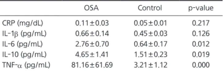
Results: Plasma TNF-α, IL-10, and IL-6 levels were significantly higher in OSA patients com- pared to controls. Total AHI showed significant positive correlations with plasma IL-6 and TNF-α levels. Percentage time of SpO
2<90 and lowest SpO
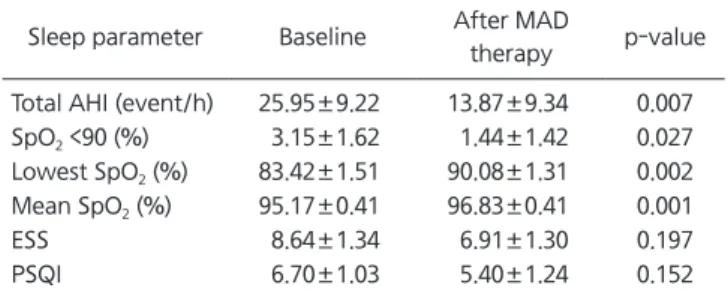
2were significantly correlated with plasma TNF-α level. ESS showed significant positive correlation with plasma IL-10 level. Total AHI, percentage time of SpO
2<90, lowest SpO
2, and mean SpO
2were significantly improved after the MAD therapy. Plasma TNF-α level was significantly decreased after MAD therapy.
Conclusions:
Conclusions: We suggest that MAD therapy is an effective treatment modality for patients with OSA and can decrease plasma cytokine level.
Key Words:
Key Words: C-reactive protein; Interleukins; Mandibular advancement device; Obstructive sleep apnea; Tumor necrosis factor-α
Correspondence to:
Jin-Woo Chung
Department of Oral Medicine and Oral Diagnosis, School of Dentistry, Seoul National University, 101 Daehak-ro, Jongno-gu, Seoul 03080, Korea Tel: +82-2-2072-3021 E-mail: [email protected] This study was supported by grant number 04-2011-0047 from the Seoul National University Dental Hospital (SNUDH) Research Fund.
JOMP
Journal of Oral Medicine and PainCopyright Ⓒ 2016 Korean Academy of Orofacial Pain and Oral Medicine. All rights reserved.
CC This is an open-access article distributed under the terms of the Creative Commons Attribution Non-Commercial License (http://creativecommons.org/licenses/by-nc/4.0/), which permits unrestricted non-commercial use, distribution, and reproduction in any medium, provided the original work is properly cited.