Received:February 3, 2014, Revised (1st) April 8, 2014, (2nd) April 28, 2014, Accepted:May 14, 2014
Corresponding to:Young Ho Lee, Division of Rheumatology, Department of Internal Medicine, Korea University Anam Hospital, Korea University College of Medicine, 73 Inchon-ro, Seongbuk-gu, Seoul 136-705, Korea. E-mail: [email protected]
pISSN: 2093-940X, eISSN: 2233-4718
Copyright ⓒ 2015 by The Korean College of Rheumatology. All rights reserved.
This is a Free Access article, which permits unrestricted non-commerical use, distribution, and reproduction in any medium, provided the original work is properly cited.
A Case of Massive Pulmonary Embolism in Systemic Lupus Erythematosus without Antiphospholipid Antibody
Jae-Hyun Jung, Min-Gu Lee, Ju-Sung Sim, Tae-Hyun Kim, Hong-Kwon Oh, Jong Dae Ji, Young Ho Lee
Division of Rheumatology, Department of Internal Medicine, Korea University College of Medicine, Seoul, Korea
Patients with systemic lupus erythematosus (SLE) are at an increased risk of developing thromboses with antiphospholipid anti- bodies (aPL). The presence of aPL is related to an increased risk of thrombotic events. However, thromboembolic events can occur in SLE patients without aPL, and pulmonary emboli are rarely reported manifestations of SLE without aPL. Here, we report on a case of massive pulmonary embolism in a 58-year-old woman with aPL-negative SLE. She presented with chest pain and dyspnea, and chest computed tomography (CT) and lung perfusion ventilation scans showed pulmonary thromboembolism.
She was administered thrombolytic agents, heparin, and warfarin. Two months later, no remarkable residual thromboembolism was observed on chest CT. (J Rheum Dis 2015;22:106-110)
Key Words. Pulmonary embolism, Systemic lupus erythematosus, Antiphospholipid antibodies
INTRODUCTION
Systemic lupus erythematosus (SLE) is a prototypic au- toimmune disease in which immune regulation is dis- rupted, leading to intense inflammation and multiple or- gan damage. Although extraordinary improvement in its prognosis and survival rate has been accomplished, throughout the SLE disease course, patients deal with possibility of disease activity, infections, irreversible organ damage, and severe clinical manifestations, i.e., thrombo- ses, which impair their quality of life and survival rate.
These thromboembolic events occur especially in patients with antiphospholipid antibodies (aPL). aPL syndrome (APS) is characterized by venous and arterial thromboses, obstetric morbidity, and the presence of aPL, including lu- pus anticoagulant, anticardiolipin antibodies (aCL), and/or antibodies primarily directed against β2-glyco- protein I (β2GPI) [1].
Among SLE patients, the prevalence of aPL is sub- stantially higher, ranging from 12% to 30% for aCL [2].
However, thromboembolic events can occur in SLE pa-
tients without aPL, and pulmonary emboli are rarely re- ported manifestations of aPL-negative SLE [3]. Though pulmonary thromboembolism is rarely reported aPL-neg- ative SLE patients, it can appear acutely and be fatal.
Pulmonary thromboembolism, however, can disappear if it is quickly diagnosed and treated, so clinicians should consider thromboembolism event when they exam all SLE patients. In this article, we describe a case of massive pulmonary embolism that developed in an aPL-negative SLE patient.
CASE REPORT
A 58-year-old female with a 3-year history of macular er- ythema and wrist and shoulder pain was admitted to the hospital emergency room with chief complains of fever and myalgia. The patient had been diagnosed with SLE approximately 2 months previously. Fluorescent anti- nuclear antibody results were positive (1:1,280, diffuse type). Anti-ds-DNA antibody titers were elevated (8.68 IU/mL). Other autoimmune markers, including anti-Sm,
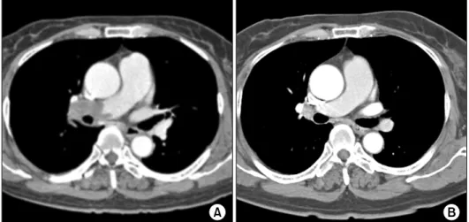
Figure 1. Spiral chest computed tomography showing pulmonary thromboembolism in the right pulmonary artery (A) and chest computed tomography showing the absence of remarkable re- sidual thromboembolism (B).
Figure 2. Perfusion ventilation scintigraphy showing segmental perfusion defects in the right lung and normal tracer dis- tribution in the left lung.
anti-RNP, anti-SSB, and anti-SSA antibodies, were all negative. She had been diagnosed with SLE on the basis of skin lesions, oral ulcer, arthritis, and presence of anti- nuclear antibodies. She was being treated with 10 mg pre- dnisolone once daily and 200 mg hydroxychloroquine per day. She was a non-smoker and her body mass index was 23.5 (control, 18.5 to 24.9). She did not have hyper- tension, diabetes mellitus, dyslipidemia or the evidences of cancer, which were risk factors of pulmonary embo- lism, but one year later she has got diabetes mellitus.
Upon presentation, the patient complained of acute on- set of fever, was diagnosed with acute pyelonephritis, was given ciprofloxacin, and the pyelonephritis symptoms improved. Her ongoing prednisolone dose was increased
to 30 mg daily owing to skin lesions and arthritis. On the 5th day of admission, she complained of dizziness. Brain magnetic resonance imaging diffusion was performed, which showed acute or recent infarction on the right low- er pons and medulla; she was therefore administered 100 mg aspirin once daily. On the 23rd day of admission, how- ever, she had developed sudden chest pain and dyspnea.
Physical examination revealed unilateral rales in the right lung base. Laboratory tests showed that the D-dimer level was increased to 3.10 μg/mL (normal, <0.5 μg/mL).
However, test results for aPL were negative: lupus anti- coagulant was negative, the aCL immunoglobulin (Ig) M level was 0.5 U/mL (normal, <7 U/mL), the aCL Ab IgG level was 1.0 U/mL (normal, <10 U/mL), the anti-phos-
pholipid Ab IgM level was <0.5 U/mL (normal, <10 U/mL), and the anti-phospholipid Ab IgG level was 0.7 U/mL (normal, <10 U/mL). The anti-β2GPI Ab was not checked in this patient. Other laboratory test results were unremarkable, except for a 34% decrease in protein S (control, 58.7% to 119.2%) and increased anti-ds-DNA of 9.5 IU/mL: the FDP level was 3.97 μg/dL (normal, <5 μg/ dL), the fibrinogen level was 868.00 mg/dL (control, 230 to 480 mg/dL), the protein C level was 81% (control, 7% to 130%), the antithrombin III level was 84%
(control, 70% to 120%), the C3 level was 86.6 mg/dL (control, 88 to 201 mg/dL), and the C4 level was 28.3 mg/dL (control, 16 to 47 mg/dL).
Chest radiography showed right pleural effusion and spiral contrast-enhanced chest computed tomography (CT) revealed large filling defects in the right main pul- monary artery that continued through the right pulmo- nary arterial branch of the upper and lower lobe (Figure 1A). Prominent large defects were noted at the bifurca- tion area. The dense parenchymal opacity in the right low- er lobe was compatible with infarction. A lung perfusion ventilation scan was performed, which showed sig- nificant perfusion defects in the right lung and normal tracer distribution in the left lung (Figure 2). On trans- thoracic echocardiography, mild mitral regurgitation was detected. Left and right ventricle function was normal.
Color Doppler sonography of the lower extremities was also performed, which revealed deep thrombosis in the right peroneal vein and the absence of deep thrombosis in the veins of both lower extremities.
Pulmonary thromboembolism was diagnosed, and the patient was administered intravenous recombinant tis- sue-type plasminogen activator (100 mg alteplase) and was started on subcutaneous low-molecular-weight hep- arin (2.0×0.6 mg enoxaparin) and 5 mg oral warfarin.
The next day, she did not complain of dyspnea and chest pain. She was discharged home with warfarin anti- coagulation (international normalized ratio [INR], 2.5 to 3.5) to be re-evaluated as an outpatient. Two months lat- er, no remarkable residual thromboembolism was ob- served on chest CT (Figure 1B).
DISCUSSION
SLE is a chronic inflammatory autoimmune disorder that affects multiple organ systems. Pulmonary involve- ment usually develops late in the disease course and can affect any part of the pulmonary system, including the air-
ways, lung parenchyma, pulmonary vasculature, pleura, and diaphragm [4].
Patients with SLE are at increased risk of venous throm- boembolism, with a prevalence of 9% [5], and patients with aPL have an even higher increased risk of 35% to 42% [6]. aPL may be present in up to two-thirds of pa- tients with lupus [7].
APS is characterized by arterial and venous thrombosis, which leads to an increased risk of thromboembolic events and recurrent fetal loss in addition to the presence of aPL or lupus anticoagulant. Pulmonary APS manifes- tations include embolism and infarction, increased pul- monary artery pressure secondary to thromboembolism, and pulmonary microvascular thrombosis. Pulmonary embolism occurs in 30% of patients and originates from the calf veins, inferior vena cava, tricuspid valve vegeta- tion, or right intracardiac thromboses in many cases.
Traditionally, elevated levels of aPLs are associated with the increased thrombotic risk that is characteristic of APL. However, as always in real clinical practice, there are often discrepancies between antibody levels and clinical disease presentation [8]. In addition, it is not unusual to find patients with clinical features suggestive of APS who are persistently negative for the criteria aCL, anti-β2GPI, and lupus anticoagulant tests [9]. Seronegative APS (SNAPS) was first introduced in 2003 by Hughes and Khamashta [8]. Rodriguez-Garcia et al. [10] reported no significant differences in the frequency of thrombotic events or obstetric morbidity in patients with SNAPS ver- sus those with seropositive APS.
Platelets are the most important components in main- taining homeostasis and play an important role in the de- velopment of arterial thrombosis and atherosclerosis.
Thrombosis can occur because of either a reactive process or manifestation of a clonal hematopoietic disorder.
Thrombocytosis usually develops secondary to a disease process such as inflammation or trauma and is accordingly termed reactive thrombocytosis, in which the platelet count increases temporarily during the active phase of the disease. Thrombosis occurs in the blood flow by the inter- action and coexistence of vascular, cellular, and humoral factors. Such factors that play important roles in the path- ophysiology of thrombosis include endothelial damage, vascular stasis, and alterations in relevant blood compo- nents, which are known as Virchow’s triad. In rheumatic disorders, both thrombocytosis and thrombosis can be ob- served and may lead to severe complications [11].
SLE is generally associated with thrombocytopenia rather
than thrombocytosis, but thrombosis can also be observed in SLE. The most common cause of thrombosis in SLE is aPL; however, thromboses in SLE patients may result from diverse mechanisms, including inflammatory, primarily thrombotic, or atherosclerotic processes. The relevance of each mechanism may vary throughout the disease course, thus atherosclerosis seems to predominate late in the evo- lution of the disease. Hereditary factors such as protein C, protein S or antithrombin III deficiency, factor V-leiden, prothrombin, or methylene tetrahydrofolate reductase may be involved in the etiology of thrombosis. Protein C, fibrinogen, D-dimer, and homocysteine levels are in- creased in SLE patients, which can cause thrombosis. In addition, inflammatory cytokines are major mediators in- volved in the activation of the coagulation cascade.
Inflammatory mediators can elevate the platelet count, platelet reactivity, and fibrinogen concentration; down- regulate natural anticoagulant mechanisms; initiate the coagulation cascade; facilitate propagation of the coagulant response; and impair fibrinolysis. Patients with auto- immune diseases, particularly SLE, are at an increased risk of developing thromboses. SLE diagnosis is the single most common variable associated with venous and arterial thromboses. Patients with SLE-related venous thromboses show vasculitis and disease activity, whereas dyslipidemia, central nervous system involvement, and disease activity are associated with arterial thromboses [12].
The patient of this case has no genetic risk factors of pul- monary embolism, which are genetic thrombophilia, an- tithrombin III deficiency, protein C deficiency, plasmi- nogen deficiency and etc. She also did not have acquired risk factors except stroke developed in hospital. Acquired risk factors of pulmonary embolism include advanced age, obesity, immobilization, major surgery, trauma, con- gestive cardiac failure, myocardial infarction, smoking, stroke, malignity, chemotherapy, central venous catheter, pregnancy, puerperality, the use of oral contraceptives and hormone replacement, previous pulmonary emboli/deep vein thrombosis, antiphospholipid syndrome, chronic ob- structive pulmonary disease and medical conditions re- quiring hospitalization. In the International Stroke Trial, the incidence of pulmonary embolism after a stroke was 0.8% at 2 weeks [13]. Her pulmonary embolism could be developed by stroke event, but cerebral infarction oc- curred during admission period and she had not any risk factors of infarction. So we can think that stroke and pul- monary embolism were affected with SLE itself.
Management of pulmonary thromboembolism in SLE
patients is the same in patients with APS as in the general population. Patients require anticoagulation treatment with heparin followed by warfarin. It is recommended that an INR ranging between 2.5 and 3.5 be maintained [14]. SLE patients are at an increased risk of developing thromboses with or without aPL. Clinicians should be aware that even if SLE patients do not have aPL, thrombo- sis can develop and be fatal.
SUMMARY
Thromboembolic events can occur in SLE patients with- out aPL, and pulmonary emboli are rarely reported mani- festations of SLE without aPL. Thus, we report a case of massive pulmonary embolism in a 58-year-old woman with aPL-negative SLE.
CONFLICT OF INTEREST
No potential conflict of interest relevant to this article was reported.
REFERENCES
1. Rezaei N, Yeganeh MZ, Ahmadi F. Antiphospholipid anti- body syndrome presenting with pulmonary embolism.
Tanaffos 2008;7:71-4.
2. Levine JS, Branch DW, Rauch J. The antiphospholipid syndrome. N Engl J Med 2002;346:752-63.
3. Matta BN, Uthman I, Taher AT, Khamashta MA. The cur- rent standing of diagnosis of antiphospholipid syndrome as- sociated with systemic lupus erythematosus. Expert Rev Clin Immunol 2013;9:659-68.
4. Gari AG, Telmesani A, Alwithenani R. Pulmonary manifes- tations of systemic lupus erythematosus. In: Almoallim H, ed. Systemic lupus erythematosus. Rijeka, Croatia, InTech, 2012, p. 313-36.
5. Gladman DD, Urowitz MB. Venous syndromes and pulmo- nary embolism in systemic lupus erythematosus. Ann Rheum Dis 1980;39:340-3.
6. Love PE, Santoro SA. Antiphospholipid antibodies: anti- cardiolipin and the lupus anticoagulant in systemic lupus erythematosus (SLE) and in non-SLE disorders. Prevalence and clinical significance. Ann Intern Med 1990;112:682-98.
7. Ruiz-Irastorza G, Egurbide MV, Ugalde J, Aguirre C. High impact of antiphospholipid syndrome on irreversible organ damage and survival of patients with systemic lupus erythematosus. Arch Intern Med 2004;164:77-82.
8. Hughes GR, Khamashta MA. Seronegative antiphos- pholipid syndrome. Ann Rheum Dis 2003;62:1127.
9. Mahler M, Norman GL, Meroni PL, Khamashta M.
Autoantibodies to domain 1 of beta 2 glycoprotein 1: a promising candidate biomarker for risk management in an- tiphospholipid syndrome. Autoimmun Rev 2012;12:313-7.
10. Rodriguez-Garcia JL, Bertolaccini ML, Cuadrado MJ, Sanna
G, Ateka-Barrutia O, Khamashta MA. Clinical manifes- tations of antiphospholipid syndrome (APS) with and with- out antiphospholipid antibodies (the so-called 'seronegative APS'). Ann Rheum Dis 2012;71:242-4.
11. Cipil H, Kargili A, Senaran H, Uz E, Kasapoglu B, Alici Ö, et al. Thrombosis and thrombocytosis in a thousand rheuma- tologic cases. New J Med 2011;28:38-42.
12. Romero-Díaz J, García-Sosa I, Sánchez-Guerrero J.
Thrombosis in systemic lupus erythematosus and other au-
toimmune diseases of recent onset. J Rheumatol 2009;36:
68-75.
13. Çobanoğlu U. Risk factor for pulmonary embolism. In:
Çobanoğlu U, ed. Pulmonary embolism. Rijeka, Croatia, InTech, 2012, p. 1-18.
14. Espinosa G, Cervera R, Font J, Asherson RA. The lung in the antiphospholipid syndrome. Ann Rheum Dis 2002;61:
195-8.