Korean J Gastroenterol Vol. 62 No. 5, 292-295 http://dx.doi.org/10.4166/kjg.2013.62.5.292 pISSN 1598-9992 eISSN 2233-6869
CASE REPORT
Korean J Gastroenterol, Vol. 62 No. 5, November 2013 www.kjg.or.kr
크론병과 동반된 헤르페스 바이러스 십이지장염 1예
이병후1,3*, 엄욱현1,3*, 전성란1,3, 김현건1,3, 이태희1,3, 김완중1,3, 김진오1,3, 진소영2
순천향대학교 의과대학 내과학교실1, 병리학교실2, 순천향대학교 서울병원 소화기병센터 소화기연구소3
Herpes Simplex Virus Duodenitis Accompanying Crohn’s Disease
Byung Hoo Lee1,3*, Wook Hyun Um1,3*, Seong Ran Jeon1,3, Hyun Gun Kim1,3, Tae Hee Lee1,3, Wan Jung Kim1,3, Jin-Oh Kim1,3 and So Young Jin2
Department of Internal Medicine1, Department of Pathology2, Soonchunhyang University College of Medicine, Institute for Digestive Research, Digestive Disease Center, Soonchunhyang University Seoul Hospital3, Seoul, Korea
Herpes simplex virus (HSV) is a recognized cause of gastrointestinal infection in immunodeficient patients. Although a few cases of HSV gastritis and colitis in immunocompromised patients have been reported, there are no reports of HSV duodenitis in patients with Crohn’s disease (CD). A 74-year-old female was admitted with general weakness and refractory epigastric pain. She had been diagnosed with CD three years ago. Esophagogastroduodenoscopy (EGD) revealed diffuse edematous and whitish mucosa with multiple erosions in the duodenum. Considering the possibility of viral co-infection, cytomegalovirus (CMV) immunohistochemical staining, PCR, and cultures of duodenal biopsies were performed, all of which were negative with the exception of the isolation of HSV in culture. After administration of intravenous acyclovir for 1 week, follow-up EGD showed almost complete resolution of the lesions and the patient’s symptoms improved. In CD patients with refractory gastro- intestinal symptoms, HSV, as well as CMV, should be considered as a possible cause of infection, so that the diagnosis of viral infection is not delayed and the appropriate antiviral treatment can be initiated. (Korean J Gastroenterol 2013;62:292-295) Key Words: Crohn disease; Herpes simplex virus; Duodenitis
Received January 20, 2013. Revised February 19, 2013. Accepted March 2, 2013.
CC This is an open access article distributed under the terms of the Creative Commons Attribution Non-Commercial License (http://creativecommons.org/licenses/
by-nc/3.0) which permits unrestricted non-commercial use, distribution, and reproduction in any medium, provided the original work is properly cited.
교신저자: 김진오, 140-743, 서울시 용산구 대사관로 59번지, 순천향대학교 서울병원 소화기병센터 소화기연구소
Correspondence to: Jin-Oh Kim, Institute for Digestive Research, Digestive Disease Center, Soonchunhyang University Seoul Hospital, 59 Daesagwan-ro, Yongsan-gu, Seoul 140-743, Korea. Tel: +82-2-709-9202, Fax: +82-2-709-9696, E-mail: jokim@schmc.ac.kr
Financial support: None. Conflict of interest: None.
*These authors contributed equally to this project and should be considered co-first authors.
INTRODUCTION
With the increasing use of immunosuppressive and bio- logical therapies, the incidence of opportunistic infections in patients with inflammatory bowel disease (IBD) is increas- ing.1 Cytomegalovirus (CMV) and Epstein-Barr virus are the most common opportunistic viral pathogens in those with IBD.2 In addition, varicella-zoster virus and human herpes vi- rus 6 have been reported in IBD patients.2
On the other hand, the prevalence of herpes simplex virus
(HSV) infection of the gastrointestinal tract is low, and the esophagus, perineum, and rectum are the most common sites of involvement.3 HSV infection is less frequent in the stomach and duodenum than in the esophagus. A few cases of HSV gastritis have been reported in immunodeficient patients. To our knowledge, there are no reports of HSV duo- denitis in Crohn’s disease (CD). We report our experience with HSV duodenitis in a patient with CD who was treated suc- cessfully with acyclovir.
Lee BH, et al. Herpes Simplex Virus Duodenitis in Crohn’s Disease 293
Vol. 62 No. 5, November 2013
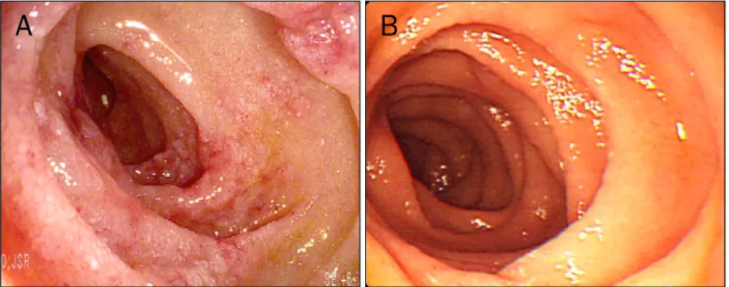
Fig. 1. Esophagogastroduodenosco- pic findings of duodenum. They showed (A) diffuse edematous and whitish mucosa with multiple ero- sions initially, and (B) nearly complete resolution of the diffuse mucosal lesions after viral treatment.
Fig. 2. Pathologic findings of the duodenal mucosa. There was blunt- ing of the villi, mononuclear cell infiltration, and mild fibrosis (H&E,
×100).
CASE REPORT
A 74-year-old female was admitted with complaints of gen- eral weakness and refractory epigastric pain for 3 months de- spite continuous proton pump inhibitor treatment. She had been diagnosed with CD involving the ileum through the ileo- cecal (IC) valve outside the hospital 3 years prior. She had been taking an oral steroid (10 mg/day) intermittently over the past 3 years, depending on her symptoms. One year ago, she started taking additional azathioprine (50 mg/day) be- cause her symptoms had become aggravated. However, these medications were discontinued due to gastrointestinal side effects including nausea and vomiting. Since then, she has been taking low-dose oral prednisolone (2.5 mg/day) and mesalazine (3 g/day). She has also been taking aspirin for coronary artery disease and a β-blocker for hypertension.
EGD conducted 6 months prior to admission showed minimal changes in the reflux esophagitis and multiple gastric ero- sions, and no specific findings were observed in the duodenum. Upon admission to our hospital, she had no other symptoms such as diarrhea, hematochezia or melena. Vital signs were stable (blood pressure 130/80 mmHg, heart rate 80 beats/min, and body temperature 36.8oC). The labo- ratory examination showed leukocytes 12,000/mm3, hemo- globin 9.2 g/dL, hematocrit 26.7%, platelet count 142,000/μL, total protein 6.5 g/dL, albumin 3.2 g/dL, total bilirubin 1.8 mg/dL, direct bilirubin 0.7 mg/dL, alkaline phosphatase 196 U/L, blood urea nitrogen 21 mg/dL, creatinine 1.16 mg/dL, glucose 112 mg/dL, sodium 138 mmol/L, potassium 4.5 mmol/L, chloride 100 mmol/L, and high-sensitivity C-re- active protein 22.80 mg/dL. Abdominal X-ray showed mild paralytic ileus. Sigmoidoscopy showed no significant change compared with the previous examination. The abdominal CT
showed no changes, such as enhanced wall thickening in the pelvic ileal loop, distal ileum or IC valve with linear ulcer formation. By EGD, the esophageal findings were unchanged and multiple gastric erosions were observed. Additionally, newly appeared multiple erosions with diffuse edematous and whitish mucosa were found in the duodenum (Fig. 1A).
We conducted duodenal biopsies. Microscopically, there were acute and chronic nonspecific inflammation without a transmural pattern of inflammation, crypts containing in- filtrating mononuclear cells, and non-caseating granulomas (Fig. 2). Due to suspicions of a viral coinfection, we conducted CMV immunohistochemical staining, PCR analysis, and cul- tures of the duodenal biopsy specimen, all of which were neg- ative, with the exception of the isolation of HSV in the culture.
Intravenous acyclovir (5 mg/kg, every 8 hour) was ad- ministered for 1 week. The epigastric discomfort improved about 3 days after the acyclovir treatment was started and
294 이병후 등. 크론병 환자에서 동반된 헤르페스 바이러스 십이지장염
The Korean Journal of Gastroenterology
she tolerated a general diet. One week after starting the drug, she underwent follow-up EGD, which showed almost com- plete resolution of the diffuse mucosal lesions (Fig. 1B), and the patient’s symptoms improved.
DISCUSSION
HSV infection is classified as primary or recurrent. Primary HSV infection causes an asymptomatic or mild oral labial (usually HSV-1) or genital (usually HSV-2) infection in im- munocompetent patients.3 Recurrent herpes infections may occur in immune-impaired patients, such as those with the human immunodeficiency virus or cancer, in whom infection may be more severe and more frequent.4 Latent HSV in the vagal ganglia might be activated as a consequence of either the general condition of the host or as a local event.5 In im- munocompromised, burned, or malnourished patients, HSV occasionally involves the visceral organs, such as the small and large intestines.6 HSV infection can affect patients with CD undergoing immunomodulatory therapy. In the current case, we believe that the duodenal herpes was a recurrent infection.
Because gastrointestinal involvement of HSV is un- common, the European Crohn’s and Colitis Organization does not recommend screening for latent HSV infection in IBD patients, even before the commencement of immuno- modulatory therapy.7 Furthermore, duodenal involvement in HSV has not been reported, and this is to our knowledge the first report of HSV duodenitis in a patient with CD.
In this patient, the endoscopic findings showed non- specific inflammation, with discrete, coalescent ulcerations in the esophagus and multiple small, raised, ulcerated pla- ques or linear, superficial ulceration in the stomach; the find- ings differed from those typical of HSV infection in the upper gastrointestinal tract.8,9 Although we considered CMV in- fection to be a more likely diagnosis, we performed duodenal biopsies and HSV tissue culture to rule out the possibility of a viral co-infection.
Histological findings such as intranuclear inclusions, mul- tinucleated giant cells, ground-glass nuclei, and ballooning degeneration of epithelial cells can establish the diagnosis, but are frequently absent in patients with HSV infection.10 Therefore, in visceral herpes, viral tissue culture is the gold standard for diagnosis.11 Nelson and Crippin10 reported that
histology and tissue cultures are essential in im- munodeficient patients and Wilcox et al.12 emphasized the analysis of tissue cultures over tissue histology for the diag- nosis of HSV disease. One study reported that HSV culture was slightly more sensitive than microscopic examination (Cowdry type A inclusions) for the diagnosis of herpes simplex esophagitis.13 The application of in situ hybridization or PCR assays in addition to immunohistochemical techniques to in- vestigate duodenal specimens might improve the diagnostic sensitivity for HSV duodenitis.3 Although HSV PCR was per- formed, we cultured the duodenal biopsy specimens and consequently confirmed the presence of HSV duodenitis.
In the setting of mucocutaneous HSV infection, intra- venous acyclovir (5 mg/kg, every 8 hour for 7 to 14 days) is effective against symptomatic HSV infection.4 In patients who are immunocompromised, experts recommend antiviral therapy for 14 to 21 days.14 In this case, antiviral therapy was discontinued after about 7 days because the patient com- plained of severe headache while on the medication, and her symptoms and endoscopic findings had improved.
In conclusion, if gastroduodenal lesions that do not re- spond to treatment are found in CD, then HSV should be con- sidered as a possible causative pathogen, so that the diag- nosis is not delayed and the appropriate antiviral treatment can be initiated promptly.
ACKNOWLEDGEMENTS
We thank the employees of Soonchunhyang University Hospital for their support and Richard Turner for editing the English.
REFERENCES
1. Viget N, Vernier-Massouille G, Salmon-Ceron D, Yazdanpanah Y, Colombel JF. Opportunistic infections in patients with in- flammatory bowel disease: prevention and diagnosis. Gut 2008;57:549-558.
2. Knösel T, Schewe C, Petersen N, Dietel M, Petersen I. Prevalence of infectious pathogens in Crohn's disease. Pathol Res Pract 2009;205:223-230.
3. Smith JO, Sterling RK, Mills AS, et al. Herpes simplex virus colitis in a patient with Crohn's disease and hepatitis B and d cirrhosis.
Gastroenterol Hepatol (N Y) 2010;6:120-122.
4. Fillet AM. Prophylaxis of herpesvirus infections in immunocom- petent and immunocompromised older patients. Drugs Aging 2002;19:343-354.
Lee BH, et al. Herpes Simplex Virus Duodenitis in Crohn’s Disease 295
Vol. 62 No. 5, November 2013 5. Löhr JM, Nelson JA, Oldstone MB. Is herpes simplex virus asso-
ciated with peptic ulcer disease? J Virol 1990;64:2168-2174.
6. Lavery EA, Coyle WJ. Herpes simplex virus and the alimentary tract. Curr Gastroenterol Rep 2008;10:417-423.
7. Rahier JF, Ben-Horin S, Chowers Y, et al; European Crohn’s and Colitis Organisation (ECCO). European evidence-based Consen- sus on the prevention, diagnosis and management of opportun- istic infections in inflammatory bowel disease. J Crohns Colitis 2009;3:47-91.
8. McBane RD, Gross JB Jr. Herpes esophagitis: clinical syndrome, endoscopic appearance, and diagnosis in 23 patients. Gastro- intest Endosc 1991;37:600-603.
9. Feldman M, Friedman LS, Sleisenger MH. Sleisenger &
Fordtran’s gastrointestinal and liver disease: pathophysiology, diagnosis, management. 7th ed. Philadelphia: Sauder, 2002.
10. Nelson AC, Crippin JS. Gastritis secondary to herpes simplex
virus. Am J Gastroenterol 1997;92:2116-2117.
11. Nahass GT, Goldstein BA, Zhu WY, Serfling U, Penneys NS, Leonardi CL. Comparison of Tzanck smear, viral culture, and DNA diagnostic methods in detection of herpes simplex and vari- cella-zoster infection. JAMA 1992;268:2541-2544.
12. Wilcox CM, Rodgers W, Lazenby A. Prospective comparison of brush cytology, viral culture, and histology for the diagnosis of ul- cerative esophagitis in AIDS. Clin Gastroenterol Hepatol 2004;
2:564-567.
13. Liebau P, Kuse E, Winkler M, et al. Management of herpes sim- plex virus type 1 pneumonia following liver transplantation.
Infection 1996;24:130-135.
14. Herpes simplex virus infection of the esophagus [Internet]. Place unknown: UpToDate; 2012 Dec 13 [cited 2012 Nov 20].
Available from: http://www.uptodate.com/contents/herpes- simplex-virus-infection-of-the- esophagus.