외고정 장치를 이용한 원위 요골 골절의 치료
허창룡・윤정로・하학승
고려대학교 의과대학 정형외과학 교실
= Abstract =
Treatment of Fractures of the Distal Radius with External Fixator
C.Y. Huh M.D., J.R. Yoon M.D., H.S. Ha M.D.
Department of Orthopaedic Surgery, Guro Hospital, College of Medicine, Korea University, Seoul, Korea
Purpose : Recently, intraarticular fractures or unstable fractures with severe comminution of the distal radius are increasing in younger ages, which are known to be difficult to treat. We designed this study to evaluate the clinical results and the prognosis of the external fixators for the treatment of the fractures of the distal radius.
Material and Method : We reviewed 16 cases of the distal radius fractures, which were treated surgically with the external fixators since January 1995 to December 1997. We applied the external fixator with closed reduction and Kirschner-wire fixation in 8 cases, and in the rest of the cases we did minimal open reduction before the application of the exteranl fixator, because it was not enough to reduce the articular fragments in those cases with closed reduction.
For the analysis of the clinincal results, we used the Demerit point rating system, and for the analysis of the radiologic parameter, we measured radial height, radial inclination, and volar tilting in comparison with the uninjured side from the anteroposterior and the lateral X-ray films which were taken at preoperative, postoperative, and last follow-up period.
Results : In clinical results by Demerit rating point system, 2 cases were excellent, 10 cases
※통신저자 : Chang Yong Huh, M.D.
Department of Orthopaedic Surgery, Korea University Guro Hospital 80 Guro-Dong, Guro-Gu, Seoul, 152-050, Korea
Tel : 82-2-818-6684 Fax : 82-2-863-4605
* This abstract was presented at the 25th annual meeting of the Korean Society of Fractures (April, 1999)
서 론
원위요골골절은정형외과영역에서가장흔히접 하는골절 중의하나로과거에는도수정복과석고부 목 고정 등의 고식적인 치료 방법으로 비교적 치료 결과가양호한것으로인식되어왔었다. 하지만최근 들어젊은연령층에서고에너지손상에의한 관절내 골절혹은분쇄를동반한불안정성골절이 증가하고 있으며이런골절들은해부학적정복이어렵고정복 후에도선열유지가어렵기때문에고식적인방법으 로는치료결과가만족스럽지못한경우가흔히있었 다. 이에 대한 해결 방안으로 관혈적정복및 내고정 술, 도수정복 또는 관혈적 정복 후 외고정술, 도수정 복 및 경피적 핀 고정술등의 수술을 이용한 보다 적 극적인치료가시도되어왔다. 이중외고정장치를이 용한수술적방법은골편의선열및요골높이유지에 용이하고조기관절운동이가능한치료방법으로제 안되고있다.
저자들은 1 9 9 5년 1월부터 1 9 9 7년 1 2월까지고려대 학교의과대학부속구로병원에서 관절침범이있거 나혹은분쇄로인해도수정복후에도불안정성을보 이는 원위 요골 골절에 대해서 도수정복 혹은 최소 절개를이용한관혈적정복후핀고정과외고정장치 를 시행하고 1년 이상 추시가 가능했던 1 6례에 대해 서임상결과를분석하였다.
연구대상 및 방법
1. 연구대상
1 9 9 5년 1월부터1 9 9 7년 1 2월까지원위요골골절로 내원한환자중골절의관절내침범이있거나분쇄로 인해도수정복후불안정한소견을보여서외고정장 치를 시행한 환자 1 6명( 1 6례)를 대상으로 하였으며 추시기간은 최하 1 2개월에서부터 최고 2 6개월까지 로 평균 1 8개월이었다. 성별은 남자가 1 4명 여자가 2 명이었고 연령은 3 0세부터 5 2세까지로 평균연령은 4 4세이었다. 이중 1 4례( 8 7 . 5 % )가 nondominant hand에 발생하였고 나머지 2례( 1 2 . 5 % )가 dominant hand에 발 생하였다. 외상의 원인은 낙상이 1 2례(75%), 교통사 고가 2례(12.5%), 압괘손상이2례( 1 2 . 5 % )로대부분고 에너지손상에의한외상이었다.
골절의 분류는 Frykman 분류법8 )에 의해 시행하였 는데그결과 Type Ⅶ이 8례(50%), Type Ⅷ이 8례( 5 0 % ) 였고, 개방성골절은2례( 1 2 . 5 % )였다.
2. 방법
8례에서 도수적 정복과 핀고정 및 외고정 장치를 시행하였고, 도수정복을 시행하였으나 정복이 만족 스럽지 않았던 8례에서는 최소 절개술을 이용한 관 혈적정복과핀고정및외고정장치를시행하였다. 골 이식은 전례에서 시행하지 않았으며 완관절은 전례 에서 중립위치에서 고정하였다. 관절운동은 평균 3 주째 시작하였고, 외고정 장치는 최하 6주에서 최고 1 2주까지 시행하였고 평균 8주간 시행 후 제거하였 다.
were good, 4 cases were fair, and none of the cases was poor. In radiologic results, the average of the radial height was 8.43㎜, the average of the radial inclination was 17.68o, and the average of the volar tilting was 3.87o.
C o n c l u s i o n : It is suggested that external fixator is one of the useful modalities in the treatment of the unstable fractures of the distal radius, and we can also improve the results of the intraarticular fractures by using the minimal open reduction technique.
Key Words: distal radius, unstable fracture, external fixator, minimal open reduction
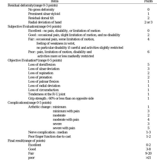
Table 1. Demerit point rating system used to evaluate end results
Items Points
Residual deformity(range 0-3 points)
No gross deformity 0
Prominent ulnar styloid 1
Residual dorsal tilt 2
Radial deviation of hand 2 or 3
Subjective Evaluation(range 0-6 points)
Excellent : no pain, disability, or limitation of motion 0 Good : occasional pain, slight limitation of motion, and no disability 2
Fair : occasional pain, some limitation of motion, 4
feeling of weakness in wrist,
no particular disability if careful and activities slightly restricted
Poor : pain, limitation of motion, disability and 6
activities more or less markedly restricted Objective Evaluation*(range 0-5 points)
Loss of dorsiflexion 5
Loss of ulnar deviation 3
Loss of supination 2
Loss of pronation 2
Loss of palmar flexion 1
Loss of radial deviation 1
Loss of circumduction 1
Tenderness st the R-U joint 1
Grip strength : 60% or less than on opposite side 1
Complications(range 0-5 points)
Arthritic change : minimum 1
minimum with pain 3
moderate 2
moderate with pain 4
severe 3
severe with pain 5
Nerve complication : median 1-3
Poor finger function due to cast 1-2
Final result(range of points)
Excellent 0-2
Good 3-8
Fair 9-20
poor >21
*The objective evaluation is based on the following ranges of motion as being the minimum for normalfunction : dorsiflexion - 45 degrees, palmar flexion - 30 degrees, radial deviation - 15 degrees, ulnar deviation - 15 degrees, pronation - 50 degrees, and supination - 50 degrees.
결 과
1. 결과판정방법
임상적인 결과의 평가 방법으로는 S a r m i e n t o1 8 )의 Demerit point rating system을이용하였는데, 크게잔여 기형, 주관적인 평가, 객관적인 평가, 합병증의 네가 지항목으로나누어각각의정도에따라점수로평가 하고 이것을종합하여 최우수, 우수, 보통, 불량의네 등급으로판정하였다(Table 1). 방사선학적계측은술 전, 술후, 최종추시시 완관절의 전후면 및 측면 방사 선 사진에서 건측과 비교하여 요골 높이, 요측 경사 및장축경사를측정하였다.
2. 결과
S a r m i e n t o의 Demerit point rating system으로 평가한 최종 결과는 최우수가 2례(12.5%), 우수가 1 0례 (62.5%), 보통이 4례( 2 5 % )이었고 불량은 한례에서도 보이지 않았으며 전체의 7 5 %에서우수이상의만족 할만한 임상결과를 보였다.(Table 2) 방사선학적으로 는건측과비교했을때평균요골높이는 8 . 4 3㎜( 3㎜- 1 1㎜), 평균요측경사는 1 7 . 6 8°( 1 0°- 2 6°), 평균장축경 사는 배측 3 . 8 7°( 0°- 1 0°)였다. 임상적으로 보통 이하 로 만족스럽지 않은 결과를 보였던 4례의 방사선학 적 계측치는 요골 높이가 3㎜- 8㎜, 요측 경사가 1 0°-
1 6°, 장축경사가장측 2°- 5°였으며이중한례는감염
이동반된경우였다.(Table 3) Table 2. Clinical Result (by Demerit point rating system)
Method\Result Excellent Good Fair Poor
C/R, pinning & E/F 2 5 1 0
minimal O/R, pinnin & E/F 2 5 1 0
Total 2 10 4 0
Table 3. Summary of end results and radiologic parameters
No. of case End result V.T.* R.H.† R.I.‡
1 Excellent 10 11 21
2 Excellent 6 10 20
3 Good 1 10 19
4 Good 0 9 20
5 Good 5 8 16
6 Good 3 9 17
7 Good 3 10 23
8 Fair 5 4 16
9 Good 3 11 22
10 Good 1 10 21
11 Good 5 9 15
12 Good 4 9 18
13 Good 2 10 15
14 Fair 4 8 10
15 Fair 2 3 14
16 Fair 4 4 16
Mean 3.87 8.43 17.68
No. 1-8 ; C/R, pinning & exteranal fixator No. 9-16 ; minimal O/R, pinning & external fixator
* volar tilting(degrees) : negative means dorsal tilting
† radial height(milimeters)
‡ radial inclination(degrees)
고 찰
원위요골 골절은정형외과영역에서가장흔하게 접하게되는골절중의하나로분쇄상이경하거나전 위가심하지않은 경우에는 보존적인 치료방법으로 만족할만한결과를얻을수있었지만고에너지손상 에의한관절내골절이나분쇄상이심한불안정성골 절의경우는치료가어렵다2 , 3 , 6 , 1 5 , 1 6 , 1 9 , 2 0 )
.
불안정성골절의경우일단정복이되었다할지라 도 다시전이가일어나거나초기에는정복이유지되 다가골유합이일어나면서서서히요골 단축이발생 하는 경우가 있다 . 이러한 경우에 인대 정복 ( l i g a m e n t o t a x i s )1 , 1 7 , 2 1 )의 개념을 이용하여 외고정 장치 를 장착시키는 방법이 조기 정복 및 정복 유지의 안 정성을얻을수있는유용한방법으로제안되고있다
1 , 5 , 7 , 1 0 , 1 1 , 1 7 , 2 1 )
. Horesh1 0 )등은 이러한 외고정 장치의 적 응증으로젊고활동적인연령의원위요골관절내골 절, 개방성골절, 다발성외상 환자및보존적치료에 실패한경우라하였으며, Cooney4 0는 (1) 장축배측경 사가 2 5°이상, 요골 단축이 1 0㎜이상인 분쇄골절, 혹
은분쇄가심한관절내골절 , (2) 보존적인치료후정 복의소실이 있을때, (3) 양측성 원위 요골 골절이라 고 하였다. 저자들은 불안정성을 보이는 원위 요골 골절 1 6례에서외고정장치를시행하였으며, 단순방 사선검사상최종추시시점까지정복의소실이나요 골단축이진행한경우는한례도없었다.
또한외고정술은관혈적정복및내고정술시행후 흔히 발생할 수 있는 연부조직의 박리로 인한 술후 관절강직을 피할 수 있으며, 도수정복및 석고 고정 시 장기간의 고정으로 인한 관절 강직을 피할 수 있 는 장점이 있다. 최근에는 술후 관절 강직을 예방하 기위해조기관절운동이시행되고있으며저자들은 이를위해 평균술후 3주째수근 관절운동을 시작하 였는데이로인한정복의소실은없었으며빠른관절 운동의회복을관찰할수있었고관절강직은감염이 동반된한례에서만나타났다.
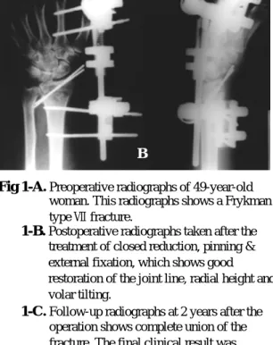
Z a g o r s k i2 2 )는 외고정 장치의 단독 사용시 요골 높 이 및 각도 등은 해부학적으로 얻을 수 있으나 만족 할만한관절면의정복은얻을수없다고하였으며실 제로관절내골절의경우외고정장치단독사용으로 Fig 1-A.Preoperative radiographs of 49-year-old
woman. This radiographs shows a Frykman type Ⅶ fracture.
Fig 1-B.Postoperative radiographs taken after the treatment of closed reduction, pinning &
external fixation, which shows good restoration of the joint line, radial height and volar tilting.
Fig 1-C.Follow-up radiographs at 2 years after the operation shows complete union of the fracture. The final clinical result was excellent.
A
C
B
는 심한 관절면의 불일치(articular incongruity)를 해결 하는데불충분했던경우가보고하였다.
저자들은 불안정성 원위 요골 골절에 대해 총 1 6 례중 8례에서도수정복, 핀고정및외고정장치를 시 행하였으며 도수정복과 외고정 장치만으로 관절면 의 정복이 만족스럽지 않았던 나머지 8례에 대해서 는 최소 피부 절개술을 이용한 정복술 후 핀고정 및 외고정장치를 시행하였다. 최소피부절개술을시행 한 8례중 5례에서 Demerit point rating system상 양호 (good) 이상의 결과를 보였으며 3례에서 보통( f a i r )의 결과를 보였다. 관절면을 침범한 원위 요골 골절에 있어서는 관절면의 정복이 중요하며 도수정복으로 관절면의 불일치를 해결할 수 없을 때는 최소 피부 절개술을통해관절면의정복을얻을수있었다.
원위 요골 골절 중 관절내 혹은 불안정성 골절의 치료에 있어서 해부학적 정복과 기능적인 결과와의 인과관계에대해서는이미알려져왔으며일부저자 들은 요골 높이 (radial height), 요측 경사 ( r a d i a l inclination) 및 장축경사(volar tilting)의회복이 예후에
중요하다고하였고1 3 ), 일부저자들은장축경사가배 측각 2 o이상이거나 요골 단축이 5㎜이상, 관절면 불 일칠가 2㎜이상일 경우 불량한 결과를 가져온다고
하였다2 1 ).
Demerit point rating system상결과가 보통(fair) 이하 로불만족스러웠던4례에대해방사선학적으로분석 한결과평균장축경사는배측 3 . 7 5°( 2°- 5°)로만족할 만한 결과를 보인 1 2례의 평균인 배측 3 . 5 8°( 0°- 1 0°) 에비해차이를보이지않았으나 , 평균요골높이는 3 례에서3 . 6 7㎜( 3㎜- 4㎜)로만족할만한결과를보인 1 2 례의 평균 9 . 6 7㎜( 8㎜- 1 1㎜)에 비해 차이를 보였으며 보통(fair) 소견을보인나머지한례에서는요골높이 가 8㎜로 유지되고 있었으나 감염이 동반된 경우였 다. 이와같이결과가불만족스러웠던경우들을분석 하여 볼 때 방사선학적인 계측 중 요골 높이가 결과 에중요한영향을미치는것을알수있었다 .
술후외고정장치유지기간에대해서는이견이있 으나 본 저자들은 최하 6주에서 최고 1 2주까지 유지 하였으며이들의평균고정기간은 8주였다.
Fig 2-A.Preoperative radiographs of 30-year-old man. This radiographs shows a Frykman type Ⅷ fracture.
Fig 2-B.Postoperative radiographs taken after the treatment of minimal open reduction, pinning & external fixation, which shows acceptable range of radial height, volar tilting and articular congruity.
Fig 2-C.Follow-up radiographs at 1 year after the operation shows complete union of the fracture. The final clinical result was good.
A
C
B
방사선학적으로 골관절염의 소견은 한례에서도 보이지 않았으나 이는 추시기간이 짧았기 때문으로 생각되며이에대해서는좀더장기적인추시가필요 하다.
결 론
1 9 9 5년 1월부터 1 9 9 7년 1 2월까지 고려대학교부속 구로병원 정형외과학교실에서 외고정장치를 이용 하여원위요골골절의치료를시행한후 1년이상추 시가가능했던 1 6례에대해연구한결과다음과같은 결론을얻었다.
1. 불안정성원위 요골 골절에있어외고정장치는 선열을유지하고정복소실을방지하는데있어서유 용한치료방법으로사료된다.
2. 도수정복및인대정복( l i g a m e n t o t a x i s )만으로만족 스럽지않았던관절내골절에서는최소 피부절개술 을 시행하였으며, 이런 경우 관혈적 정복으로 인한 관절 강직을 최소화하면서 관절면의 정복을 향상시 킴으로써결과를호전시킬수있었다.
R E F E R E N C E S
1) Agee JM : External fixation, Technical advances based upon multiplanar ligamentotaxis. Orthop Clin North Am, 24: 265-274,1993.
2) Axelord TS and McMurty RY: Open reduction and internal fixation of comminuted intraarticular fractures of the distal radius. J Hand Surg, 15-A:
1,1990.
3) Bradway JK, Amadio PC and Cooney WP: Open reduction and internal fixation of displaced, comminuted intraarticular fractures of the distal end of the radius. J Bone Joint Surg, 71-A: 839-847,1989.
4) Cooney WP: External fixation of distal radius fractures. Clin Orthop, 180: 44-49,1983
5) Cooney WP, Linscheid RL and Dobyns JH : External pin fixation for unstable Colles’fracture. J Bone Joint Surg, 61-A: 840-845,1979.
6) Fernandez DL and Geisslorr WB: Treatment of displaced articular fractures of the radius. J Hand S u r g, 16-A: 375,1991.
7) Frederic S, William P, Cooney WP and Franz B:
Small external fixation devices for the hand and wrist. Clin Orthop, 293: 77-82,1993.
8) Frykman G: Fracture of the distal radius including sequelae shoulder-hand-finger syndrome.
Disturbance in the distal radioulnar joint and impairment of nerve function : A clinical and experimental study(supplementum). Acta Orthop S c a n d, 108:1-155,1967.
9) Green DP : Pins and plaster treatment of comminuted fractures of the distal end of the radius. J Bone Joint Surg, 57-A: 304-310,1975.
10) Horesh Z, Volpin G, Hoerer C and Stein H: The surgical treatment of severe comminuted intraarticular fractures of the distal radius with small AO external fixation device. Clin Orthop, 263: 147- 1 5 3 , 1 9 9 1 .
11) Howard PW, Steward HD, Hind RE and Burke F D: External fixation or plaster for severely displaced comminuted Colles’fractures?. J Bone Joint Surg, 71-B: 68-73,1989.
12) Jenkins NH, Jones DG and Johnson SR: External fixation of Colles’fractures. J Bone Joint Surg, 69- B: 207-211, 1987.
13) McMurtry RY and Jupiter JB: Fracturese of the distal radius. In: Browner BD, Jupiter JB, Levine AM, Trafton PG eds. Skeletal trauma, vol 2.
Saunders, P h i l a d e l p h i a, pp 1063-1094,1992 14) Leung KS, Shen WY and Tsang HK : An
effective treatment of comminuted fractures of the distal radius. J Hand Surg, 15-A: 11,1990.
15) Melone CP Jr: Open treatment for displaced articular fractures of the distal radius. Clin Orthop, 202: 103,1986.
16) Raskin KB and Melone CP Jr: : Unstable articular fractures of the distal radius. Comparative techniques of ligamentotaxis, Orthop Clin North A m, 24: 275-286,1993.
17) Sarmiento A, Pratt GW, Berry IVC and Sinclair W F: Colles’fractures. Functional bracing in supination. J Bone Joint Surg, 57-A: 311-317,1975.
18) Seitz WH, Putnam MD and Dick HML: Limited open surgical approach for external fixation of the distal radius fractures. J Hand Surg , 15-A:
2 2 8 , 1 9 9 0 .
19) Vaughan PA, Lui SM and Harrington IJ : Treatment of unstable fractures of the distal radius. J Hand Surg, 67-B: 385,1985.
20) Wiliam H and Seitz Jr. : External fixation of diatal radius fractures. Indication and technical principles.
Orthop Clin North Am, 24: 255-264,1993.
21) Weber ER : A rational approach for the recognition and treatment of Colles’fractures. Hand Clin, 3:
1 3 - 2 1 , 1 9 8 7 .
22) Zagorski JB : Comminuted fractures of the distal r a d i u s . Instructional Course Lectures, A A O S , M o s b y Co, 39: 225-263,1990.