J Korean Soc Radiol 2016;75(2):138-142 http://dx.doi.org/10.3348/jksr.2016.75.2.138
INTRODUCTION
Granular cell tumor (GCT) is an extremely rare mesenchymal soft tissue neoplasm of Schwann cell origin. GCT may occur throughout the body, usually in the head and neck, trunk and upper extremities; rarely in the abdominal wall. They are usual- ly benign and solitary; however, approximately 0.5–2% of GCTs are malignant tumors, and 5–10% of GCTs occur in the form of multiple lesions (1-4). While the benign GCTs are smaller and slow-growing tumors, malignant GCTs (MGCTs) are larger and fast-growing tumors, and exhibit a high rate of metastasis and short survival. The radiologic findings of the lesion have been described only in a few reports. In this article, we report a case of 66-year-old man with pathologically proven MGCT aris- ing in the abdominal wall muscles, illustrate the computed to-
mography (CT) findings of the lesion, and discuss imaging dif- ferential diagnosis.
CASE REPORT
A 66-year-old man visited our hospital with a 30-year history of a palpable and recently growing soft tissue mass in the left anterolateral abdominal wall. Physical examination revealed a hard and well-circumscribed mass with no tenderness. The mass was covered by apparently normal skin. The patient had no history of abdominal trauma or surgery.
CT (LightSpeed 16, GE Healthcare, Milwaukee, WI, USA) demonstrated a relatively homogeneous enhancing solid mass in the left anterolateral abdominal wall, arising from the muscle lay- er (Fig. 1A). A subtle ill-defined low attenuated portion was ap-
Malignant Granular Cell Tumor of the Abdominal Wall Mimicking Desmoid Tumor: A Case Report with CT Imaging Findings and Literature Review
복벽을 침범한 악성 과립세포종의 증례와 CT 영상소견 보고 및 문헌 고찰
Jehong Yoon, MD, Sung Eun Ahn, MD*, Dong Ho Lee, MD, Seong Jin Park, MD, Sung Kyoung Moon, MD, Joo Won Lim, MD
Department of Radiology, Kyung Hee University Hospital, Kyung Hee University School of Medicine, Seoul, Korea
Granular cell tumors (GCTs) are extremely rare mesenchymal neoplasms of Schwann cell origin. Malignant GCTs (MGCTs) comprise 0.5–2% of all GCTs. In the present report, we describe a case of a 66-year-old man with MGCT of the abdominal wall. The patient visited our hospital due to a recently growing palpable soft tissue mass in the abdomi- nal wall. Computed tomography scan revealed a 4.3 × 4.1 × 2.9 cm sized mass arising from the left abdominal wall, which was contemplated as a desmoid tumor before sur- gical excision. Histopathological examination confirmed MGCT.
Index terms Granular Cell Tumor Fibromatosis Abdominal Wall
Received December 17, 2015 Revised January 27, 2016 Accepted March 28, 2016
*Corresponding author: Sung Eun Ahn, MD
Department of Radiology, Kyung Hee University Hospital, Kyung Hee University School of Medicine,
23 Kyungheedae-ro, Dongdaemun-gu, Seoul 02447, Korea.
Tel. 82-2-958-9502 Fax. 82-2-968-0787 E-mail: [email protected]
This is an Open Access article distributed under the terms of the Creative Commons Attribution Non-Commercial License (http://creativecommons.org/licenses/by-nc/3.0) which permits unrestricted non-commercial use, distri- bution, and reproduction in any medium, provided the original work is properly cited.
parent within the mass and the mass measured 4.3 × 4.1 × 2.9 cm. The mass did not show extension to the parietal peritoneum or subcutaneous fat layer. The mass was well-defined, but focally ill-defined at the inferior aspect (Fig. 1B). There was no calcifica- tion or cystic portion within the mass.
Initially, the CT findings suggested the presence of a benign soft tissue mass, which was considered as a desmoid tumor. Sub- sequently, surgical mass excision was performed. The mass showed adhesions to the adjacent subcutaneous fat tissue. On macroscopic inspection, a partially ill-demarcated round mass,
measuring 4.5 × 3.4 × 3.0 cm was found. On histological exami- nation, tumor cells with abundant cytoplasm and intracytoplas- mic granules (Fig. 1C) were observed and they were found to be positive for periodic acid-Schiff staining reaction (Fig. 1D) and S-100 (Fig. 1E). Up to 3 mitotic figures were counted in 10 high- power fields. The histologic report confirmed the diagnosis of malignant GCT. The patient was discharged after the surgery without any complications. There was no evidence of local recur- rence or metastasis on follow up imaging studies after 30 months.
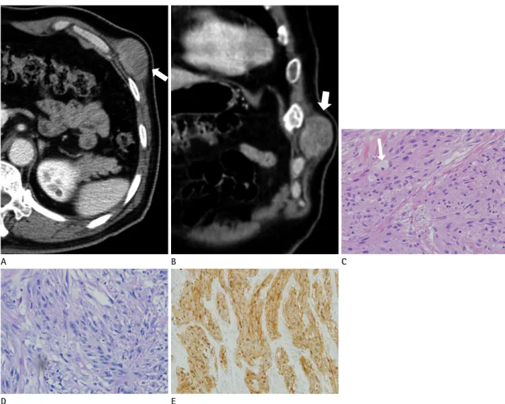
Fig. 1. A 66-year-old man with a malignant granular cell tumor of the abdominal wall muscle.
A. The axial contrast-enhanced abdominal CT scan reveals a well-defined, relatively homogeneous enhancing solid mass in the left anterolateral abdominal wall, arising from the left internal oblique and transverse abdominal muscles. A focal ill-defined low attenuated portion can be seen in the mass (arrow).
B. The coronal image demonstrates a focally ill-defined mass at the inferior edge (arrow).
C. Hematoxylin-eosin staining (× 400) shows tumor cells with abundant cytoplasm and intracytoplasmic granules (arrow).
D. Periodic acid-Schiff stain (× 400) shows tumor cells with positive purple-magenta staining.
E. Staining for S-100 (× 150) shows tumor cells with positive red-brick color staining.
A B C
D E
DISCUSSION
GCTs are rare and almost invariably benign masses, which usually develop as painless masses with normal overlying skin.
GCTs occur in a wide variety of visceral sites, and skin and sub- cutaneous tissue. In majority of the cases, the head, neck and oral cavity (30%), especially the tongue, are involved. McGuire et al. (5) reported that MGCTs are twice as likely to occur in fe- males as in males in the age range of 30–50 years. The neoplasm is typically slow-growing, well-circumscribed, firm and round- ed, with a diameter ranging from 5–20 mm, although larger tu- mors may be seen. The skin surrounding the tumor is usually normal, although it may be thickened, hyperpigmented, and on
rare occasions, it may be ulcerated (4, 6).
Most of the GCTs exist as a benign solitary nodule with very scarce cases of MGCTs in the form of a high-grade sarcoma of Schwann cell origin and they are also reported as Abrikossoff’s tumor (7). Although GCT was initially reported as myoblastoma of the tongue based on the presence of signs of infiltration be- tween the striated muscles, indicating a muscular origin, recent studies have indicated that it has a peripheral neuroectodermal origin as the tumor cells are strongly positive for S-100 protein, similar to the Schwann cells in the peripheral nerve (4, 8).
Histologically, benign GCTs are composed of large polygonal cells containing numerous eosinophilic granules. The nuclei are relatively small and mildly pleomorphic with prominent nucle-
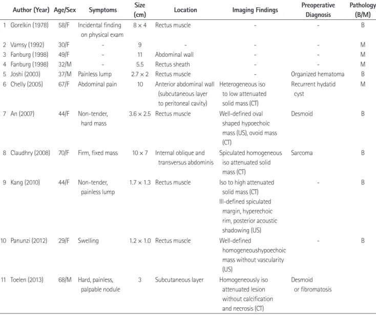
Table 1. Summary of the 11 Cases of Reported Granular Cell Tumors of the Abdominal Wall Author (Year) Age/Sex Symptoms Size
(cm) Location Imaging Findings Preoperative
Diagnosis
Pathology (B/M) 1 Gorelkin (1978) 58/F Incidental finding
on physical exam
8 × 4 Rectus muscle - - B
2 Vamsy (1992) 30/F - 9 - - - M
3 Fanburg (1998) 49/F - 11 Abdominal wall - - M
4 Fanburg (1998) 32/M - 5.5 Rectus sheath - - M
5 Joshi (2003) 37/M Painless lump 2.7 × 2 Rectus muscle - Organized hematoma B
6 Chelly (2005) 67/F Abdominal pain 10 Anterior abdominal wall (subcutaneous layer to peritoneal cavity)
Heterogeneous iso to low attenuated solid mass (CT)
Recurrent hydatid cyst
M
7 An (2007) 44/F Non-tender, hard mass
3.6 × 2.5 Rectus muscle Well-defined oval shaped hypoechoic mass (US), ovoid mass (CT)
Desmoid B
8 Claudhry (2008) 70/F Firm, fixed mass 10 × 7 Internal oblique and transversus abdominis
Spiculated homogeneous iso attenuated solid mass (CT)
Sarcoma B
9 Kang (2010) 44/F Non-tender, painless lump
1.7 × 1.3 Rectus muscle Iso to high attenuated solid mass (CT) Ill-defined spiculated margin, hyperechoic rim, posterior acoustic shadowing (US)
- B
10 Panunzi (2012) 29/F Swelling 1.2 × 1.0 Rectus muscle Well-defined
homogeneoushypoechoic mass without vascularity (US)
- B
11 Toelen (2013) 68/M Hard, painless, palpable nodule
3 Subcutaneous layer Homogeneously iso attenuated lesion without calcification and necrosis (CT)
Desmoid or fibromatosis
B = benign, M = malignant, US = ultrasonography
oli (3). Approximately 0.5–2% of GCTs are malignant, and they have the following characteristics which indicate more likelihood of malignancy; mass size larger than 5 cm, nuclear pleomorphism, metastatic lymph nodes, aggressive clinical behavior, rapid growth, and ulceration (1-4).
In order to diagnose MGCT, Fanburg et al. devised a classifi- cation system by analyzing 73 atypical MGCTs using the fol- lowing six parameters: increased nuclear-to-cytoplasmic ratio, nuclear pleomorphism, necrosis, spindling of tumor cells, vesicu- lar nuclei with prominent nucleoli, and a mitotic count of more than two in 10 high-power fields (200 × field). According to the Fanburg criteria, GCT was classified as atypical if it satisfied 1–2 criteria, and it was classified as malignant if it satisfied 3–6 cri- teria. Although rare, complete surgical resection of all GCTs is recommended because of its malignant potential and possibility of local recurrence (9).
In our study, the lesion was confirmed to be MGCT and we observed a mass with soft tissue attenuation located in the ab- dominal wall muscle with relatively homogeneous attenuation including a subtle internal low attenuated portion, which is con- sidered to reflect the pathologically necrotic portion. This lesion was first thought to be a desmoid tumor. Typically, 28–69% of desmoid tumors have been reported to present as an intra-ab- dominal or an abdominal wall mass in various studies, and since a desmoid tumor manifests as a soft tissue mass with variable at- tenuation and enhancement on CT, it is difficult to differentiate it from GCT due to overlap of many imaging features (10). The ra- diologic characteristics of MGCTs are unclear since there are few reports on the imaging findings, but generally, since the disease presents as a mass with soft tissue attenuation, differential diag- nosis with soft tissue tumorous lesions is necessary. If the lesion appears heterogeneous or has a part showing low attenuation, the possibility of MGCT should be considered.
GCTs rarely develop in the abdominal wall. There are 10 cases of GCTs of the abdominal wall in the English literature and one case in the Korean literature (11). These cases are summarized in Table 1, including patient characteristics such as age, sex, symptoms, locations, preoperative diagnosis, imaging findings, and pathologic group. According to the table, four out of the eleven cases were confirmed to be of MGCT; therefore, in GCTs with abdominal wall involvement, a higher prevalence of malignancy is more likely than in the previously known cases.
All the four previously reported MGCTs of the abdominal wall were more than 5 cm in terms of mass size, but the lesion in our case was smaller than 5 cm. Also, two of the six benign ab- dominal GCTs measured 8 cm and 10 cm, respectively. This proves that it is difficult to differentiate between benign and malignant lesions based on the size alone.
There is only one case report describing an abdominal MGCT.
The reported CT imaging finding of MGCTs was a heteroge- neously low attenuated mass within the subcutaneous layer of the abdominal wall extending into the peritoneal cavity. Howev- er, since our case presented as a well-defined, relatively homo- geneous enhancing solid mass despite being a MGCT, the im- aging findings of MGCTs can be considered to be non-specific.
Therefore, it is difficult to differentiate between benign and oth- er malignant lesions such as sarcoma or desmoid tumor based on the imaging findings alone or the size criteria.
In conclusion, although GCTs of the abdominal wall are ex- tremely rare, they should be considered in the differential diagno- sis of an abdominal wall mass arising from the abdominal mus- cles. Furthermore, even if the lesion is less than 5 cm and presents as a homogeneously enhancing solid mass, it is difficult to differ- entiate benign lesions from malignant lesions based on the size or imaging findings alone. Therefore, it is necessary to confirm the diagnosis through biopsy or to perform complete surgical resec- tion of a mass with abdominal wall involvement.
REfERENCES
1. Chen J, Wang L, Xu J, Pan T, Shen J, Hu W, et al. Malignant granular cell tumor with breast metastasis: a case report and review of the literature. Oncol Lett 2012;4:63-66 2. Curtis BV, Calcaterra TC, Coulson WF. Multiple granular cell
tumor: a case report and review of the literature. Head Neck 1997;19:634-637
3. Marangi GF, Toto V, Poccia I, Gigliofiorito P, Brunetti B, Persichetti P. Multiple localization of granular cell tumour:
a case report. Cases J 2009;2:8751
4. Tan TJ, Alassiri AH, Ng TL, Mallinson PI, Munk PL. Malignant granular cell tumor of the foot-multimodality imaging findings and literature review. Clin Imaging 2015;39:543- 546
5. McGuire LS, Yakoub D, Möller MG, Rosenberg A, Livingstone
A. Malignant granular cell tumor of the back: a case report and review of the literature. Case Rep Med 2014;2014:
794648
6. Qureshi NA, Tahir M, Carmichael AR. Granular cell tumour of the soft tissues: a case report and literature review. Int Semin Surg Oncol 2006;3:21
7. Abrikossoff AL. Uber myome, ausgehend von der querg- estreiften willkürlichen muskulatur. Virchows Arch Path Anat 1926;260:215-233
8. Stefansson K, Wollmann RL. S-100 protein in granular cell tumors (granular cell myoblastomas). Cancer 1982;49:
1834-1838
9. Chen WS, Zheng XL, Jin L, Pan XJ, Ye MF. Novel diagnosis and treatment of esophageal granular cell tumor: report of 14 cases and review of the literature. Ann Thorac Surg 2014;
97:296-302
10. Shinagare AB, Ramaiya NH, Jagannathan JP, Krajewski KM, Giardino AA, Butrynski JE, et al. A to Z of desmoid tu- mors. AJR Am J Roentgenol 2011;197:W1008-W1014 11. Kang E, Lee YH, Park SH, Choi SS. Granular cell tumor oc-
curring at the abdominal wall: a case report. J Korean Soc Radiol 2010;63:547-550
복벽을 침범한 악성 과립세포종의 증례와 CT 영상소견 보고 및 문헌 고찰
윤제홍 · 안성은* · 이동호 · 박성진 · 문성경 · 임주원
과립세포종(granular cell tumor)은 Schwann 세포에서 기원하는 매우 드문 종류의 중간엽 세포성 종양이다. 악성 과립세 포종은 전체 과립세포종의 약 0.5~2%를 차지하는 것으로 알려져 있는데, 본 증례 보고에서는 66세 남성에서 복벽을 침범 한 악성 과립세포종의 사례를 다루고 있다. 환자는 최근 들어 좌측 복벽에서 촉지 가능하면서 지속적으로 크기 증가를 보이 는 종괴를 주소로 내원하였으며, CT에서 좌측 복벽의 약 4.3 × 4.1 × 2.9 cm 크기의 종괴가 확인되어 섬유모세포 종양 (desmoid tumor) 의심하에 수술적 제거를 시행하였으나 최종 병리소견에서 악성 과립세포종으로 확인되었다. 이에 저자 들은 복벽을 침범한 악성 과립세포종의 영상 및 병리소견과 함께 관련 문헌 고찰을 보고하고자 한다.
경희대학교 의학전문대학원/의과대학 경희대학교병원 영상의학과