Bronchiectasis is an important cause of chronic cough, recurrent infection, and hemoptysis (1). For optimal treatment of a patient with bronchiectasis, its extent must be determined, especially when surgical resection is considered (2). Because tubular dilatation of airways can be seen in asthma, and bronchial dilatation associat- ed with acute infection may subside, it is also important to know the type of bronchiectasis.
B ronchiectasis : Diagnostic Ac c u ra cy of Chest Computed Radiogra p hy1
Eung Yeop Kim, M.D., Boo-Kyung Han, M.D., Tae Sung Kim, M.D., Jung Hwa Hwang, M.D., Jung Hwan Yoon, M.D., Chul H. Paik, Ph.D., Kyung Soo Lee, M.D., Jae Min Cho, M.D.,
Sang Hee Choi, M.D., Hye - Kyung Yoon, M.D.
Purpose : The aim of this study was to assess the diagnostic accuracy of chest comput- ed radiography for the detection of bronchiectasis diagnosed by high-resolution CT.
Materials and Methods : Our study included 100 consecutive patients with bronchiec- tasis and 20 normal subjects, all seen on high-resolution CT. Two independent ob- s e r vers analyzed chest computed radiographs and recorded the presence and type of b r o n c h i e c t a s i s, and the invo l ved lobe.
Results : On high-resolution CT, bronchiectasis was seen in one lobe in 29 patients, t wo lobes in 29, three lobes in 16, four lobes in 14, five lobes in 10, and six lobes in t wo. The bronchiectasis was tubular in 55 patients, mixed tubular and cystic in 29, and cystic in 16. For observer 1, the sensitivity, specificity, and accuracy of chest com- puted radiography was 95%, 85%, and 93%, respective l y, while for observer 2, the corresponding figures were 93%, 85%, and 92%. Sensitivity and specificity for observ- er 1 were 33% and 96% for the right upper lobe (46% and 95% for observer 2), 68%
and 86% for the right middle lobe (76% and 86%), 70% and 78% for the right lowe r lobe (48% and 83%), 50% and 100% for the left upper lobe (50% and 97%), 63% and 90% for the lingular segment (49% and 93%), and 87% and 75% for the left lower lobe (75% and 90%), respective l y. Tubular bronchiectasis involving a single lobe was the most common source of false negative readings based on the findings of chest com- puted radiography.
Conclusion : Because chest computed radiography is not inferior to high-resolution CT for the detection of bronchiectasis, the routine use of chest computed radiography in screening for bronchiectasis is feasible. Howeve r, due to its low sensitivity in detect- ing bronchiectasis in a specific lobe, preoperative high-resolution CT exa m i n a t i o n m ay be needed.
Index words : B r o n c h i e c t a s i s
Ra d i o g r a p hy, computer-assisted
Computed tomography (CT), thin-section
1Department of Radiology, Samsung Medical Center, Sungkyunkwan University School of Medicine
Received August 4, 1998 ; Accepted January 15, 1999
Kyung Soo Lee, M.D., Department of Radiology, Samsung Medical Center, Sungkyunkwan University School of Medicine
#50, Ilwon-Dong, Kangnam-Ku, Seoul, 135-710, Korea.
Tel. 82-2-3410-2511, FAX. 82-2-3410-2559, e-mail. [email protected]
Because radiographic features are nonspecific, it is dif- ficult to make a definitive diagnosis of bronchiectasis on the basis of chest radiographic findings. The reported accuracy of chest radiographs for the diagnosis of bronchiectasis has varied between 63% and 93% (3,4).
At present, high-resolution CT with a reported diag- nostic accuracy of 87-95%, is the modality of choice for the diagnosis of bronchiectasis (5,6). Because it is cur- rently the diagnostic gold standard for the detection of bronchiectasis, few studies have compared its diagnos- tic accuracy with that of chest radiography (7).
The purposes of our study were to assess the overall diagnostic accuracy of chest computed radiography for the detection of bronchiectasis diagnosed by high-reso- lution CT and to test the feasibility of chest computed radiography for the depiction of bronchiectasis in each lobe of the lung.
Materials and Methods
This study included 100 consecutive patients in whom bronchiectasis had been diagnosed and 20 nor- mal subjects. All findings were based on the results of high-resolution CT scans obtained between September 1995 and August 1997. Fifty-eight patients were men and 42 were women, ranging in age from 18 to 80 years (mean, 44 years). Age-adjusted normal subjects (range, 2 1-69 years; mean, 46 years) were those in whom high- resolution CT scans revealed no abnormalities of the thorax. In normal subjects, CT scans were performed on suspicion of either bronchiectasis or interstitial pneu- monia. The cause of bronchiectasis was postinfectious in all patients except one, in whom it was caused by cil- iary dyskinesia syndrome. The patients (n = 100) com- plained of cough (n = 100), sputum (n = 100), hemopty- sis (n = 47), and dyspnea (n = 27). Thirty one (31/100, 31%) patients were smokers (mean, 16-pack years) and seven (7/20, 35%) normal subjects were smokers (mean, 17-pack years). Chest computed radiographs and CT s- cans were obtained within 0 to 239 days of each other (mean, 14.5 days; in 52% of patients, studies were ob- tained within one week of each other). When two or more CT scans were obtained in one patient, the most recent scans were selected for evaluation and compari- son with chest computed radiographs.
All CT scans were obtained using a HiSpeed Advantage Scanner (GE Medical Systems, Milwaukee, Wis., U.S.A.). Thin-section (1-mm collimation) CT scans were obtained through the thorax at 10-mm intervals
and at end inspiration. Additional expiratory thin-sec- tion (1-mm collimation) CT scans were obtained at 30mm intervals through the thorax for the evaluation of air trapping. The scans were reconstructed using a bone algorithm. Both mediastinal (width, 400 HU; level, -5 0 HU) and lung (width, 1,500 HU; level, -700 HU) win- dow images were printed. All chest computed radi- ographs were obtained using a Fuji computed radiogra- phy system (FCR 9000; Fuji, Tokyo, Japan) and, image parameters for computed radiography were as follows:
183-cm film-focus distance, 12:1 oscillating grid, and phototimed exposure. The default mode of image pro- cessing was used, including dynamic range compres- sion, gradation enhancement, and edge enhancement;
gradient amount (GA)=0.9, gradient type (GT)=E, gra- dient center (GC)=1.6, gradient shift (GS)=-0.2, en- hancement frequency (RN) = 4, enhancement type (RT)=R, enhancement factor (RE)=0.5, DR frequency (DRN)=2, DR type (DRT)=B, DR factor (DRE)=0.6.
Chest images were depicted as single format by a 14”×
1 7”laser-printed hard copy. Both posteroanterior (PA) and lateral radiographs were obtained in 102 patients and PA radiographs only in 18.
High-resolution CT scans were evaluated retrospec- tively by two experienced chest radiologists who took no part in the assessment of chest computed radiographs.
These radiologists assessed the CT scans together and decisions were reached by consensus. They recorded the presence, type, and location of bronchiectasis, which was considered to be present when at least one of the followings was noted: lack of tapering of a bronchus, dilated bronchi abutting the mediastinal pleu- ra, internal diameter of bronchi greater than that of the adjacent pulmonary artery, visualized bronchi within 1- cm of the parietal pleura, and mucus-filled dilated bronchi (8). When bronchiectasis was present, it was di- vided into three types: tubular, cystic, or mixed tubular and cystic. Its location was classified into six lobes with the lingular segment of the left upper lobe as a separate lobe.
Chest computed radiographs were observed by two independent chest radiologists who did not participate in CT evaluation. One observer (observer 1) had four years experience of interpreting chest computed radi- ographs, and the other (observer 2) had two years expe- rience. They recorded the presence and type of bronchiectasis, and the involved lobe. The lingular seg- ment of left upper lobe was also considered as a sepa- rate lobe. Bronchiectasis was considered to be present
when at least one of the following findings was noted:
bronchovascular bundle crowding associated with lobar or segmental volume loss, tram-track appearance of di- lated bronchi, mucoid impaction, cystic lung lesion with or without air-fluid level, and ring-like lesions (4).
Interobserver agreement as to the presence of bronchi- ectasis in each patient and each lobe was analyzed using Kappa statistics. For each set of readings, sensitivity, specificity and accuracy of chest computed radiography for the diagnosis of bronchiectasis were calculated, as were diagnostic accuracy in each lobe and the frequen- cy of findings suggestive of bronchiectasis, as seen on chest computed radiographs.
R e s u l t s
High-resolution CT showed that bronchiectasis was p- resent in one lobe in 29 patients, two lobes in 29, three lobes in 16, four lobes in 14, five lobes in 10, and six lobes in two on high-resolution CT. It was of tubular type in 55 patients, mixed tubular and cystic in 29, and cystic in 16.
The most common finding of bronchiectasis observed on chest computed radiographs was bronchovascular bundle crowding associated with lobar or segmental volume loss (91% [181/200]). The other findings were
tram-track appearance of dilated bronchi (49% [98/200]) (Fig. 1), ring-like lesions (39% [77/200]), cystic lung le- sion with or without air-fluid level (29% [58/200]), and mucoid impaction (8% [16/200]).
For observer 1, the sensitivity, specificity, and accura- cy of chest computed radiography were 95%, 85%, and 93%, respectively; for observer 2, 93%, 85%, and 92%, respectively. Interobserver agreement as to the pres- ence of bronchiectasis in a given patient was good (κ=
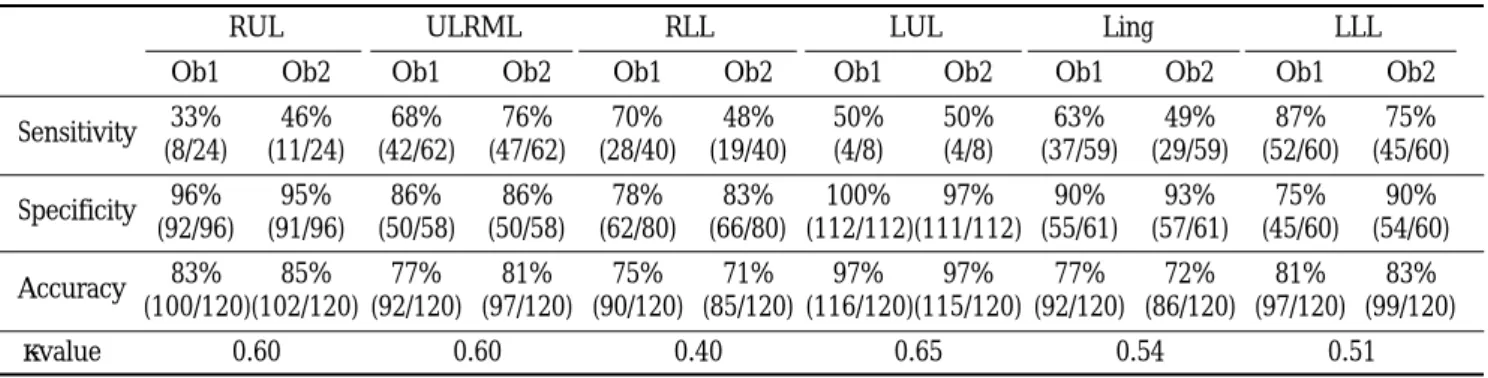
0.61) and was also good in a given lobe (κ= 0 . 4 0-0 . 6 5 ) (Table 1). Sensitivity and specificity for observer 1 were 33% and 96% for the right upper lobe (46% and 95% for observer 2), 68% and 86% for the right middle lobe (76% and 86%), 70% and 78% for the right lower lobe (48% and 83%), 50% and 100% for the left upper lobe (50% and 97%), 63% and 90% for the lingular segment (49% and 93%), and 87% and 75% for the left lower lobe (75% and 90%), respectively (Table 1).
A total of 14 undetected bronchiectatic lesions were p- resent in one lobe in eight patients (57%), in two lobes in five (36%), and in three lobes in one (7%). Twelve (86%) of the 14 false-negative readings occurred in cases of tubular bronchiectasis and the remaining two (14%) in cases of cystic bronchiectasis. On chest computed ra- diographs, tubular bronchiectasis involving a single lobe (eight of 14 false negative readings) was the most diffi-
Fig. 1. Typical feature of tubular bronchiectasis on chest computed radiograph in 49-year-old man
A. Posteroanterior radiograph shows tram lines (arrows) in the left retrocardiac area. Both observers read radiograph as positive for bronchiectasis in the left lower lobe.
B. Lung window of thin-section (1-mm collimation) CT scan obtained below the level of inferior pulmonary vein shows slightly dilated bronchi in the left lower lobe (arrow) and the lingular segment of the left upper lobe (open arrow). Both observers missed bronchiectasis in the lingular segment of the left upper lobe.
A
B
cult to detect (Fig. 2). One of the two missed cystic bronchiectatic lesions was present in the right middle lobe with surrounding pulmonary emphysema. The other cystic bronchiectatic lesion was localized in the lingular segment of the left upper lobe.
Eight (20%) of 40 readings in six normal subjects were false positive for bronchiectasis. In these cases, the in- tervals between chest computed radiographs and HRCT scans were 0 day in 4 normal subjects, 3 days in one, and 81 days in one. For both radiologists, readings in t- wo normal subjects were false positive (no interval be- tween chest computed radiographs and HRCT scans).
In one there were parenchymal bands (Fig. 3), and in one, both hemidiaphragms were elevated. In the re- maining four false-positive readings in four normal sub- jects, the lungs were normal on high-resolution CT and the causes of false positivity were unclear.
D i s c u s s i o n
Prior to the advent of high-resolution CT, which is currently the first choice of diagnostic modality for the detection of bronchiectasis, bronchography had been considered the gold standard. Common plain radi- ographic findings are peribronchial fibrosis, increased size and number of bronchovascular markings by re- tained secretions, bronchial crowding, loss of lobar or lung volume, ring-like lesions (honeycombing), cystic lung lesion with or without air-fluid level, and mucoid impaction (4). However, these findings except cystic lung lesion and mucoid impaction are nonspecific (9). In our study, the most common chest computed radi- ographic finding was bronchovascular bundle crowding associated with lobar or segmental volume loss (91%).
Table 1. Sensitivity, Specificity, and Accuracy of Chest Computed Radiography in the Detection of Bronchiectasis for Each Lobe
R U L U L R M L R L L L U L L i n g L L L
O b 1 O b 2 O b 1 O b 2 O b 1 O b 2 O b 1 O b 2 O b 1 O b 2 O b 1 O b 2
S e n s i t i v i t y 3 3 % 4 6 % 6 8 % 7 6 % 7 0 % 4 8 % 5 0 % 5 0 % 6 3 % 4 9 % 8 7 % 7 5 %
( 8 / 2 4 ) ( 1 1 / 2 4 ) ( 4 2 / 6 2 ) ( 4 7 / 6 2 ) ( 2 8 / 4 0 ) ( 1 9 / 4 0 ) ( 4 / 8 ) ( 4 / 8 ) ( 3 7 / 5 9 ) ( 2 9 / 5 9 ) ( 5 2 / 6 0 ) ( 4 5 / 6 0 ) S p e c i f i c i t y 9 6 % 9 5 % 8 6 % 8 6 % 7 8 % 8 3 % 1 0 0 % 9 7 % 9 0 % 9 3 % 7 5 % 9 0 %
( 9 2 / 9 6 ) ( 9 1 / 9 6 ) ( 5 0 / 5 8 ) ( 5 0 / 5 8 ) ( 6 2 / 8 0 ) ( 6 6 / 8 0 ) ( 1 1 2 / 1 1 2 )( 1 1 1 / 1 1 2 ) ( 5 5 / 6 1 ) ( 5 7 / 6 1 ) ( 4 5 / 6 0 ) ( 5 4 / 6 0 )
A c c u r a c y 8 3 % 8 5 % 7 7 % 8 1 % 7 5 % 7 1 % 9 7 % 9 7 % 7 7 % 7 2 % 8 1 % 8 3 %
( 1 0 0 / 1 2 0 )( 1 0 2 / 1 2 0 ) ( 9 2 / 1 2 0 ) ( 9 7 / 1 2 0 ) ( 9 0 / 1 2 0 ) ( 8 5 / 1 2 0 ) ( 1 1 6 / 1 2 0 )( 1 1 5 / 1 2 0 ) ( 9 2 / 1 2 0 ) ( 8 6 / 1 2 0 ) ( 9 7 / 1 2 0 ) ( 9 9 / 1 2 0 )
κ- v a l u e 0 . 6 0 0 . 6 0 0 . 4 0 0 . 6 5 0 . 5 4 0 . 5 1
Note-RUL : right upper lobe, RML : right middle lobe, RLL : right lower lobe, LUL : left upper lobe (upper division only), Ling : lingular segment, LLL : left lower lobe, Ob1 : observer 1, Ob2 : observer 2, κ-value: interobserver agreement
Fig. 2. False-negative reading on chest computed radiograph in 30-year-old man with tubular bronchiectasis A,B. Posteroanterior (A) and lateral (B) radiographs show no evidence of bronchiectasis.
C. Lung window of thin-section (1-mm collimation) CT scan obtained at level of liver dome shows mucoid impaction in the dilated bronchus (arrow) in the posterior basal segment of the right lower lobe.
A B C
This is nonspecific and can be seen in other diseases in- cluding simple atelectasis without bronchiectasis. Tram- track appearance of dilated bronchi (49%), ring-like le- sions (36%), cystic lung lesion with or without air-fluid level (29%), and mucoid impaction (8%) were also seen.
The reported diagnostic accuracy of chest radiography for the detection of bronchiectasis varies. Cooke et al.
(10) found that the sensitivity of chest radiography, compared with bronchography, was only 37%, and the specificity, 95%. In a study by Currie et al. (3), the sensi- tivity, specificity, and diagnostic accuracy of chest radi- ography in the detection of bronchographically-diag- nosed bronchiectasis were 47%, 100%, and 63%, re- spectively. Recently, van der Bruggen-Bogaarts et al.
(11) reported a sensitivity of 88% and specificity of 74%
in chest radiography for the detection of high-resolu- tion-CT-diagnosed bronchiectasis. In their study, chest radiographs were obtained using a high-kilovoltage technique (120 to 140 kV). Conversely, using a low-kilo- voltage technique, Gudbjerg et al. (4) found that the di- agnostic accuracy of chest radiography was 93%. They found a normal chest radiograph in only 7% of 112 bronchographically proven cases of bronchiectasis.
Given the inherent problem of the low-kilovoltage tech- nique (narrow dynamic range), however, we do not be- lieve that such a high accuracy can be achieved.
Sensitivity (observer 1 and observer 2, 93% and 95%, respectively), specificity (85% and 85%), and accuracy
(92% and 93%) of chest computed radiography in the detection of bronchiectasis in our study are, to the best of our knowledge, higher than in previous reports (3,11), and this may be partly due to the computed radi- ographic system used. In computed chest radiography, we applied postprocessing of dynamic range compres- sion. In this type of compression, both peripheral pul- monary markings and mediastinal structures are clearly and simultaneously visualized on radiographs (12). The positive effect of dynamic range compression may lead to increases in the detection rate of bronchiectasis, espe- cially in the retrocardiac portion of both lower lobes.
Corroborative evidence of this is the fact that when the high-kilovoltage technique is used, the diagnostic accu- racy of chest radiography is high. As with dynamic range compression, retrocardiac lower lobes are clearly visualized.
The high sensitivity of chest radiography noted in the studies of Gudbjerg (4) and van der Bruggen-Bogaarts et al. (11), and in ours, may suggest that chest computed radiography can be used as a screening tool in the diag- nosis of bronchiectasis. Patients suffering from this con- dition usually undergo medical treatment, and diagnosis per se is important in the treatment of the disease. If, however, symptoms are severe or persistent and the disease is confined to a few bronchi, resection may be considered (13). Because of its low sensitivity in the de- tection of bronchiectasis in a specific lobe in our study, Fig. 3. False-positive reading on chest computed radiograph in healthy 44-year-old m a n .
A. Lateral chest computed radiograph shows crowding of bronchovascular mark- ings in the left lower lobe and posterior displacement of the left major fissure. Both observers read radiograph as positive for bronchiectasis in the left lower lobe.
B. Lung window of thin-section (1-mm collimation) CT scan obtained at level of liv- er dome shows a parenchymal band (arrows) in the left lower lobe which caused crowding of bronchovascular markings on chest computed radiograph.
A
B
the use of high-resolution CT for preoperative evalua- tion may be justified.
Bronchiectasis of the right middle and left lower lobe was most easily detected on chest computed radi- ographs (Table 1). Because the right middle lobe, sur- rounded by both major and minor fissures, is well de- fined anatomically, volume decrease or morphological change may be easily detected on chest computed radi- ographs. The enhanced visibility of left lower lobar vol- ume through the heart shadow, as seen on computed chest radiographs, may have increased the detectability of bronchiectasis in this lobe.
In our study, the radiologists missed bronchiectasis in 14 of 200 readings (7%). The 14 false-negative readings were in cases involving one lobe in eight patients (57%), two lobes in five (36%), and three lobes in one (7%). In addition, in 12/14 false-negative readings, bronchiecta- sis was of the tubular type, and tubular bronchiectasis involving a single lobe is therefore the most common source of false-negative readings in chest computed ra- d i o g r a p h y .
Four of eight false-positive readings occurred in cases in which a parenchymal band was present or both hemidiaphragms were elevated. These results suggest that a parenchymal band or elevation of a hemidi- aphragm, as seen on chest radiographs, can simulate bronchiectasis due to bronchovascular crowding with or without volume loss.
Our study suffers from certain limitations. One is the fact that the radiologists analyzed the radiographic find- ings of CT-diagnosed rather than pathologically diag- nosed cases of bronchiectasis. Because CT scans are not a perfect diagnostic modality (sensitivity, 94%-9 6 % ; specificity, 93%-100%) (6,14), our results might not have reflected the true accuracy of chest computed radi- ography in the diagnosis of bronchiectasis. However, because surgery is performed in only a limited number of patients with bronchiectasis, and high-resolution CT is currently the diagnostic gold standard, our results may have some validity in clinical situations. A second limitation is that because only bronchiectatic and nor- mal patients were observed, differentiation between normal and abnormal radiographs may have been much easier. The inclusion of other radiographically similar conditions such as pulmonary fibrosis, atelecta-
sis, chronic bronchitis, emphysema, or interstitial lung disease might have resulted in different levels of diag- nostic accuracy.
In conclusion, chest computed radiography is not infe- rior to high-resolution CT in the detection of bronchiec- tasis. Because of its high accuracy, chest computed radi- ography can be used as a routine examination in the screening of patients with bronchiectasis, but its low sensitivity for a given specific lobe, compared to CT, may justify preoperative high-resolution CT examina- t i o n .
R e f e r e n c e s
1. Millar AB, Boothroyd AE, Edwards D, Hetzel MR. The role of computed tomography (CT) in the investigation of unex- plained hemoptysis. Respir Med 1992;86:39-44
2. McGuinness G, Naidich DP, Leitman BS, McCauley DI.
Bronchiectasis: CT evaluation. AJR 1993;160:253-259 3. Etienne T, Spiliopoulos A, Megevend R. Bronchiectasis: indi-
cation and timing for surgery. Ann Chir 1993;47:729-735 4. Currie DC, Cooke JC, Morgan AD, et al. Interpretation of
bronchograms and chest radiographs in patients with chronic sputum production. Thorax 1987;42:278-284
5. Gudbjerg CE. Roentgenologic diagnosis of bronchiectasis: an analysis of 112 cases. Acta Radiol 1955;43:209-226
6. Kang EY, Miller RR, Muller NL. Bronchiectasis: comparison of preoperative thin-section CT and pathologic findings in re- sected specimens. Radiology 1995;195:649-654
7. Grenier P, Maurice F, Musset D, Menu Y, Nahum H. Bronchi- ectasis: assessment by thin-section CT. Radiology 1986; 161:95- 99
8. Kim JS, Muller NL, Park CS, Grenier P, Herold CJ. Cylindrical bronchiectasis: diagnostic findings on thin-section CT. A J R 1997;168:751-754
9. Stanford W, Galvin JR. The diagnosis of bronchiectasis. C l i n Chest Med1988;9:691-699
10. Cooke JC, Currie DC, Morgan AD, et al. Role of computed to- mography in diagnosis of bronchiectasis. T h o r a x 1 9 8 7 ; 4 2 : 2 7 2 - 277
11. van der Bruggen-Bogaarts BA, van der Bruggen HM, van Waes PF, Lammers JJ. Screening for bronchiectasis: a comparative s- tudy between chest radiography and high-resolution CT. C h e s t 1996;109:608-611
12. Goodman LR, Wilson CR, Foley WD. Digital radiography of the chest: promises and problems. AJR 1988;155:1241-1252 13. Phillips MS, Williams MP, Flower CD. How useful is comput-
ed tomography in the diagnosis and assessment of bronchiec- tasis? Clin Radiol 1986; 37:321-325.
14. Joharjy IA, Bashi SA, Adbullah AK. Value of medium thick- ness CT in the diagnosis of bronchiectasis. A J R 1987; 149:
1133-1137
기관지확장증 :
흉부전산화방사선촬영의 진단정확도1성균관대학교 의과대학 삼성서울병원 진단방사선과
김응엽・한부경・김태성・황정화・윤정환・백철화・이경수・조재민・최상희・윤혜경
목적 : 고해상전산화단층촬영 (HRCT) 에서 진단된 기관지확장증에 대한 흉부 전산화 방사선촬영 (CR) 의 진
단 정확도를 알아보고자 하였다.
대상 및 방법 : H R C T에서 진단된 기관지확장증 환자 1 0 0명과 정상 2 0명을 대상으로 하였다. 두 명의 방사선과
의사가 독자적으로 흉부 C R을 분석하고 기관지확장증의 존재유무, 종류, 침범엽을 기록하였다.
결과 : H R C T에서 진단한 기관지확장증은 2 9예에서 폐의 1엽, 29예에서 2엽, 16예에서 3엽, 14예에서 4엽, 10예에
서 5엽, 2예에서 6엽에 있었다. 기관지확장증은 5 5예에서 관상형, 29예에서 관상형과 낭성 혼합형, 16예에서 낭성 이었다. 기관지확장증의 진단에 있어서 흉부 C R의 전체적인 민감도, 특이도, 정확도는 제1관찰자에서 각각 9 5 % , 83%, 93%였고, 제2관찰자에서 각각 93%, 85%, 92%였다. 우상엽의 경우 민감도와 특이도는 제1관찰자에서 각각 3 3 %와 9 6 %였으며(제2관찰자에서 각각 46%, 95%), 우중엽의 경우 6 8 %와 86% (76%, 86%), 우하엽의 경우 7 0 % 와 78%(48%, 83%), 좌상엽의 경우 5 0 %와 100% (50%, 97%), 설분절(lingular segment)의 경우 6 3 %와 9 0 % (49%, 93%), 좌하엽의 경우 8 7 %와 75% (75%, 90%)였다. 한 엽을 침범한 관상형 기관지확장증의 경우가 흉부 C R에서 가장 흔한 가음성 판독의 원인이었다.
결론 : 기관지확장증의 진단에 있어서 흉부 C R은 H R C T의 진단 정확도에 근접하는 높은 정확도를 가지므로,
선별검사 목적으로 사용될 수 있다. 하지만, 특정 엽에서 진단의 민감도가 낮으므로 수술 전 H R C T는 필요하다 고 생각한다.
대한방사선의학회지 1 9 99;40: 8 71- 8 7 7