서 론
인체 조직들에서 항상성을 유지하기 위한 세포자멸사(apoptosis)
가 슬관절 분야에서도 연구가 이루어져 왔으며1-5) 전방십자인대 조직에 대해서도 특정 인자에 의해 발생된 세포자멸사를 동물 검 체를 이용해 연구가 이루어진 바 있으나,6,7) 인체 전방십자인대의 노화(aging process)에 의한 퇴행성 변화에서 일어나는 세포자멸 사 연구는 이루어진 바가 없다. 임상적으로 퇴행성 관절염 환자 의 슬관절 전치환술 시 전방십자인대의 파열 및 결손 소견을 자 주 관찰할 수 있는데,8) 저자들은 이런 현상이 노화와 관련되어 전 방십자인대에서 세포자멸사가 일어나며, 연령이 증가함에 따라 세포자멸사는 증가하고 동시에 세포증식은 감소할 것이라는 가
Copyright © 2015 by The Korean Orthopaedic Association
“This is an Open Access article distributed under the terms of the Creative Commons Attribution Non-Commercial License (http://creativecommons.org/licenses/by-nc/4.0/) which permits unrestricted non-commercial use, distribution, and reproduction in any medium, provided the original work is properly cited.”
The Journal of the Korean Orthopaedic Association Volume 50 Number 4 2015 Received January 27, 2015 Revised April 1, 2015 Accepted April 27, 2015 Correspondence to: Hwa Sung Lee, M.D.
Department of Orthopaedic Surgery, Yeouido St. Mary’s Hospital, College of Medicine, The Catholic University of Korea, 10 63-ro, Yeongdeungpo-gu, Seoul 07345, Korea TEL: +82-2-3779-1192 FAX: +82-2-783-0252 E-mail: [email protected] Seo Won Jeong’s current affiliation: Institute of Clinical Medicine Research, Yeouido St. Mary’s Hospital, Seoul, Korea.
인체 슬관절 퇴행성 관절염 환자에서 전방십자인대 노화와 관련된 세포자멸사 및 세포증식에 대한 예비조사
현낙민 • 김기원 • 정서원 • 정진화 • 이화성
가톨릭대학교 의과대학 여의도성모병원 정형외과학교실
Preliminary Examination for Apoptosis and Cell Proliferation Related to Aging Process in the Anterior Cruciate Ligament Cells
of the Human Degenerative Arthritic Knee Joint
Rak Min Hyun, M.D., Ki Won Kim, M.D., Seo Won Jeong, M.Sc., Jin Wha Chung, M.D., and Hwa Sung Lee, M.D.
Department of Orthopaedic Surgery, Yeouido St. Mary’s Hospital, College of Medicine, The Catholic University of Korea, Seoul, Korea
Purpose: We investigated the association between aging-induced apoptosis and cell proliferation in the anterior cruciate ligament (ACL) of
aged patients.Materials and Methods: Twenty patients with osteoarthritis who underwent total knee replacement arthroplasty were enrolled in the
study. They were divided into three groups according to age: group A (≤65 years), group B (66-75 years), and group C (≥76 years). The ACL tissue was obtained intraoperatively and subjected to terminal deoxynucleotidyl transferase dUTP nick-end labeling staining and immunohistochemistry to quantify the apoptosis and cell proliferation indices.Results: Apoptosis occurred in all groups, with the highest apoptosis index found in group C, followed by that in groups B and A. A
statistically significant positive linear correlation was observed, with a 1-year increase in age resulting in an average increase of 1.49 in the apoptotic index. The lowest cell proliferation index was observed in group C, followed by that in group B and group A, with a 1-year increase in age resulting in an average decrease of 1.0 in the cell proliferation index, which was a statistically significant negative linear correlation. Consequently, a statistically significant negative correlation was confirmed between the apoptosis and cell proliferation indices, whereby an increase of 1.0 in the apoptosis index was concurrent with a decrease of 0.45 in the cell proliferation index.Conclusion: Apoptosis occurred in the ACL of human knee joint. With increasing age, apoptosis increased and cell proliferation decreased.
Key words: knee joint, anterior cruciate ligament, elderly, apoptosis, cell proliferation
설하에 퇴행성 관절염 환자에서 세포자멸사의 발현 여부와 세포 증식과의 상호 관계성을 알아보고자 하였다.
대상 및 방법
1. 연구재료
Kellgren and Lawrence grade IV의 퇴행성 관절염으로 슬관절 전 치환술을 받은 환자 총 20명을 대상으로 하였으며, 본 연구는 여 의도성모병원의 임상시험 심사위원회(institutional review board) 의 승인하에 시행되었다. 남자가 2예, 여자가 18예였으며, 평균 연 령은 70.5세(61-79세)였다. 연령별로 65세 이하는 5예로 A군, 66세 에서 75세까지는 9예로 B군, 76세 이상은 6예로 C군으로 분류하 였다. 수술 전 진료기록 및 영상을 고찰하여 슬관절 외상이 없었 음을 확인하였고, 수술 중 제거되는 전방십자인대를 실험을 위해 채취하였으며, 조직 채취 시 더 이상의 손상이 가해지지 않도록 하면서 칼을 이용해 남아있는 전방십자인대 실질부를 채취하였 다. 채취된 조직은 -70oC로 급속 냉동하였다.
2. 연구방법
세포자멸사를 관찰하는 방법으로 손상된 DNA를 염색하여 관찰 하는 terminal deoxynucleotidyl transferase dUTP nick-end labeling (TUNEL) 분석, 항체에 있는 형광의 정도를 파악하는 fluores- cence-activated cell sorter (FACS) 분석, 세포자멸사에 따른 DNA 분절화를 이용하는 전기영동법(electrophoresis) 등이 있으며, 본 연구에서는 TUNEL 방법을 이용하여 세포자멸사를 관찰하였다.
수술 중 채취된 전방십자인대를 이용하여 TUNEL 방법 및 세 포증식을 알아보기 위한 Ki-67 면역조직화학염색법(immunohis- tochemistry)을 실시하였으며, 먼저 각 조직에서 세포자멸사의 발 생 여부를 조사하였다. 이의 확인 후 광학현미경으로 고배율(×
200)에서 임의로 10군데를 선택하여 총 세포 수와 TUNEL 방법과 Ki-67 면역조직화학검사에서 양성으로 염색된 세포를 백분율로 계산하여 세포자멸사 지수 및 세포증식 지수로 정량화하였다. 세 포자멸사와 세포증식의 전체평균을 계산하였으며, 각 군에 따른 세포자멸사 비율을 조사하여 통계적 처리 후 상대 비교를 하였 고, 세포증식에 대한 조사도 병행하여 세포자멸사의 연령별 수치 와 상관관계를 알아보았다.
1) TUNEL 분석
본 연구에서는 ApopTag-peroxydase in situ apoptosis detection kit (Intergene, Purchase, NY, USA)를 이용하여 TUNEL 방법을 시행 하였다. 박절한 조직을 xylene과 알코올로 탈파라핀 및 함수과정 을 거친 후 37oC로 가열한 phosphate buffered saline (PBS)에 30분 간 넣어두었다. Proteinase K (20 μm/ml)와 함께 실온에서 15분간 처리한 후 증류수로 세척하고 3% 과산화수소 용액에 5분간 처리
하여 내인성(endogenous) peroxidase을 차단하였다. Terminal de- oxynucleotidyl transferase와 37oC에서 1시간 동안 반응시키고, 차 단용액(blocking solution)을 실온에서 10분간 처리 후 peroxidase- conjugated streptavidin으로 실온에서 30분간 반응시켰다. 그후 PBS로 수세하고, 3,3'-diaminobenzidine hydrochloride로 실온에서 발색시켰으며, 대조염색은 methyl green으로 시행하였다.
2) 면역조직화학적 검사(immunohistochemistry)
세포자멸사의 외재 및 내재 경로에서 발현되는 단백질 효소군 의 항체(Fas, Fas ligand [FasL], Caspase 8, 9, 10 [Santa Cruz Bio- technology, Santa Cruz, CA, USA]; Ccaspase 3 [Labvision, Fremont, CA, USA])와 세포증식과 관련된 Ki-67의 항체(Labvision)를 준 비하였다. 적출한 전방십자인대 조직을 10% 중성 포르말린 용액 에 고정하여 파라핀 블록을 만든 후 5 μm로 박절하여 연속절편 을 얻었다. Aminoprophyltriethoxysilane이 도포된 슬라이드에 부 착시키고 60oC에서 1시간 파라핀을 녹인 후 100% xylene에서 10 분간 2회 탈파라핀 과정과 100%, 90%, 80%, 70% 알코올로 30분간 함수과정을 거친 후 증류수로 세척하였다. 항원성 회복을 위하여 10% citrate buffer (pH 6.0)에서 15분 동안 가열 처리한 후 메탄올 과 30% 과산화수소가 9:1의 비율로 섞인 용액에 10분간 처리하여 내인성 peroxidase을 차단하였다. 이후 연관된 일차 항체를 4oC에 서 16시간 동안 반응시킨 후 streptavidin-biotin-peroxidase com- plex와 실온에서 30분 반응시키고 3,3
’
-diaminobenzidine hydro- chloride으로 20분간 실온에서 발색시켰다. 대조염색은 Mayer’
s hematoxylin으로 시행하였다.
3) 통계적 방법
통계처리는 SAS ver 9.2 (SAS Institute, Cary, NC, USA) 프로그 램을 이용하였다. 각 군 간의 비교는 Mann-Whitney U test 방법 으로 통계처리하였고, p<0.05 인 경우에 통계적 의미가 있는 것 으로 평가하였다. 연속형 변수(continuous variable)에 대해서는 mean±standard deviation으로, 범주형 변수(categorical variable)에 대해서는 n (%)로 표현하였다. Ki-67과 TUNEL, 연령의 관련성은 단순선형 회귀분석(simple linear regression)을 통해 모형을 제시하 고, 회귀계수(regression coefficient)와 r2를 제시하였다.
결 과
1. 세포자멸사와 세포증식
채취된 모든 전방십자인대 검체에서 Fas와 FasL이 발현되는 것 을 확인할 수 있었으며, 세포자멸사 발생 관련 하류경로에서도 caspase 3, 8, 9가 모두 발현되는 것이 관찰되었다(Fig. 1). TUNEL 방법과 Ki-67 단백질에 대한 면역조직학적 검사에서는 세포자멸 사와 세포증식이 일어나는 것이 관찰되었으며(Fig. 2), 전체 평균
은 TUNEL 양성반응이 48.1%±25.8%, Ki-67 양성반응이 22.9%±
14.9% 소견을 보였다.
2. 연령대별 전방십자인대 세포자멸사 및 세포증식
Ki-67과 TUNEL에 대해서 성별과 나이별로 각 그룹의 평균을 비
교한 결과, 성별에 따른 통계적 유의성은 없었으며, 세포자멸사는 A군보다 B군에서 높고, B보다 C군이 높았으며, A군과 B군은 전 체 평균보다 낮고, C군은 높았다. 세포증식지수는 A군보다 B군 이 낮고, B군보다 C군에서 낮았으며, 전체평균보다 A군과 B군은 높고, C군은 낮았다(Table 1).
A B
C D
E
Figure 1. Anterior cruciate ligament cells show expression of Fas (A) and FasL (B). In the downstream of the apoptosis pathway, caspase 8 (C), caspase 9 (D), and caspase 3 (E) are expressed. Arrows indicate positive ligament cells. Counterstaining method: Mayer’s hematoxylin; ×400.
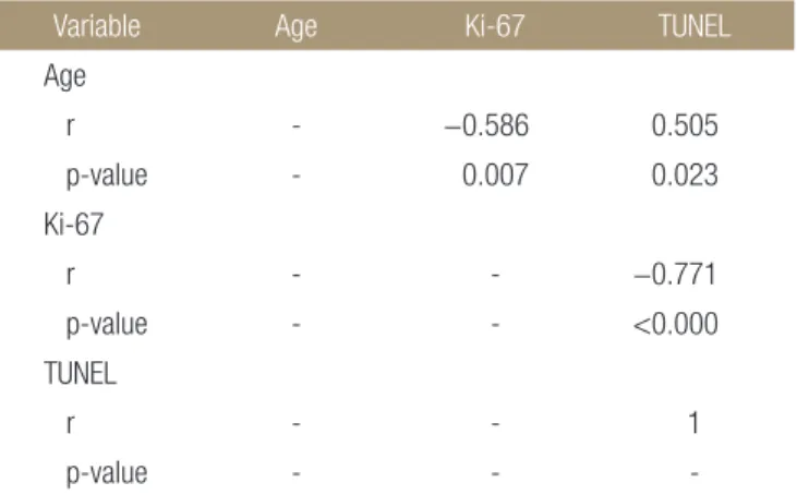
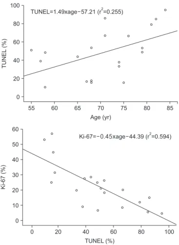
3. 연령과 세포자멸사 지수 및 세포증식 지수의 상관관계 세포자멸사 지수는 연령이 증가할수록 증가하여 연령이 1세 증 가 시 평균적으로 1.49 증가하였고, 상관 분석상 연령에 대해 통 계적으로 유의한 직선적 양의 상관관계를 보였다. 세포증식 지수 는 연령이 증가할수록 감소하여 연령이 1세 증가 시 평균적으로 1.0만큼 감소하였고, 상관 분석상 연령에 대해 통계적으로 유의한 직선적 음의 상관관계를 보였다. 결과적으로 세포자멸사 지수와 세포증식 지수는 상당히 높은 음의 상관관계를 보였으며, 세포자 멸사지수가 1.0 증가 시 세포증식 지수는 0.45 감소하였다(Table 2, Fig. 3).
고 찰
세포자멸사는 세포를 죽음으로 유도하여 손상되거나 더 이상 기 능하지 못하는 세포를 제거함으로써 인체의 항상성을 유지하는 생리적 반응으로 유핵 세포 죽음의 가장 흔한 형태이며, 인체 여 러 조직의 항상성 유지 및 정상적인 조직의 회전에 필요하다. 세 포자멸사는 세포 내, 외의 환경 변화로 기인하는 다양한 신호들, 즉 노화, 이온화 방사선(ionizing radiation), 바이러스 감염에 의한 세포 손상, 세포외 환경, 세포간 상호작용, 호르몬, 약물 등에 의해 활성 또는 억제된다.9,10) 이렇게 다양한 자극 및 신호들에 의해 활 성화되는 세포자멸사에 대한 연구가 인체의 여러 조직을 대상으 로 이루어지고 있으며, 정형외과 영역에서도 여러 근골격 조직에 대한 연구가 이루어지고 있다. 대표적으로 관절 연골세포에서 세
A B
Figure 2. Immunohistochemical staining of Ki-67 protein (A) and terminal deoxynucleotidyl transferase dUTP nick-end labeling (TUNEL) (B) show positive ligament cells, indicated by arrows. Counterstaining method: hematoxylin for Ki-67 protein and methyl green for TUNEL; ×400.
Table 1. Mean Values of the Levels of Staining in the Ki-67 and TUNEL Analyses according to Sex and Age
Variable Ki-67 TUNEL
Sex
Male 35.7±24.5 31.9±30.5 Female 21.5±13.9 49.9±25.6
p-value 0.213 0.363
Age group (yr)
≤65 31.0±12.6 38.5±16.4 66–75 26.7±15.3 38.0±25.7
≥76 10.5±8.2 71.3±18.2
p-value 0.035 0.021
Values are presented as mean±standard deviation. TUNEL, terminal deoxynucleotidyl transferase dUTP nick-end labeling.
Table 2. Correlations between Age and the Levels of Staining for Ki- 67 and TUNEL
Variable Age Ki-67 TUNEL
Age
r - −0.586 0.505
p-value - 0.007 0.023
Ki-67
r - - −0.771
p-value - - <0.000
TUNEL
r - - 1
p-value - - -
TUNEL, terminal deoxynucleotidyl transferase dUTP nick-end labeling.
포자멸사를 유도하는 것들로 nitric oxide와 세포막의 Fas 수용체 와 FasL이 보고되었으며,11,12) 반월상 연골의 경우 nitrotyrosine의 발현이 정상에 비해서 퇴행성 관절염의 반월상 연골에서 의미있 게 증가되어 있고, 그 정도는 조직의 퇴행성 변화가 심할수록 더 욱 현저하다고 밝혀졌다.5) 슬관절 전방십자인대에서는 특정 인 자에 의해 일어나는 세포자멸사를 동물실험을 통해 고찰한 일부 논문들이 제시되어 있으나,6,7) 인체의 노화에 의해 일어나는 세포 자멸사의 자연경과에 대하여는 연구된 바가 없다. 특히 슬관절을 이루고 있는 반월상 연골 및 관절 연골세포의 세포자멸사에 대한 연구에서 연령 증가와 세포자멸사는 밀접한 관련이 있다는 연구 결과1,3)와 고령이라도 퇴행성 관절염이 없다면 세포자멸사가 증 가되어 있지 않았다는 결과를 제시하며 연령 증가와 세포자멸사 의 상관관계는 크지 않다2,5)는 의견을 보이는 서로 상반된 연구결 과들이 함께 있어 퇴행성 정도가 아닌 노화와 세포자멸사와의 관 계성 및 비율에 대한 지속적인 연구가 필요할 것으로 생각한다.
세포자멸사를 관찰하는 방법으로는 TUNEL 방법, FACS로 분 석하는 방법, DNA 전기영동법 등 여러 가지 방법이 있으며, 이들 중 TUNEL 방법은 조직 자체 내에서 세포자멸사를 관찰하는 것 으로 위양성률을 줄이고 조직의 위치에 따른 분석이 가능하여,13) 본 연구에서는 TUNEL 방법을 이용하여 인체 조직에서 수술 중 채취한 전방십자인대 조직으로 연령 증가에 따른 세포자멸사를
관찰하였다. 연구 결과 연령이 증가함에 따라 세포자멸사가 증가 한다는 것을 보여주었으며, 퇴행성 정도와의 관계에서는 비록 본 연구에서 연령에 따른 전방십자인대의 조직학적 퇴행성 정도를 조사하지 않았으나 기존의 연구에서 연령이 증가함에 따라 조직 학적 퇴행성 정도가 정비례 관계로 일정하게 증가한다고 보고되 어8) 연령 증가와 퇴행성 변화 그리고 세포자멸사가 증가하는 정 비례 관계성을 가지고 있다고 판단된다.
Ki-67 단백질은 유사분열(mitosis)뿐만 아니라 세포분열주기 (cell division cycle)의 G1, S, G2단계에서 핵 내에 나타난다.14) G0 단계의 휴지기 또는 정지기의 세포에서는 Ki-67이 발현되지 않 아 세포들의 주어진 세포들 집단에서 증식 부분을 결정하는 좋은 표식자이다.14,15) 본 연구에서는 연령이 증가함에 따라 세포자멸사 증가와 함께 세포증식은 일정하게 감소하는 것으로 관찰되었는 데, 노화과정에서 전방십자인대의 세포자멸사와 세포증식은 서 로 독립되어 발생하는 것이 아니라 일정한 방향성을 가지고 상호 조절되며 이루어지고 있다는 것으로 생각된다. 종양학 분야에서 는 이렇게 상호 관련성을 가지는 세포자멸사와 세포증식을 하나 의 지표로 apoptosis index (AI)/proliferation index (PI)를 구해 임상 예후, 조직학적 등급, 세포에 대한 약물치료 설정 등 다양한 요소 와의 관계에 대한 연구가 이루어지고 있어,16,17) 정형외과 영역에 서도 다양한 근골격근 조직에 대한 여러 요소들과 AI/PI와의 관
0 20 40 60 80 100
60 50 40 30 20 10
Ki-67(%)
TUNEL (%) 0
Ki-67= a ge 44.39 (r =0.594)
255 60 65 70 75 80 85
100
80
60
40
20
TUNEL(%)
Age (yr) 0
TUNEL=1.49xage 57.21 (r =0.255)
255 60 65 70 75 80 85
60 50 40 30 20 10
Ki-67(%)
Age (yr) 0
Ki-67= 1.00xage+93.721 (r =0.343)
2Figure 3. Graph shows a positive correlation between age and terminal deoxynucleotidyl transferase dUTP nick-end labeling (TUNEL), a negative correlation between age and Ki-67, and a negative correlation between TUNEL and Ki-67.
련성 연구가 필요할 것으로 생각된다.
세포자멸사를 유발하는 인자들에 대한 연구뿐만 아니라 세포 자멸사가 어떤 경로에 의해 이행되고 있는지에 대한 연구도 중 요하다. 세포자멸사는 세포막의 Fas 수용체에 가해지는 자극에 의한 외재(extrinsic) 경로 또는 mitochondria 자극에 의한 내재 (intrinsic) 경로로 발생한다.18-20) 본 연구에서는 각 경로에 관여하 는 단백질 효소군 및 관련인자에 대한 항체를 이용한 면역조직화 학적 검사를 시행하여 두 경로에서 모두 발현되어 있다는 것을 확인하였으나 어느 경로가 주된 역할을 하는 것인지에 대해서는 연구가 이루어지지 않아 각 경로의 역할 및 비율에 대한 후속 연 구가 이루어져야 할 것이다. 그 외에 본 연구의 한계점으로 고령 의 심한 퇴행성 관절염 환자의 인체 조직을 이용한 연구로 사체 를 이용하여 특정 질환에 이환되지 않은 조직을 이용하였다면 노 화와의 관련성을 보다 뚜렷이 증명할 수 있겠으나 중증의 관절염 환자의 조직을 이용하였기에 관찰된 결과의 해석에 제한이 있다 는 점과 표본의 크기가 크지 않다는 점이 있다. 그러나 기존의 연 구들이 동물실험이나 사체연구에서 채취한 조직을 이용해 특정 조건을 인위적으로 발생시켜 실험을 한 것과 달리 본 연구는 인 체조직에서 채취한 검체를 이용한 예비 조사 결과로서 향후 이루 어질 세포자멸사 연구에 도움이 될 수 있을 것이라는 점에서 의 미있는 연구로 생각된다.
결 론
인체의 전방십자인대는 다른 외적 독립변수가 발생하지 않는다 면 연령이 증가함에 따라 세포자멸사가 일정하게 증가하며, 세포 증식은 일정하게 감소한다. 이렇게 상호작용으로 서로 균형을 이 루며 일정한 방향성을 가지고 일어나는 세포자멸사와 세포증식 을 단일 지수화하고 이를 하나의 지표로서 여러 가지 요소들과의 상관관계를 연구한다면 추후 임상적으로 사용할 수 있는 기초가 될 것이라고 생각된다.
CONFLICTS OF INTEREST
The authors have nothing to disclose.
REFERENCES
1. Adams CS, Horton WE Jr. Chondrocyte apoptosis increases with age in the articular cartilage of adult animals. Anat Rec.
1998;250:418-25.
2. Aigner T, Hemmel M, Neureiter D, et al. Apoptotic cell death is not a widespread phenomenon in normal aging and osteoarthritis human articular knee cartilage: a study of pro-
liferation, programmed cell death (apoptosis), and viability of chondrocytes in normal and osteoarthritic human knee cartilage. Arthritis Rheum. 2001;44:1304-12.
3. Hashimoto S, Ochs RL, Komiya S, Lotz M. Linkage of chon- drocyte apoptosis and cartilage degradation in human osteo- arthritis. Arthritis Rheum. 1998;41:1632-8.
4. Uysal M, Akpinar S, Bolat F, Cekin N, Cinar M, Cesur N.
Apoptosis in the traumatic and degenerative tears of hu- man meniscus. Knee Surg Sports Traumatol Arthrosc.
2008;16:666-9.
5. Yoon HK, Seo SS, Choi JS, Park JK, Kim YJ. Apoptosis in the meniscus of human osteoarthritic knee. J Korean Orthop Res Soc. 2002;5:43-54.
6. Gyger O, Botteron C, Doherr M, Zurbriggen A, Schawalder P, Spreng D. Detection and distribution of apoptotic cell death in normal and diseased canine cranial cruciate ligaments. Vet J. 2007;174:371-7.
7. Murakami H, Shinomiya N, Kikuchi T, Yoshihara Y, Nemoto K. Upregulated expression of inducible nitric oxide synthase plays a key role in early apoptosis after anterior cruciate liga- ment injury. J Orthop Res. 2006;24:1521-34.
8. Hasegawa A, Otsuki S, Pauli C, et al. Anterior cruciate liga- ment changes in the human knee joint in aging and osteoar- thritis. Arthritis Rheum. 2012;64:696-704.
9. Krammer PH. CD95's deadly mission in the immune system.
Nature. 2000;407:789-95.
10. Waldherr K, Zurbriggen A, Spreng DE, Forterre S. In vi- tro cytoprotective effects of acetylsalicylic acid, carprofen, meloxicam, or robenacoxib against apoptosis induced by sodium nitroprusside in canine cruciate ligament cells. Am J Vet Res. 2012;73:1752-8.
11. Blanco FJ, Ochs RL, Schwarz H, Lotz M. Chondrocyte apop- tosis induced by nitric oxide. Am J Pathol. 1995;146:75-85.
12. Hashimoto S, Setareh M, Ochs RL, Lotz M. Fas/Fas ligand expression and induction of apoptosis in chondrocytes. Ar- thritis Rheum. 1997;40:1749-55.
13. Gavrieli Y, Sherman Y, Ben-Sasson SA. Identification of pro- grammed cell death in situ via specific labeling of nuclear DNA fragmentation. J Cell Biol. 1992;119:493-501.
14. Scholzen T, Gerdes J. The Ki-67 protein: from the known and the unknown. J Cell Physiol. 2000;182:311-22.
15. Bonfil RD, Gonzalez AD, Siguelboim D, et al. Immunohis- tochemical analysis of Ki-67, p21waf1/cip1 and apoptosis in marker lesions from patients with superficial bladder
tumours treated with vinorelbine intravesical therapy in a preliminary phase I trial. BJU Int. 2001;88:425-31.
16. Kimura T, Tanaka S, Haruma K, et al. Clinical significance of MUC1 and E-cadherin expression, cellular proliferation, and angiogenesis at the deepest invasive portion of colorectal cancer. Int J Oncol. 2000;16:55-64.
17. Michael-Robinson JM, Reid LE, Purdie DM, et al. Prolif- eration, apoptosis, and survival in high-level microsatel- lite instability sporadic colorectal cancer. Clin Cancer Res.
2001;7:2347-56.
18. Kim KW, Kim YS, Ha KY, et al. An autocrine or paracrine Fas-mediated counterattack: a potential mechanism for apop- tosis of notochordal cells in intact rat nucleus pulposus. Spine (Phila Pa 1976). 2005;30:1247-51.
19. Scaffidi C, Fulda S, Srinivasan A, et al. Two CD95 (APO-1/
Fas) signaling pathways. EMBO J. 1998;17:1675-87.
20. Scaffidi C, Schmitz I, Zha J, Korsmeyer SJ, Krammer PH, Peter ME. Differential modulation of apoptosis sensitivity in CD95 type I and type II cells. J Biol Chem. 1999;274:22532-8.
인체 슬관절 퇴행성 관절염 환자에서 전방십자인대 노화와 관련된 세포자멸사 및 세포증식에 대한 예비조사
현낙민 • 김기원 • 정서원 • 정진화 • 이화성
가톨릭대학교 의과대학 여의도성모병원 정형외과학교실
목적: 고령 환자에서 노화에 의한 슬관절 전방십자인대의 세포자멸사 및 세포증식과의 연관성을 알아보고자 하였다.
대상 및 방법: 퇴행성 관절염으로 슬관절 전치환술을 받은 환자 20명을 대상으로 하였으며, 연령별로 65세 이하는 A군, 66세에서 75세까지는 B군, 76세 이상은 C군으로 분류하였다. 수술 중 채취된 전방십자인대를 이용하여 terminal deoxynucleotidyl transferase dUTP nick-end labeling 방법 및 면역조직화학염색법을 하였고, 세포자멸사 지수 및 세포증식 지수로 정량화하였다.
결과: 모든 실험군에서 세포자멸사가 발현하는 것을 확인하였으며, 세포자멸사 지수는 A군보다 B군에서 높고, B보다 C군이 높았다.
상관분석에서 연령이 1세 증가 시 평균적으로 1.49 증가하였고, 통계적으로 유의한 직선적 양의 관계를 보였다. 세포증식 지수는 A 군보다 B군이 낮고, B군보다 C군에서 낮았으며, 연령이 1세 증가 시 평균적으로 1.0 감소하였고, 통계적으로 유의한 직선적 음의 상 관관계를 보였다. 세포자멸사 지수와 세포증식 지수는 통계적으로 유의한 음의 상관관계를 보였으며, 세포자멸사 지수가 1.0 증가 시 세포증식 지수는 0.45 감소하였다.
결론: 슬관절 전방십자인대에서 세포자멸사를 확인할 수 있었으며, 연령이 증가함에 따라 세포자멸사가 증가되고, 세포증식은 감소 하였다.
색인단어: 슬관절, 전방십자인대, 고령, 세포자멸사, 세포증식
접수일 2015년 1월 27일 수정일 2015년 4월 1일 게재확정일 2015년 4월 27일 책임저자 이화성
07345, 서울시 영등포구 63로 10, 가톨릭대학교 의과대학 여의도성모병원 정형외과학교실 TEL 02-3779-1192, FAX 02-783-0252, E-mail [email protected] 정서원 저자 현 소속: 여의도성모병원 임상의학연구소.
Copyright © 2015 by The Korean Orthopaedic Association
“This is an Open Access article distributed under the terms of the Creative Commons Attribution Non-Commercial License (http://creativecommons.org/licenses/by-nc/4.0/) which permits unrestricted non-commercial use, distribution, and reproduction in any medium, provided the original work is properly cited.”