343
Copyrights © 2013 The Korean Society of Radiology
INTRODUCTION
Reversible cerebral vasoconstriction syndrome (RCVS) is characterized by the association of severe headaches with or without focal neurological deficit, and a reversible cerebral vaso- constriction assessed by an initial angiography (MR, CT or con- ventional angiography), with the disappearance of arterial ab- normalities within less than 3 months demonstrated by a follow- up angiography (1). RCVS usually occurs in middle-aged women.
Recurrent thunderclap headaches, defined as severe headaches peaking at less than 1 min, are the clinical hallmark of RCVS (1- 4). Postpartum, exposure to vasoactive drugs, and catechol- amine-secreting tumors are well known precipitating causes of RCVS (1). We report a RCVS case of a 46-year-old woman pa- tient. In this case, the precipitating cause for RCVS was not dis- closed; however, a small bowel gastrointestinal stromal tumor (GIST) with gastrointestinal (GI) bleeding was incidentally de- tected. This report describes the clinical and imaging features of
this case, and the questionable relationship between RCVS and GI bleeding will be briefly discussed.
CASE REPORT
A 46-year-old previously healthy woman experienced sudden severe headache (visual analog scale score = 7) with vomiting and a sensation of a hammer beating inside the head. She visited the hospital and received a brain CT scan. The CT showed no abnormality, and she was conservatively managed with analge- sics. However, she experienced a waxing and waning headache every other day. 17 days after the first episode of headache, a sud- den right side low extremity weakness developed, and she was admitted to a tertiary hospital. Neurological examination re- vealed a right lower-extremity monoplegia (grade 2) and sensory loss to pain and touch in the right lower extremity. Otherwise, her vital signs and the results of physical and laboratory exami- nations, except for the value of hemoglobin and hematocrit,
Case Report
pISSN 1738-2637 / eISSN 2288-2928 J Korean Soc Radiol 2013;69(5):343-346 http://dx.doi.org/10.3348/jksr.2013.69.5.343
Received June 23, 2013; Accepted August 20, 2013 Corresponding author: Ji Kang Park, MD Department of Radiology, Jeju National University Hospital, Jeju National University School of Medicine, 15 Aran 13-gil, Jeju 690-767, Korea.
Tel. 82-64-717-1371 Fax. 82-64-757-8276 E-mail: [email protected]
This is an Open Access article distributed under the terms of the Creative Commons Attribution Non-Commercial License (http://creativecommons.org/licenses/by-nc/3.0) which permits unrestricted non-commercial use, distri- bution, and reproduction in any medium, provided the original work is properly cited.
This report was supported by the 2013 scientific promo- tion program funded by Jeju National University.
We report a 46-year-old woman patient with reversible cerebral vasoconstriction syndrome (RCVS). She presented with severe headache, multiple cerebral infarction, and multifocal severe stenosis in the intracranial arteries on magnetic resonance angiography (MRA). One month after the episode, a small bowel gastrointestinal stromal tumor (GIST) was incidentally detected during the evaluation of severe ane- mia and GIST was removed. Follow-up MRA was performed 3 months and 1 year after an initial attack of headache, and multifocal severe intracranial arterial stenot- ic lesions were completely resolved, she did not experience any episode of RCVS during the 2 years.
Index terms Headache
Reversible Cerebral Vasoconstriction Syndrome Stroke
Reversible Cerebral Vasoconstriction Syndrome: A Case Report
소장의 기스트 종양에서 생긴 장 출혈이 있던 환자에서 발생한 가역성 뇌혈관수축 증후군Jeong Sub Lee, MD, Ji Kang Park, MD, Seung Hyoung Kim, MD, Sun Young Jeong, MD
Department of Radiology, Jeju National University Hospital, Jeju National University School of Medicine, Jeju, Korea
RCVS in a Patient with Bleeding from a GIST
344
J Korean Soc Radiol 2013;69(5):343-346 jksronline.orgshe experienced a syncope and dizziness, and severe anemia was detected (hemoglobin 6.4 g/dL, hematocrit 21.1%). She was ad- mitted to our hospital for evaluation of anemia 1 month after the previous ischemic event. Endoscopy for stomach, duodenum, rectum, and colon was normal. Enhanced CT scan revealed a 6.9 cm sized solid enhancing mass abutting small bowel loop in the lower abdomen, and a small bowel GIST or lymphoma was sus- pected. The mass was excised and diagnosed as a GIST with a low mitotic index. After the operation, she recovered well and her he- moglobin level was elevated to 12.4 g/dL at the time of discharge.
A follow-up MRA was performed 3 months after the initial at- tack of headache, multifocal severe stenosis of intracranial arter- ies was completely resolved, and the intracranial arteries ap- peared as normal (Fig. 2). Follow-up MRA was repeated 1 year were within a normal range. Magnetic resonance imaging (MRI)
revealed a subacute left side anterior cerebral artery (ACA) and right posterior cerebral artery territory (PCA) infarction with hemorrhage (Fig. 1). Magnetic resonance angiography (MRA) revealed bilateral multifocal severe stenosis and a beaded ap- pearance in ACA, middle cerebral artery (MCA), PCA, distal vertebral artery, and basilar artery (Fig. 1). She was conserva- tively managed with aspirin and Gliatilin (choline alfoscerate), and her right side weakness and sensory loss were improved.
During admission, anemia was detected with a fluctuating he- moglobin level (hemoglobin level 8.9→8.3→ 7.4→10.4 g/dL) over 3 days; this was conservatively managed with blood transfusion.
She was discharged and received a rehabilitation treatment in a local rehabilitation hospital. During a rehabilitation treatment,
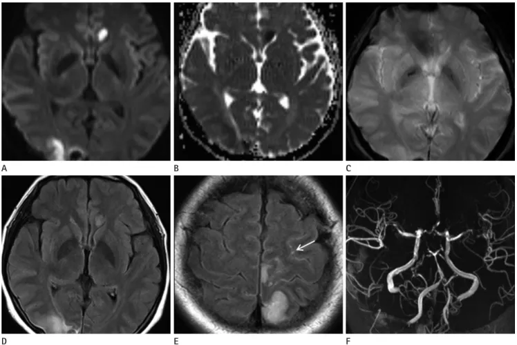
Fig. 1. MR imaging features of RCVS in a 46-year-old woman.
A-E. Diffusion-weighted imaging (A), apparent diffusion coefficient map (B), and FLAIR (D, E) show early subacute infarctions in the right PCA and left ACA territories. T2*-gradient echo imaging (C) shows early subacute ICH in the right occipital lobe. FLAIR (E) shows subtle sulcal hyper- intensity (arrow) that is suspicious for convexity SAH.
F. Time-of-flight MRA shows multifocal segmental severe narrowing with or without adjacent segmental dilatation in bilateral MCA, ACA, PCA, basilar artery, and distal vertebral artery.
Note.-ACA = anterior cerebral artery, FLAIR = fluid attenuated inversion recovery, ICH = intracerebral hemorrhage, MRA = magnetic resonance angiography, PCA = posterior cerebral artery, RCVS = reversible cerebral vasoconstriction syndrome, SAH = subarachnoid hemorrhage
E B
D A
F C
Jeong Sub Lee, et al
345
jksronline.org J Korean Soc Radiol 2013;69(5):343-346
include the postpartum period, vasoactive drugs such as canna- bis, cocaine, antidepressants, nasal decongestants, and ergot al- kaloid, catecholamine-secreting tumor such as pheochromocy- toma, glomus or carcinoid tumor, and immunosupressants or blood products such as transfusion (1, 5, 6). Although the exact precipitating cause was not determined, GI bleeding from GIST might be a possible precipitant of RCVS in our case. GI bleeding or GIST was not associated with RCVS in the previous reports.
Paraneoplastic hypoglycemia induced by an insulin-like growth factor II from GIST has been reported in some patients (7). How- ever, there have been no reports of any GIST producing catechol- amine. In this case, we did not evaluate the level of blood cate- cholamine. Therefore, there may not be sufficient evidence to suggest that GIST may be a precipitant of RCVS. In our opinion, GI bleeding might be the precipitating cause of RCVS. Although the hemoglobin level at the initial attack of headache was not available, the available data of the hemoglobin level suggested that intermittent and considerable GI bleeding might have been present before the event of RCVS. Intermittent and considerable GI bleeding may induce an increased sympathetic tone by sym- pathetic reflex compensation (8). Therefore, we presume that GI bleeding from the GIST may be a possible precipitant of RCVS in this patient.
The differential diagnosis of RCVS is cerebral vasculitis, in- cluding primary angiitis of central nervous system and vaso- after the 2nd MRA examination, and the intracranial arteries
appeared as normal. During the 2 year follow-up period, she ex- perienced no episode of RCVS since the removal of GIST.
DISCUSSION
Initial presentation of RCVS is usually a severe headache, and a moderate intermittent headache can develop between the epi- sodes of severe headache. All significant headaches usually dis- appear 3 weeks after the initial attack of headache (1). Imaging abnormalities in RCVS include convexity subarachnoid hemor- rhage (SAH), reversible brain edema, intracerebral hemorrhage (ICH), and cerebral infarction (2-4). In all cases, reversible vaso- constriction of cerebral arteries should be documented by angi- ographic imaging (1). Imaging abnormalities on CT or MRI have been reported in 37-81% of RCVS patients, and a combi- nation of lesions can be present (3, 4). Convexity SAH and re- versible edema are early imaging manifestations and usually can be seen within the first week of headache onset. Convexity SAH has been reported in 30-34% of RCVS patients and it is seen in a few cases of sulci near convexity (3, 4). Reversible brain edema has been reported in 8-38% (2-4) and it may have a similar dis- tribution to that of posterior reversible encephalopathy syndrome (2-4). ICH has been reported in 12-20% of RCVS patients (3, 4) and it is more frequently single than multiple and more lobar than deep, and is usually associated with other types of abnor- malities such as convexity SAH or infarction (3, 4). ICH can oc- cur early in the course of RCVS (3). Ischemic strokes have been reported in 6-39% of RCVS patients (2-4) and they usually oc- cur later in the course of RCVS. In the previous reports, infarc- tions usually occurred about 10 days after the first thunderclap headache (2-4). For diagnosis of RCVS, a cerebral angiography is needed to show segmental narrowing and dilatation (string of beads) of one or more intracranial arteries (1). The initial angio- gram may be normal if it is performed within one week of the initial attack. In such cases, a second angiogram several days lat- er may be diagnostic (2). Maximum vasoconstriction of the branches of the MCAs is reached at a mean of 16 days after clini- cal onset (2). Although no pathophysiology has been established, the unpredictable and transient dysregulation of cerebral arterial tone with sympathetic overactivity seems to have a role in the development of RCVS (1). Precipitants of RCVS are various and
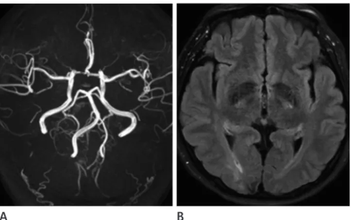
Fig. 2. Follow-up MR imaging 3 months after initial attack of RCVS.
A. Multifocal severe stenosis disappears on time-of-flight MRA and intracranial arteries looks as normal.
B. FLAIR imaging shows resolution of the lesions with subtle residual high signal intensity in the infarction area and low signal intensity in the ICH area.
Note.-FLAIR = fluid attenuated inversion recovery, ICH = intracere- bral hemorrhage, MRA = magnetic resonance angiography, RCVS = reversible cerebral vasoconstriction syndrome
A B
RCVS in a Patient with Bleeding from a GIST
346
J Korean Soc Radiol 2013;69(5):343-346 jksronline.org3. Ducros A, Fiedler U, Porcher R, Boukobza M, Stapf C, Bouss- er MG. Hemorrhagic manifestations of reversible cerebral vasoconstriction syndrome: frequency, features, and risk factors. Stroke 2010;41:2505-2511
4. Singhal AB, Hajj-Ali RA, Topcuoglu MA, Fok J, Bena J, Yang D, et al. Reversible cerebral vasoconstriction syndromes:
analysis of 139 cases. Arch Neurol 2011;68:1005-1012 5. Calabrese LH, Dodick DW, Schwedt TJ, Singhal AB. Narrative
review: reversible cerebral vasoconstriction syndromes. Ann Intern Med 2007;146:34-44
6. Ducros A, Boukobza M, Porcher R, Sarov M, Valade D, Bousser MG. The clinical and radiological spectrum of re- versible cerebral vasoconstriction syndrome. A prospective series of 67 patients. Brain 2007;130(Pt 12):3091-3101 7. Escobar GA, Robinson WA, Nydam TL, Heiple DC, Weiss GJ,
Buckley L, et al. Severe paraneoplastic hypoglycemia in a patient with a gastrointestinal stromal tumor with an exon 9 mutation: a case report. BMC Cancer 2007;7:13
8. Guyton AC, Hall JE. Textbook of medical physiology, 11th ed. Philadelphia: Elsevier Saunders, 2006:279-281
9. French KF, Hoesch RE, Allred J, Wilder M, Smith AG, Digre KB, et al. Repetitive use of intra-arterial verapamil in the treatment of reversible cerebral vasoconstriction syndrome.
J Clin Neurosci 2012;19:174-176 spasm induced by aneurysmal SAH (a-SAH). The initial presen-
tation of this case mimicked a-SAH; however, the amount of SAH was not sufficiently significant to suspect aneurysm rupture, and the clinical course completely differed to that of a-SAH. Vasculitis may mimic the vasospasm and parenchymal changes of RCVS.
Usage of steroids is usually avoided in RCVS because steroids may worsen the clinical course of RCVS (4, 9). Therefore, differ- ential diagnosis with cerebral vasculitis may be important. In ce- rebral vasculitis, the headache is usually not severe and depend- ing on the clinical course, with the thunderclap type headache, the patient may have recurrent episodes of attack. In vasculitis, arterial abnormalities do not improve as rapidly (1).
In conclusion, we report a RCVS case in a patient with GI bleeding from a small bowel GIST. Although the causality for RCVS may not be definite, considerable GI bleeding from GIST may be a suspicious precipitant of RCVS in this case.
REFERENCES
1. Ducros A. Reversible cerebral vasoconstriction syndrome.
Lancet Neurol 2012;11:906-917
2. Chen SP, Fuh JL, Wang SJ, Chang FC, Lirng JF, Fang YC, et al. Magnetic resonance angiography in reversible cerebral vasoconstriction syndromes. Ann Neurol 2010;67:648-656
소장의 기스트 종양에서 생긴 장 출혈이 있던 환자에서 발생한 가역성 뇌혈관수축 증후군
이정섭 · 박지강 · 김승형 · 정선영
저자들은 46세 여자 환자에서 발생한 가역성 뇌혈관수축 증후군 1예를 보고하고자 한다. 환자는 심한 두통, 다발성 뇌경 색 및 뇌동맥의 수축을 보였고, 뇌혈관수축 증후군 발생 1개월 후 심한 빈혈에 대한 검사 중 우연히 소장의 위장관 기질종 양이 진단되어 수술적 치료를 받았다. 뇌혈관수축 삽화 3개월 및 1년 후의 자기공명 혈관영상에서 이전에 보였던 뇌혈관수 축은 완전히 소실되었고 2년의 추적기간 동안 가역성 뇌혈관수축 증후군의 재발은 없었다.
제주대학교 의과대학 제주대학교병원 영상의학과