submit.radiology.or.kr J Korean Soc Radiol 2013;68(1):1-4
1 INTRODUCTION
Anatomical and morphological variation of vertebral arteries is important in neurointervention, surgery and other non-inva- sive procedures (1). The left vertebral artery almost always aris- es as the first branch of the left subclavian artery. However, in approximately 5% of all cases, the left vertebral artery has been reported to arise at the aortic arch (2-4). In some of these cases, duplication of the vertebral artery occurs, although this is very rare and has only been reported a few times (2-4). Anecdotal evidence describes clinical symptoms, such as dizziness or ver- tigo, in patients with anomalous vertebral artery origins, al- though there is no conclusive evidence of an association with cerebrovascular accidents (2); there has also been much contro- versy over the supposed negative effects of vertebral artery du- plication. An effect on the hemodynamics of the intracranial component is a possibility (2). In this study, we describe a pa- tient with left vertebral artery duplication which was detected incidentally.
CASE REPORT
A dual origin of the left vertebral artery and an aneurysm whose origin was shared with the left anterior temporal artery were incidentally detected by magnetic resonance angiography (MRA) and computed tomography angiography (CTA) in a 51-year-old female who visited our institute for health screen- ing (Fig. 1). After the examination, conventional angiography was performed on the patient. The anomalous vessel was dis- covered to be the third branch of the aortic arch, and it opaci- fied the left vertebral artery due to the retrograde flow into the second limb, which arose as the first branch of the left subclavi- an artery (Fig. 2). Selective left vertebral angiography through the usual vertebral artery origin, which is the left subclavian ar- tery, revealed retrograde flow of the left vertebral artery into a hypoplastic limb arising from the aortic arch. CTA, MRA and angiography showed that the proximal duplicated limbs of the left vertebral artery united to form the distal part of the left ver- tebral artery at the C4 level.
Case Report
pISSN 1738-2637
J Korean Soc Radiol 2013;68(1):1-4
Received September 9, 2012; Accepted October 15, 2012 Corresponding author: Dong Woo Park, MD Department of Radiology, College of Medicine, Hanyang University, Hanyang University Guri Hospital, 153 Gyeongchun-ro, Guri 471-701, Korea.
Tel. 82-31-560-2543 Fax. 82-31-560-2551 E-mail: [email protected]
Copyrights © 2013 The Korean Society of Radiology
Duplication of vertebral arteries is a very rare but clinically important condition. A duplicated vertebral artery origin can influence hemodynamics, pathogenesis of vascular lesions and treatment options. In cases of vertebral artery duplication, the vertebral arteries generally enter the transverse foramen higher up than normal.
Awareness of these vertebral artery variants before procedures, such as neurointer- vention or surgery, may be beneficial. Here, we describe a case of a 51-year-old fe- male patient with left vertebral artery duplication which was detected incidentally.
Index terms Vertebral Artery Origin
Duplication
Duplication of the Left Vertebral Artery Origin: A Case Report
좌측 추골동맥의 중복 기원: 증례 보고Sang-Wook Shin, MD, Dong Woo Park, MD, Choong Ki Park, MD, Young Jun Lee, MD, Seung Ro Lee, MD
Department of Radiology, College of Medicine, Hanyang University, Hanyang University Guri Hospital, Guri, Korea
Duplication of the Left Vertebral Artery Origin
submit.radiology.or.kr
J Korean Soc Radiol 2013;68(1):1-4
2
ing at the site of bifurcation of the innominate artery into the right subclavian and right common carotid arteries was reported in 1.11% of all specimens (7 of 693). However, duplication of ver- tebral arteries is extremely rare. Only three cases of a duplicated left vertebral artery arising from the aortic arch and the left sub- clavian artery have been reported (2-4). A hypoplastic duplicated limb was reported in two of these cases; in one case, it was the branch from the aortic arch and in the other, the branch from the left subclavian artery. In the case reported here, the hypoplastic limb was a direct branch from the aorta.
Right vertebral artery duplication has occasionally been re- ported. Goddard et al. (5) reported two cases of duplication of the right vertebral artery. In one case, the duplication was prox- imal, with both origins located in the right subclavian artery
DISCUSSION
The term “vertebral artery duplication” is applied to a verte- bral artery that has two origins with fusion at the neck level.
The duplicated segments almost always fuse at C4-6 (5). Verte- bral artery duplication occurs in cases in which an artery origi- nates in an unusual location. A number of studies have reported the frequency of anomalous vertebral artery origins. Panicker et al. (6) reported an abnormal left vertebral artery origin in 1 out of 20 middle-aged female cadavers during the period 1998- 2002. In a study by Bergman et al. (7) on 693 laboratory speci- mens, a left vertebral artery arising at the aortic arch between the left common carotid and left subclavian arteries was reported in 2.46% of all specimens (17 of 693). A right vertebral artery aris-
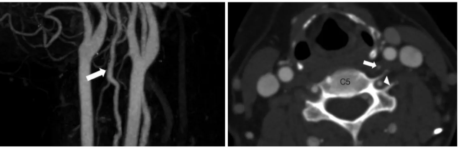
Fig. 1. Duplication of the left vertebral artery origin in a 51-year-old female patient (magnetic resonance angiogram not shown).
A. Computed tomography angiography (CTA) maximum intensity projection image shows that the duplicated limbs of the left vertebral artery unite to form the distal part of the left vertebral artery (arrow).
B. CTA axial source image shows a hypoplastic limb (arrow) arising from the aortic arch and a normal limb (arrowhead) arising from the left sub- clavian artery at the C5 level.
A B
A B C
Fig. 2. Duplication of the left vertebral artery origin in a 51-year-old female patient.
A. Left subclavian angiographic image shows the normal limb of the duplicated left vertebral artery (arrow) arising from the left subclavian ar- tery.
B, C. Selective angiography of the anomalous hypoplastic limb (arrowhead) shows it is the third branch of the aortic arch, and opacifies the left vertebral artery as a result of the retrograde flow into the normal limb (arrow).
Sang-Wook Shin, et al
submit.radiology.or.kr J Korean Soc Radiol 2013;68(1):1-4
3
REFERENCES
1. Matula C, Trattnig S, Tschabitscher M, Day JD, Koos WT.
The course of the prevertebral segment of the vertebral artery: anatomy and clinical significance. Surg Neurol 1997;48:125-131
2. Satti SR, Cerniglia CA, Koenigsberg RA. Cervical vertebral artery variations: an anatomic study. AJNR Am J Neurora- diol 2007;28:976-980
3. Koenigsberg RA, Pereira L, Nair B, McCormick D, Schwartz- man R. Unusual vertebral artery origins: examples and re- lated pathology. Catheter Cardiovasc Interv 2003;59:244- 250
4. Kendi AT, Brace JR. Vertebral artery duplication and aneu- rysms: 64-slice multidetector CT findings. Br J Radiol 2009;82:e216-e218
5. Goddard AJ, Annesley-Williams D, Guthrie JA, Weston M.
Duplication of the vertebral artery: report of two cases and review of the literature. Neuroradiology 2001;43:477-480 6. Panicker HK, Tarnekar A, Dhawane V, Ghosh SK. Anomalous
origin of left vertebral artery - embryological basis and ap- plied aspects - A case report. J Anat Soc India 2002;51:234- 235
7. Bergman RA, Afifi AF, Miyauchi R. Illustrated Encyclopedia of Human Anatomic Variation. Available at: http://www.
anatomyatlases.org/Anatomic Variants/Cardiovsacular/Im- ages0001/0095.shtml
8. Bruneau M, Cornelius JF, Marneffe V, Triffaux M, George B.
Anatomical variations of the V2 segment of the vertebral artery. Neurosurgery 2006;59(1 Suppl 1):ONS20-ONS24;
discussion ONS20-ONS24
9. Goray VB, Joshi AR, Garg A, Merchant S, Yadav B, Mahesh- wari P. Aortic arch variation: a unique case with anoma- lous origin of both vertebral arteries as additional branch- es of the aortic arch distal to left subclavian artery. AJNR Am J Neuroradiol 2005;26:93-95
10. Komiyama M, Morikawa T, Nakajima H, Nishikawa M, Yasui T. High incidence of arterial dissection associated with left vertebral artery of aortic origin. Neurol Med Chir (Tokyo) 2001;41:8-11; discussion 11-12
(the origins were not described in the other case). Satti et al. (2) also reported a case in which the right vertebral artery was par- tially duplicated with a common origin as the second branch of the right subclavian artery. However, a duplicated vertebral ar- tery origin appears to be more common on the left side.
Embryologically, the vertebral arteries typically originate bi- laterally from the distal end of the seventh dorsal intersegmen- tal arteries. A duplicated vertebral artery occurs if additional cervical intersegmental branches arise from the descending aorta (2, 3). This is due to the failure of the right or left fifth in- tersegmental artery to regress (5).
A complete understanding of the origins of anomalous verte- bral arteries may be important. Vertebral arteries almost always enter the C6 transverse foramen (8). However, in cases where the left vertebral artery arises from the aortic arch between the left common carotid artery and the left subclavian artery, it generally enters the transverse foramen at the level of C4-C5 rather than C6 (9). Therefore, the risk of atherosclerosis in ver- tebral arteries arising from the aortic arch may be higher than in those with other configurations due to their long preverte- bral segments. In addition, a left vertebral artery of the aortic origin is thought to be associated with a higher incidence of vertebral artery dissection than a left vertebral artery of the left subclavian artery origin (10). Anomalous vertebral artery ori- gins affect hemodynamics and may lead to intracranial malfor- mation. In 2009, Kendi and Brace (4) reported a case of a dupli- cated vertebral artery with an intracranial aneurysm. However, there is no conclusive evidence that this congenital anomaly predisposes to cerebrovascular accidents (2); thus, more studies are needed to determine its consequences.
The presence of a vertebral artery segment outside the bony cervical spine as high as C4 (the level of the carotid bifurcation in the majority of patients) is important for neurointervention or spinal surgery. The knowledge of the potential left vertebral artery origin variants may influence the choice and route of the endovascular treatment (7).
In conclusion, duplication of vertebral artery origins is ex- tremely rare, but may have serious implications, with particular regards to hemodynamics, angiography, endovascular and sur- gical procedures.
Duplication of the Left Vertebral Artery Origin
submit.radiology.or.kr
J Korean Soc Radiol 2013;68(1):1-4
4
좌측 추골동맥의 중복 기원: 증례 보고
신상욱 · 박동우 · 박충기 · 이영준 · 이승로
추골동맥의 중복 기원은 매우 드물지만 임상적으로 중요하다. 추골동맥의 중복 기원은 혈관병변의 혈역학적인 영향을 끼 칠 수 있으며, 현기증이나 현훈과 같은 환자의 임상 증상과의 관련성도 보고된다. 또한 환자의 치료 결정과도 관련이 있을 수 있으며, 이러한 증례에서 보통 추골동맥은 정상보다 높은 위치에서 경추의 횡돌기공에 진입하게 된다. 신경계 중재나 수술 전에 이러한 변이에 대해 미리 인식하는 것은 시술에 도움을 줄 수 있다. 우연히 좌측 추골동맥의 중복 기원이 발견된 51세 여성의 증례를 보고한다.
한양대학교 의과대학 구리병원 영상의학과