신경모세포종(neuroblstoma), 신경절모세포종(ganglio- neuroblastoma), 그리고 신경절세포종(ganglioneuroma)은 원시 신경능선(primordial neural crest)에서 기원한 다양한 조직학적 성숙도를 가지는 종양들이다. 이들 종양은 교감신경 조직이 있는 어디에서나 발생할 수 있으며, 흔히 발생하는 곳 은 복강 내, 부신, 종격동 등으로 중추신경계 발생은 흔하지 않 다(1-3). 중추신경계에 발생한 경우, 대부분 대뇌반구에 있으 며, 송과체, 척수, 그리고 소뇌에 발생한 증례들이 소수 발표되 었다(2-7). 저자들이 알기에는 시상하부에 발생한 신경절모세 포종의 증례는 아직 보고된 적이 없었다. 최근 저자들은 시상 하부에 발생한 신경절모세포종의 증례를 경험하였기에, 병리 소견과 영상소견을 함께 보고한다.
증례 보고
4세 남아가 상기도 감염증상 후 3주 전부터 발생한 두통, 구 토, 보행장애와 말더듬증(dysarthria)으로 본원 응급실에 내 원하였다. 환자는 출생 전 또는 출생 시 합병증의 기왕력이 없 었다. 내원 당시 신경학적 검사에서 양쪽 상, 하지의 근력은 4 정도로 측정되었으며, 말할 때 발음이 정확하지 않았다. 혈청 과 요 검사에서 이상소견은 관찰되지 않았다.
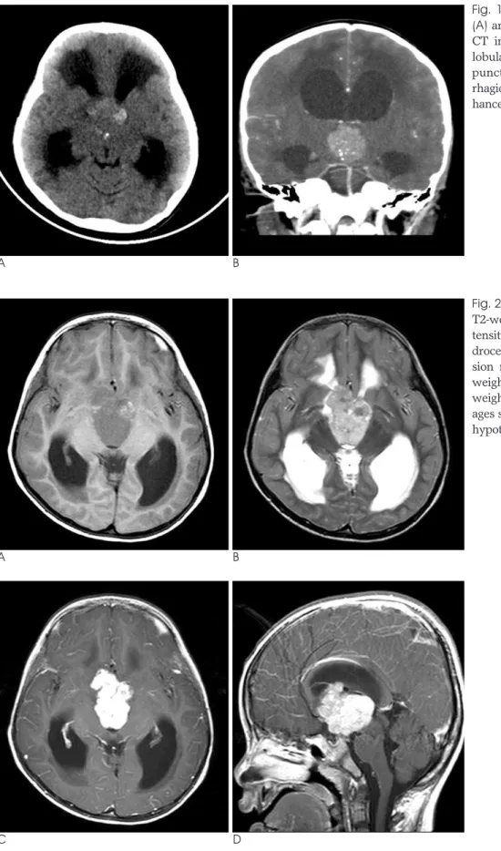
뇌 전산화단층촬영(Computed Tomography: CT)과 자기 공명영상(Magnetic Resonance Imaging: MRI)을 시행하였 다. CT 상 안장위(suprasellar), 제3 뇌실 부위에 석회화와 출혈을 동반한 고체 종괴(solid mass)가 관찰되었다. 이 병변 은 비교적 균질한 조영증강을 보이고, 수두증이 동반되어 있었 다(Fig. 1A, B). MRI에서 병변은 비교적 경계가 명확한 소엽 상의 종괴로, 제 3뇌실 바닥부 또는 시상하부에서 기시한 것으 로 생각되었다. T1 강조영상과 T2 강조영상에서 석회화와 출
혈부위를 제외한 부위에서는 비교적 회색질과 동등한 정도의 균일한 신호강도를 보였고, T1 강조영상에서 출혈부위는 고 신호강도로 관찰되었다(Fig. 2A, B). CT에서 보였던 석회화 와 출혈 부위는 경사 에코 영상(gradient echo image)에서 낮은 신호강도를 보였다. 조영증강 하였을 때 강하고 균질한 조영증강을 보였으며, 종괴에 의한 폐쇄성 수두증을 동반하였 다(Fig. 2C, D).
환자는 우측 전두 - 두정엽 개두술과 종괴의 부분 절제술을 시행하였다. 수술 소견상 출혈을 동반한 매우 단단한 종괴가 관찰되었고 종괴에 의해 제 3뇌실의 천장부(roof)가 밀려 올라 가 있었다. 수술 중 병변은 과혈관성 종양과 같이 비교적 출혈 을 많이 하는 양상이었다.
병리소견상 신경절세포 성분(ganglioneuromatous component)과 신경모세포종 성분(neuroblastomatous component)이 혼합된 비교적 전형적인 신경절모세포종의 소 견을 보였다(Fig. 3).
고 찰
배아종양(embryonal tumors)은 다음과 같이 3가지로 분 류된다: 원시성 신경외배엽성 종양(PNET: primitive neuroectodermal tumor), 신경모세포종, 신경절모세포종 (1-3). 이 중 신경모세포종과 신경절모세포종은 신경모세포의 분화 정도에 따라 분류되는데, 신경절모세포종은 50% 이상 차지하는 신경절세포 성분에 신경모세포종 성분이 혼합된 형 태이고, 반대로 신경모세포종은 50% 미만의 신경절세포 성분 을 포함한 형태이다. 따라서 더 미분화된 종양 형태인 신경모 세포종에 비해 신경절모세포종은 국소적인 종양이며 더 좋은 예후를 보인다(3-6).
신경절모세포종의 가장 흔한 발생 부위는 부신 수질, 후복막 강, 그리고 후 종격동이고, 이보다 덜 흔한 부위로 목과 골반강
─ 149 ─ 대한영상의학회지 2009;60:149-152
시상하부에 발생한 신경절신경모세포종:
영상과 조직학적 소견: 증례 보고
1손 영 준∙전 세 정∙최 시 성
중추신경계의 신경절 세포 종양은 흔하지 않다. 소수의 증례가 보고되었고, 발생한 위치는 척 수, 송과체, 대뇌반구, 그리고 소뇌였다. 최근 저자들은 4세 남아 환자에서 발생한 시상하부의 신경절모세포종을 경험하였고, 저자들이 알기로, 시상하부에 발생한 첫 번째 증례이다. 따라서 저자들은 증례를 영상과 병리학적 소견과 함께 보고하고자 한다.
1원광대학병원 영상의학과
이 논문은 2008년 9월 7일 접수하여 2008년 10월 20일에 채택되었음.
을 들 수 있다. 반면, 중추신경계 발생은 드문데, 대부분 대뇌 반구에 발생하며, 소뇌반구, 척수, 그리고 송과체에서 발생한 예가 소수 보고되었다(2-6). 임상양상은 발생한 부위에 따라
다양하게 나타나며, 중추신경계에 발생한 경우, 경련, 두통, 보 행장애, 그리고 운동마비 등의 증상을 보일 수 있다(3, 4, 6).
다른 부위에 발생한 경우와 마찬가지로, 영상의학적 소견은
─ 150 ─
손영준 외: 시상하부에 발생한 신경절신경모세포종
A B
Fig. 1. A, B. The non-enhanced axial (A) and contrast enhanced coronal (B) CT images show a well-demarcated, lobulated, solid mass with multiple punctate calcifications and hemor- rhagic foci. The mass is strongly en- hanced.
A B
Fig. 2. A-D. Axial T1-weighted (A) and T2-weighted images (B) show an isoin- tensity solid mass with obstructive hy- drocephalus. Focal hyperintensity le- sion means hemorrhagic foci on T1- weighted image. The postcontrast T1- weighted axial (C) and sagittal (D) im- ages show well enhancing mass in the hypothalamus.
C D
비교적 경계가 잘 지워지는 고형종괴로 보이며, 내부에 석회화 를 보일 수 있고 출혈이나 괴사가 흔하게 동반되어 비균질한 조영증강 소견을 보인다. 병리학적으로는 출혈이 생긴 부위에 신경모세포종 성분이 우세하게 보인다고 한다(2). 본 증례는 종괴 내부에 괴사 등으로 말미암은 낭성병변을 거의 포함하지 않은 고체 종괴로 보였으며, 조영증강이 잘되고 일부 출혈과 석회화가 관찰되었다. 따라서 저자들은 소아에서 제 3뇌실 근 처에서 발생하며, 위와 같은 영상소견을 보일 수 있는 질환인 두개인두종(craniopharyngioma), 배아종(germ cell tumor), 그리고 시상하부에서 발생한 신경교종(glioma)을 감 별진단에 포함하였다.
본 증례가 이전 증례들과 다른 점은 종양의 발생위치였는데, 본 증례는 신경절모세포종양이 시상하부에 있었고, 저자들이 알기에는 이것이 첫 번째 증례이다.
신경절모세포종의 예후에 대해서는 정확히 알려지진 않았지 만, 면역 조직화학 검사(immunohistochemistry) 에서 Mib- 1(Ki-67 antigen: proliferation antigen) 표지 지표 (labeling index)가 낮을수록 상대적으로 예후가 좋은 것으로
알려져 있다(5, 8). 치료는 수술적 절제를 원칙으로 하고 있으 며, 필요에 따라서 항암치료나 방사선 치료를 병행할 수 있다 (2-5). 본 증례도 종양을 완전히 절제할 수 없었기 때문에 수 술 후 항암치료를 병행하였다.
결론적으로, 저자들은 이전에 보고된 적이 없었던 신경절모 세포종의 증례를 경험하였기에 보고하는 바이다.
참 고 문 헌
1. Rorke LB, Hart MN, McLendon RE. Supratentorial primitive neu- roectodermal tumor (PNET). In Kleihues P, Cavenee WK. Pathology and genetics of tumours of the nervous system. 2nd ed. Lyon: IARC Press, 2000:141-144
2. Lonergan GJ, Schwab CM, Suarez ES, Carlson CL. Neuroblastoma, ganglioneuroblastoma, and ganglioneuroma: radiologic-pathologic correlation. Radiographics 2002;22:911-934
3. Gasparetto EL, Rosemberg S, Matushita H, Leite Cda C.
Ganglioneuroblastoma of the cerebellum: neuroimaging and pathological features of a case. Arq Neuropsiquiatr 2007;65:338-340 4. Nakazato Y, Hosaka N. A 32-year-old man with left temporal lobe
─ 151 ─ 대한영상의학회지 2009;60:149-152
A
B
C Fig. 3. Histopathologic findings.
A. The tumor shows dual morphologic features consisting of highly cellular primitive small round cells (neuroblastoma compo- nent) in the upper corner and a comparatively low cellular and differentiated glial cells in the lower corner (ganglioglioma compo- nents) (H & E ×200).
B. The neuroblastomatous component consists of compacted primitive small round cells surrounded by fibrils (H & E ×400).
C. The gangilogliomatous component consists of differentiating glial cells and multinucleated ganglion cells (H & E ×400).
tumor. Neuropahtology 2004;24:261-262
5. Tanaka M, Shibui S, Nomura K, Nakanishi Y. Pineal ganglioneu- roblastoma in an adult. J Neurooncol 1999;44:169-173
6. Sibilla L, Martelli A, Farina L, Uggetti C, Zappoli F, Seassa F, et al.
Ganglioneuroblastoma of the spinal cord. AJNR Am J Neuroradiol 1995;16(4 Suppl):875-877
7. Durity FA, Dolman CL, Moyes PD. Ganglioneuroblastoma of the cerebellum: case report. J Neurosurg 1968;28:270-273
8. Nishihara H, Ozaki Y, Ito T, Yoshinaga T, Tabu K, Tanino M, et al.
A case of cerebral ganglioneuronal tumor in the parietal lobe of an adult. Brain Tumor Pathol 2008;25:45-49
─ 152 ─
손영준 외: 시상하부에 발생한 신경절신경모세포종
J Korean Soc Radiol 2009;60:149-152
Address reprint requests to : See Sung Choi, M.D., Department of Radiology, Wonkwang University Hospital, 344-2 Shinyong-dong, Iksan, Chunbuk 570-711, Korea.
Tel. 82-63-859-1920 Fax. 82-63-851-4749 E-mail: [email protected]
Ganglioneuroblastoma of the Hypothalamus:
Radiologic and Pathological Findings of a Case
1Young Jun Sohn, M.D., Se Jeong Jeon, M.D., See Sung Choi, M.D.
1Department of Radiology, Wonkwang University Hospital
Ganglion cell tumors of the central nervous system (CNS) are uncommon. There have been few reports in the literature about ganglion cell tumors that arise from the spinal cord, pineal gland, cerebral hemisphere or cerebellum. We recently experienced a case of ganglioneuroblastoma that developed from the hypothalamus in 4-year-old boy. To the best of our knowledge, this is the first reported case of ganglioneuroblastoma in the hypothalamus. We report on this case and we present the neuroimaging and pathologic findings.
Index words :Ganglioneuroblastoma Hypothalamus Neoplasms Tomography