www.jkfas.org pISSN 1738-3757 eISSN 2288-8551 J Korean Foot Ankle Soc 2019;23(3):139-142 https://doi.org/10.14193/jkfas.2019.23.3.139
나 활액막 절제술 후 잔류 병변으로 인한 높은 재발률로 인해 활액 막 전 절제술을 이용한 병변의 완전한 절제가 중요하다.4) 이에 저 자들은 발목관절에 발생한 미만형 색소 융모 결절성 활액막염 환 자에 관절경적 및 개방적 활액막 절제술을 이용하여 수술적 치료 를 시행한 증례를 경험하여 이를 문헌 고찰과 함께 보고하고자 한 다. 본 연구는 분당제생병원 기관생명윤리심의위원회(Institutional Review Board)의 승인을 받았다.
증례 보고
52세 남자 환자가 좌측 발목관절 동통 및 종창을 주소로 내원하 였다. 특이 과거력은 없었으나 내원 2개월 전부터 동통 및 종창이 시작되어 이후 점차 악화되었다. 내원 당시 이학적 소견상 좌측 발 목관절에 통증을 동반한 종창 소견을 보였으나 운동 범위의 제한 은 없었다. 단순 방사선 검사상 특이 소견 없었으며, 혈액 검사상 감염 및 염증 등의 이상 소견은 보이지 않았다. 자기공명영상 촬영 색소 융모 결절성 활액막염(pigmented villonodular synovitis)은
원인이 확실하지 않고 관절이나 건초의 활액막조직의 국소적인 결 절성 병변을 형성하고 융모의 증식을 가져오는 질환으로 연간 100 만 명당 1.8명 정도로 발생하는 매우 드문 질환이다.1) Granowitz와 Mankin2)은 색소 융모 결절성 활액막염을 국소형과 미만형으로 구 분하였다. 국소형은 활액막의 일부에 결절이나 자루형 종물이 특 징적으로 관찰되며 미만형은 병변이 전체 활액막을 침범하는 것으 로 알려져 있고 국소형보다 재발이 흔한 것으로 알려져 있다.3)
특히 미만형 색소 융모 결절성 활액막염은 국소 병변 절제술이
Case Report
This is an Open Access article distributed under the terms of the Creative Commons Attribution Non-Commercial License (http://creativecommons.org/licenses/CC
by-nc/4.0) which permits unrestricted non-commercial use, distribution, and reproduction in any medium, provided the original work is properly cited.
Copyright 2019 Korean Foot and Ankle Society. All rights reserved.ⓒ
Pigmented villonodular synovitis (PVNS) is a rare proliferative disease involving the synovial membranes. Complete excision with a total synovectomy is important for diffuse type PVNS because of its high recurrence rate. In the ankle, complete excision of diffuse type PVNS is difficult due to the anatomical structure of the ankle joint. This paper reports the author’s experience of surgical treatment with combined open and arthroscopic synovectomy. In this manner, it is expected that the complications of the open procedure and the re- currence rate of arthroscopic procedure can be reduced.
Key Words: Pigmented villonodular synovitis, Ankle, Synovectomy
발목관절의 미만형 색소 융모 결절성 활액막염에서 관절경적 및 개방적 활액막 절제술을 이용한 수술적 치료: 증례 보고
최준철, 송우석, 변찬웅, 김 진, 한은미*
분당제생병원 정형외과, 병리과*
Surgical Treatment for Diffuse Pigmented Villonodular Synovitis of the Ankle by A Combined Open and Arthroscopic Synovectomy:
A Case Report
Jun-Cheol Choi, Woo-Suk Song, Chan-Woong Byun, Jin Kim, Eun-Mee Han*
Departments of Orthopedic Surgery and *Pathology, Bundang Jesaeng General Hospital, Daejin Medical Center, Seongnam, Korea
Received May 14, 2019 Revised June 5, 2019 Accepted June 25, 2019 Corresponding Author: Woo-Suk Song
Department of Orthopedic Surgery, Bundang Jesaeng General Hospital, Daejin Medical Center, 20 Seohyeon-ro 180beon-gil, Bundang-gu, Seongnam 13590, Korea
Tel: 82-31-779-0175, Fax: 82-31-779-0176, E-mail: [email protected] ORCID: https://orcid.org/0000-0003-2749-6803
Financial support: None.
Conflict of interest: None.
140 Vol. 23 No. 3, September 2019
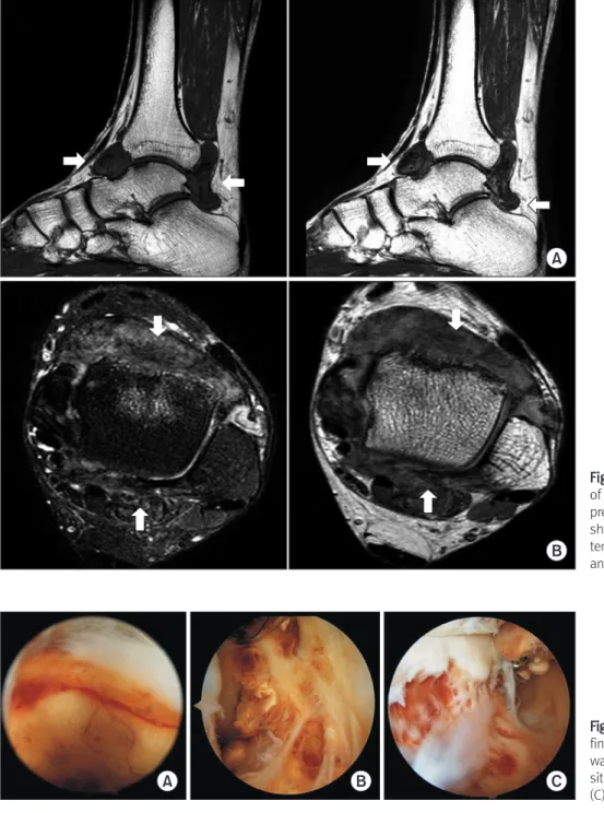
검사상 경거 관절(tibiotalar joint) 및 내, 외측구(medial and lateral gutter)를 포함한 발목관절을 광범위하게 침범한 종괴 및 활액막의 융모성 비후가 관찰되었다. 종괴와 비후된 활액막은 혈철소 침착 에 의해 T1, T2 강조영상 모두에서 근육보다 매우 낮은 신호강도를 보였다(Fig. 1). 병변의 위치 및 자기공명영상 촬영 검사 소견을 토 대로 관절낭내 종괴 및 색소 융모 결절성 활액막염 등의 양성 병변 을 우선 고려하였으며 명확한 동통 및 압통을 호소하여 수술을 시 행하였다.
발목관절 전방부에 대해 관절경적 활액막 전 절제술을 시행하였 고 발목관절 후방부의 종괴 및 잔류 병변에 대해 개방적 활액막 전
절제술 및 종괴 절제술을 시행하였다. 관절경 소견상 미만성 양상 의 광범위한 활액막 비후와 혈철소의 침착 소견 및 출혈 병변이 여 러 군데 관찰되었고 거골 경부의 골 미란 소견이 보였다(Fig. 2).
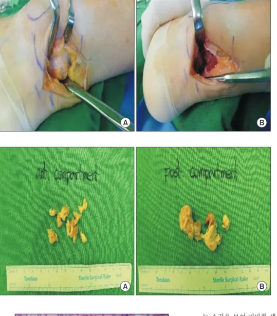
후방부에서 아킬레스건 후외측으로 7 cm 크기의 종절개 후 병변 주변의 연부조직을 박리하자 거골하 관절의 후내측으로 이어지는 5 cm×3 cm×1.2 cm 크기의 황갈색 관절낭내 종괴성 병변이 관찰 되어 주변 연부조직을 포함한 광범위 절제를 시행하였고, 추가적 으로 발목관절의 내, 외측구 및 비골근 주위의 병변들을 모두 제거 하였다(Fig. 3). 절제된 조직(Fig. 4) 및 종괴는 조직학적으로 활액 막 증식 및 섬유화, 혈철소 침착, 그리고 조직구의 침윤이 관찰되
A
B
Figure 1. Magnetic resonance imaging of left ankle. T1-, T2-weighted fat sup- pressed sagittal (A) and axial (B) images show large sized mass of low signal in- tensity in anterior and posterior aspect of ankle (arrows).
A B C
Figure 2. Anterior arthroscopic approach findings. (A) Diffuse synovial hypertrophy was found. (B) Diffuse hemosiderin depo- sition and hemorrhagic lesion were found.
(C) Bony erosion of talar neck was found.
www.jkfas.org 141 Jun-Cheol Choi, et al. Combined Open and Arthroscopic Synovectomy
는 소견을 보여 미만형 색소 융모 결절성 활액막염을 진단하였다 (Fig. 5).
수술 직후부터 환자는 좌측 발목관절의 통증은 호전되었다. 3년 간 추시관찰한 결과 수술 전 발목관절의 동통 및 종창은 관찰되지 않았으며, 수술 후 발생 가능한 관절강직 등의 후유증 없이 일상 생활 및 스포츠 활동에 불편감이 없었다.
고 찰
색소 융모 결절성 활액막염은 무릎 관절을 침범하는 경우가 가 장 흔하며, 발목관절을 침범하는 경우는 대략 2.5%의 발생률로 드 물게 보고되고 있다.5) Saxena와 Perez6)는 건초염, 골 연골 병변, 부 골 증후군 및 힘줄 손상과 구분하긴 힘든 증상이 있는 발목관절의 색소 융모 결절성 활액막염 환자 10명에 대해 활액막 절제술, 건삭 절제술, 골 병변에 대한 골이식술 등 다양한 치료법의 결과를 보고 했다.
종괴 절제술 및 활액막 절제술이 일차적 치료 방법이지만, 미만
A B
Figure 3. Posterior approach showing a pigmented villonodular synovitis mass (A) after through debridement (B).
A B
Figure 4. The yellowish-brown pigmented villonodular synovitis mass tumor speci- men after synovectomy. Anterior com- partment (A) and posterior compartment (B).
Figure 5. The tumor consists of mononuclear cells, a few multinucle- ated giant cells and some foamy macrophages (H&E stain, x40).
142 Vol. 23 No. 3, September 2019
형 색소 융모 결절성 활액막염의 경우 활액막 전 절제술을 시행함 에도 높은 재발률을 보이고 있다. Flandry 등7)은 26명의 환자에서 적극적인 개방적 활액막 전 절제술을 통해 2명의 재발과 24명의 환 자에서 관절 기능의 만족을 얻을 수 있었다고 하였으나 수술 후 조 기에 관절강직이 나타난 경우가 2명 있었다. Chin 등8)은 38명의 환자 중 적극적인 개방적 활액막 절제술 후 조기 관절강직이 나타 난 경우는 3명이었고 35명의 환자에서 관절기능의 만족을 얻었다 고 하였으나, 수술 후 3개월째 시행한 자기공명영상에서 잔류 병변 이 관찰된 경우가 13명이었다. 이 중 11명에서는 방사성동위원소 를 이용한 관절 내 방사선치료를 시행하였고 2명에서는 외부방사 선치료를 시행하였으나 각각 5명과 2명에서 재발이 관찰되었다.
이처럼 수술 후 관절 강직을 야기하는 관절의 미란 및 퇴행성 변화 를 방지하기 위해 연부조직 및 연골 손상을 최소화한 관절경적 활 액막 전 절제술을 시도하여 후유증을 감소시키고 회복기간을 줄일 수 있다는 보고가 있으나, 발이나 발목관절같이 해부학적 구조가 복잡한 경우에는 완전 절제를 위해 광범위한 주위 조직에 접근하 기가 쉽지 않아 불완전 절제시 역시 재발 가능성이 높다.
Stephan 등9)에 따르면 미만형 색소 융모 결절성 활액막염의 치 료법에서 개방적 활액막 절제술의 재발률은 19%∼33%로 보고되 었고 수술 후 심부 감염, 관절 강직, 절개상 관련 합병증이 동반하 였다. 관절경적 활액막 절제술만 시행했을 때의 합병증 발생률은 그보다 적으나 재발률은 92%∼94%로 보고되었으며, 아울러 관절 경적 및 개방적 활액막 절제술을 동반으로 시행했을 때 재발률이 9%∼25%로 개방적 활액막 절제술만 시행했을 때와 비슷하게 낮 은 재발률을 보였다.
현재까지 광범위하게 발생한 발목관절의 미만형 색소 융모 결절 성 활액막염의 경우 후내측 및 후외측으로 절개하여 적극적인 개 방적 활액막 전 절제술을 시행했다.10) 본 증례에서는 발목관절 전 방으로 관절경을 사용하여 발목관절의 연부조직 및 연골 손상을 최소화 하면서 최대한의 관절경적 활액막 전 절제술을 시행하였고 그 후 잔류 병소의 추가적인 완전 절제를 위해 후방의 아킬레스건 후외측으로 절개하여 관절 후방으로 내외측구와 경거관절 및 거 골하 관절의 병변 모두 관찰 가능한 시야를 확보할 수 있었다. 또 한 이 방법으로 기존의 적극적인 개방적 술식에서 발생 가능한 심 부감염, 관절강직 및 절개상 관련 합병증의 감소를 기대할 수 있었 다.
발목관절에서 발생한 미만형 색소 융모 결절성 활액막염에서 개 방적 활액막 전 절제술로 인한 술 후 합병증, 관절경적 활액막 전 절제술 후 불완전한 절제로 인한 높은 재발율을 고려해볼 때 관절
경적 및 개방적 활액막 절제술을 전후방 접근으로 복합적으로 이 용한다면 각 수술법에 따른 합병증 및 재발률을 줄일 수 있는 효과 적인 치료방법이 될 수 있다고 생각한다.
REFERENCES
1. Myers BW, Masi AT. Pigmented villonodular synovitis and te- nosynovitis: a clinical epidemiologic study of 166 cases and literature review. Medicine (Baltimore). 1980;59:223-38. doi:
10.1097/00005792-198005000-00004.
2. Granowitz SP, Mankin HJ. Localized pigmented villonodular synovitis of the knee. Report of five cases. J Bone Joint Surg Am.
1967;49:122-8. doi: 10.2106/00004623-196749010-00010.
3. Jaffe HL, Lichtenstein L, Sutro CJ. Pigmented villonodular syno- vitis, bursitis and tenosynovitis: a discussion of the synovial and bursal equivalents of the tenosynovial lesion commonly denoted as xanthoma, xanthogranuloma, giant cell tumor or myelo- plaxoma of the tendon sheath, with some consideration of this tendon sheath lesion itself. Arch Pathol. 1941;31:731-65.
4. Byers PD, Cotton RE, Deacon OW, Lowy M, Newman PH, Sis- sons HA, et al. The diagnosis and treatment of pigmented vil- lonodular synovitis. J Bone Joint Surg Br. 1968;50:290-305. doi:
10.1302/0301-620X.50B2.290.
5. Ushijima M, Hashimoto H, Tsuneyoshi M, Enjoji M. Pig- mented villonodular synovitis. A clinicopathologic study of 52 cases. Acta Pathol Jpn. 1986;36:317-26. doi: 10.1111/j.1440- 1827.1986.tb01022.x.
6. Saxena A, Perez H. Pigmented villonodular synovitis about the ankle: a review of the literature and presentation in 10 athletic patients. Foot Ankle Int. 2004;25:819-26. doi:
10.1177/107110070402501112.
7. Flandry FC, Hughston JC, Jacobson KE, Barrack RL, McCann SB, Kurtz DM. Surgical treatment of diffuse pigmented villonodular synovitis of the knee. Clin Orthop Relat Res. 1994;(300):183-92.
doi: 10.1097/00003086-199403000-00024.
8. Chin KR, Barr SJ, Winalski C, Zurakowski D, Brick GW. Treat- ment of advanced primary and recurrent diffuse pigmented villonodular synovitis of the knee. J Bone Joint Surg Am.
2002;84:2192-202. doi: 10.2106/00004623-200212000-00011.
9. Stephan SR, Shallop B, Lackman R, Kim TW, Mulcahey MK.
Pigmented villonodular synovitis: a comprehensive review and proposed treatment algorithm. JBJS Rev. 2016;4:01874474- 201607000-00005. doi: 10.2106/JBJS.RVW.15.00086.
10. Brien EW, Sacoman DM, Mirra JM. Pigmented villonodular syno- vitis of the foot and ankle. Foot Ankle Int. 2004;25:908-13. doi:
10.1177/107110070402501211.