Activated Protein C Protects Myocardium Via Activation of Anti-apoptotic Pathways of Survival in Ischemia-reperfused Rat Heart
Activated protein C (APC) is known to be beneficial on ischemia reperfusion injury in myocardium. However, the protection mechanism of APC is not fully understood. The purpose of this study was to investigate the effects and possible mechanisms of APC on myocardial ischemic damage. Artificially ventilated anaesthetized Sprague-Dawley rats were subjected to a 30 min of left anterior descending coronary artery occlusion followed by 2 hr of reperfusion. Rats were randomly divided into four groups; Sham, I/R, APC preconditioning and postconditioning group. Myocardial infarct size, apoptosis index, the phosphorylation of ERK1/2, Bcl-2, Bax and cytochrome c genes and proteins were assessed.
In APC-administrated rat hearts, regardless of the timing of administration, infarct size was consistently reduced compared to ischemia/reperfusion (I/R) rats. APC improved the expression of ERK1/2 and anti-apoptotic protein Bcl-2 which were significantly reduced in the I/R rats. APC reduced the expression of pro-apoptotic genes, Bax and cytochrome c.
These findings suggest that APC produces cardioprotective effect by preserving the expression of proteins and genes involved in anti-apoptotic pathways, regardless of the timing of administration.
Key Words: Activated Protein C; Cardioprotection; Reperfusion Injury, ERK1/2; bcl-2- Associated X Protein
Jia-Wang Ding1, Xiao-Hong Tong1, Jun Yang1, Zhao-Qi Liu3, Yan Zhang1, Jian Yang1, Song Li2, and Li Li1 Department of Cardiology1, the First College of Clinical Medical Science, China Three Gorges University, Yichang, Hubei; Department of Cardiology2, Yichang Central People’s Hospital, Yichang, Hubei; The Institute of Molecular Biology3, China Three Gorges University, Yichang, Hubei, China
Received: 24 April 2010 Accepted: 24 May 2010 Address for Correspondence:
Xiao-Hong Tong, M.D.
Department of Cardiology, The First College of Clinical Medical Science, Yichang 443003, Hubei, China
Tel: 15071754676 Email: [email protected]
DOI: 10.3346/jkms.2010.25.11.1609 • J Korean Med Sci 2010; 25: 1609-1615
INTRODUCTION
Myocardial ischemia-reperfusion injury (MIRI) is a complex pathophysiological event, resulting in serious acute and chron- ic heart damage. Although the precise mechanism responsible for MIRI remains unclear, it is widely accepted that apoptotic cell death is involved in the development of ischemic heart dam- age. Activated protein C (APC) is an important physiological anticoagulant that is produced from protein C by the action of the thrombin–thrombomodulin complex on endothelial cells (1). APC plays a significant role in the regulation of anti-inflam- matory processes by inhibiting cytokine production by mono- cytes and has been reported to attenuate tissue or organ injury in various pathological conditions (2-5). It has been well dem- onstrated that APC can reduce hypoxia-induced apoptosis in neuron and cerebral endothelial cells (2). In myocardial isch- emia, APC has been reported to have beneficial effects in isch- emia-reperfusion injury with its inhibitory effects on the myo- cardial apoptosis and inflammation (1).
Apoptosis is a crucial event in various physiologic processes, such as embryogenesis, organ development, and cell prolifera- tion, as well as in pathologic processes, which contributes sig- nificantly to post-ischemic cardiomyocyte death (6). But there
are no evidence regarding the effect of activated protein C on anti-apoptotic pathways in ischemia/reperfusion (I/R) injury.
In mammalian cells, among three major mitogen activated pro- tein kinases (MAPKs) signaling pathways, activation of extracel- lular signal related protein kinases (ERK1/2) exerts beneficial effect on post-ischemic myocardial apoptosis (7). Thus, we eval- uated the effects of activated protein C against I/R injury on myo- cardial infarction in rat in terms of the expression of proteins in- volved in apoptosis signaling cascades.
MATERIALS AND METHODS Animals
Male Sprague–Dawley (SD) rats (SPF grade, 220-250 g) were purchased from Tongji Medical School, Huazhong University of Science and Technology (HUST), China. Animals were main- tained under standard laboratory conditions at 25±2°C, relative humidity of 50±15%, and normal photoperiod (12 hr dark and 12 hr light). The procedures for experiments and animal care were approved by the institutional animal care and use committee of HUST (approval number: 2004-0007), and conformed to the guide for the care and use of laboratory animals by the National Institutes of Health (NIH Publication No. 80-23).
Drugs and preconditioning
APC was purchased from Sigma-Aldrich (Oakvile, ON, Canada), and dissolved in distilled water on the day of administration.
The 48 rats were randomly divided into four groups: the sham operation group (sham); I/R group (I/R); APC-preconditioning group (Pre-APC) and APC-postconditioning group (Post-APC).
In the sham operation and I/R groups, 0.9 percent saline solu- tion (2 mL/kg) was injected 5 min prior to the reperfusion pro- tocol. In APC-preconditioning group, APC (2 mg/kg) was given also 5 min prior to the reperfusion protocol. In APC-postcondi- tioning group, APC was administrated 5 min after reperfusion.
Experimental protocol
The rats were anesthetized with an intraperitoneal injection of pentobarbital sodium (30 mg/kg). After endotracheal intubation, ventilation was provided by a rodent respirator at a respiratory rate of 50/min with a tidal volume of 20 mL/kg body weight (8).
A left parasternal incision was made through the third and fourth ribs, and the pericardium was gently opened to expose the heart.
The lateral anterior descending artery (LAD) was ligated using a 6-0 silk suture. Additionally, a medical latex tube (socket, inner diameter, 1.5 mm) was placed between the ligature and the LAD.
Myocardial ischemia was induced by compressing the LAD by tightening the ligature around the latex tube. The ECG was mon- itored for changes in the ST-T segment caused by tightening or loosing the ligation. After 30-min ischemia, the latex tube was removed to reperfuse the myocardium by restoring the coronary circulation. After 2-hr reperfusion, rats were killed, and parts of the anterior wall of the left ventricular myocardium near the cardiac apex were obtained for further analysis. The sham con- trol group underwent the same procedures, with the exception of the induction of myocardial ischemia/reperfusion.
Hemodynamic measurements
We assessed cardiac hemodynamic function in vivo. The right common carotid artery of rat was isolated and cannulated with a 2-Fr micromanometer. Left ventricular (LV) pressure and LV end-diastolic pressure (LVEDP) were measured by the catheter advanced into the LV cavity, and data were recorded and ana- lyzed. These parameters as well as LV maximum positive change in pressure over time (+dP/dtmax), as a measure of cardiac con- tractility, and LV minimum negative change in pressure over time (–dP/dtmin), as a measure of relaxation, were recorded at baseline and after administration of APC.
Determination of LV area at risk and infarct size
At the end of the perfusion protocols, the coronary artery was reoccluded and 4 mL of 0.1% Evans blue dye was injected into the aorta to delineate the zone at risk. Stained hearts were fro- zen, sliced, and incubated at 37°C in 1% triphenyltetrazolium chloride to delineate the infarcted tissue. Slices were then fixed
and quantified by planimetry using the computer-based image analyzer SigmaScan Pro 5.0 (SPSS Science, Chicago, IL, USA).
Infarct size was expressed as a percentage of the area at risk zone.
The area at risk was calculated as a percentage of the total ven- tricular area (9).
Immunoblot analysis
LV specimens were pulverized and dissolved in lysis buffer. The solution was vigorously homogenized and centrifuged at 12,000 g for 10 min at 4°C. The supernatant was separated in a 12% SDS- polyacrylamide gel and transferred to PVDF membrane. After blocking the membrane with Tris-buffered saline-Tween 20 (TBS- T, 0.1% Tween 20) containing 5% non-fat dried milk for 1 hr at room temperature, membranes were washed twice with TBS-T and incubated with primary antibodies for 1 hr at room temper- ature or for overnight at 4°C. The following primary antibodies were used: rabbit anti-extracellular signal-regulated kinases (ERK1/2), mouse anti-phospho ERK1/2 and mouse anti-β actin antibodies (Sigma, St. Louis, MO, USA). The membranes were washed three times with TBS-T for 10 min, and then incubated for 1 hr at room temperature with horseradish peroxidase (HRP)- conjugated secondary antibodies. After extensive washing, the bands were visualized by an enhanced chemiluminescence system (10).
RT-PCR analysis
The total RNA from the cardiac muscle samples was extracted and purified using the Trizol reagent kit (Invitrogen, CA, USA).
Total RNA was reversely transcribed into complementary DNA (cDNA) using the cDNA synthesis kit (Takara Bionic, Otsu, Japan) according to the manufacturer’s protocol. The opticon-2 real- time PCR reactor (MJ Research, Fremont, CA, USA) and real- time PCR kit (SYBR Premix Ex TaqTM, TaKaRa, Japan) were em- ployed based on the manufacturer’s instruction. A 20 µL reverse transcription reaction mixture was incubated at 42°C for 15 min, heated to 95°C for 2 min. The PCR condition was at 95°C for 10 sec, then 40 cycles of 95°C for 5 sec, and 60°C for 30 sec and 72°C for 1 min. GAPDH and β-actin were used as the housekeeping genes. The mRNA levels were calculated based on the method of 2-ΔΔCt (11, 12). The primers were as follows: Bax, sense primer 5´-AGACACCTGAGCTGACCTTGGAG-3´, antisense primer 5´-GTTGAAGTTGCCATCAGCAAACA-3´. Bcl-2, sense primer 5´-TGAACCGGCATCTGCACAC-3´, antisense primer 5´-CGT- CTTCAGAGACAGCCAGGAG-3´. cytochrome c, sense primer 5´-CATAAGACTGGACCAAACC-3´, antisense primer 5´-TCT- GCCCTTTCTCCCT-3´. β-actin, sense primer 5´-GTCCCTCA- CCCTCCCAAAAG-3´, antisense primer 5´-GCTGCCTCAACA- CCTCAACCC-3´. GAPDH, sense primer 5´-ACCACAGTCCAT- GCCATCAC-3´ and antisense primer 5´-TCCACCACCCTGTT- GCTGTA-3´.
Immunohistochemistry analysis
The myocardial tissues were fixed with 10% neutral-buffered formalin, dehydrated in increasing concentrations of ethanol, embedded in paraffin, and sectioned into 4-μm slices. After de- paraffinization, the sections were immersed in 3% hydrogen peroxide in methanol for 30 min, placed in a 500-W microwave for 5 min and incubated with normal goat serum for 10 min.
The primary antibody was diluted appropriately and incubated with the sections overnight at 4°C. Biotinylated secondary anti- body was added followed by the addition of HRP-Streptavidin complex, and then the sections were stained with DAB solution and counterstained with hematoxylin. Brown immunoprecipi- tates occurred in positive cytoplasm after staining. In immuno- histochemical analysis of quantity, five random microscopic fields from each section were examined at ×400 magnification with a micro-image collecting system. The average grey values were analyzed using LEICA Q55OIW image analysis software.
Determination of myocardial apoptosis
Myocardial tissue samples were obtained at the end of the reper- fusion, fixed in 10% paraformaldehyde, paraffin-embedded, and sectioned. TUNEL staining was performed in deparaffinized and rehydrated sections according to the demands of TUNEL assay kit. After TUNEL staining, the sections were counterstained with hematoxylin. Total cardiomyocytes and TUNEL positive cells in the specimens were counted under light microscopic analysis. Five high-power fields (×400) of each section were ran- domly selected from the marginal and the central parts of the ischemic zone. The ratio of TUNEL-positive cells to total cardio- myocytes was apoptosis index (AI).
Statistical analysis
Quantitative data were expressed as mean±SD. For experiments with three or more groups of animals, one-way analysis of vari- ance (ANOVA) was performed, and Student–Newman–Keuls (SNK)-qtest was used for selected groups. A P value of less than 0.05 was considered as statistically significant. Analysis was car-
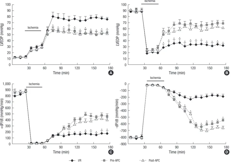
Fig. 1. The changes of the hemodynamic parameters before and during myocardial I/R. (A) Left ventricular end-diastolic pressure (LVEDP) during ischemia-reperfusion injury. (B) Left ventricular developed pressure (LVDP). (C) Contractility (+dP/dt) during ischemia-reperfusion injury. dP/dtmin during ischemia-reperfusion injury. (D) Compliance (-dP/dt) during ischemia-reperfusion injury. All values are means±SE (n=6).
*P<0.05 for Pre-APC and Post-APC vs the I/R group.
LVEDP (mmHg)+dP/dt (mmHg/min) LVEDP (mmHg)-dP/dt (mmHg/min)
Time (min)
Time (min)
Time (min)
Time (min)
Ischemia
Ischemia
30 60 90 120 150 180
30 60 90 120 150 180
30 60 90 120 150 180
30 60 90 120 150 180
100 90 80 70 60 50 40 30 20 10 0
1,000 900 800 700 600 500 400 300 200 100 0
100 90 80 70 60 50 40 30 20 10 0
0 -100 -200 -300 -400 -500 -600 -700 -800 -900
Ischemia
Ischemia
* * *
*
*
*
*
*
*
*
*
*
*
*
*
*
*
*
*
*
*
*
*
*
*
*
*
*
* *
*
* *
*
* *
* *
* *
*
A B
C D
I/R Pre-APC Post-APC
ried out using Statistical Product and Service Solutions (SPSS 11.0).
Student–Newman–Keuls (SNK)-qtest is a kind of method to made multiple comparison.
Statistical Product and Service Solutions (SPSS11.0) is Statis- tical software.
RESULTS
The effects of APC on cardiac function
Parameters were studied both at baseline and in response to APC administration and included LVDP, LVEDP, and LV+dP/dtmax
and LV–dP/dtmin as measures of ventricular contractility and re- laxation, respectively. Rats undergoing I/R had significantly im- paired LV contractile function. Treatment with APC significant- ly preserved impaired LV contractility and relaxation. Obviously improvements in LV +dP/dtmax and LV –dP/dtmin were also seen
in animals treated with APC. No differences existed in LVEDP, LVDP, +dP/dt, and –dP/dt between the APC groups during isch- emia and reperfusion (Fig. 1).
The effects of APC on infarct size
Mean infarct size of the AAR in the I/R group was 58.7±3.1%. In the groups administered with APC, infarct sizes were signifi- cantly reduced to 31.4±2.6% and 28.4±2.8% in the Pre-APC and Post-APC group, respectively (all P<0.01). No significant differ- ences were observed between the APC-administrated groups (Fig. 2).
Western blotting analysis
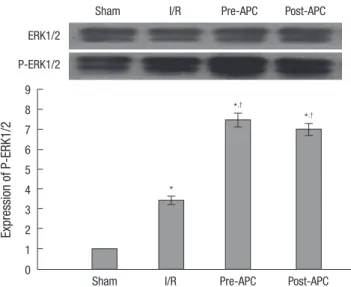
Western blot analysis of ERK1/2 is shown in Fig. 3. I/R injury obviously increased P-ERK1/2 expression (3.4±0.21; respective- ly) compared to the sham as measured densitometrically (fold relative to sham). In contrast to the I/R group, APC treatment significantly increased P-ERK1/2 expression (7.45±0.34 vs 3.4±
0.21, P<0.01 and 6.98±0.31 vs 3.4±0.21, P<0.01). Phosphoryla- tions of ERK1/2 were similar between the APC-administrated groups (Fig. 3).
The expression of Bax, Bcl-2 and cytochrome c
Expression levels of pro-apoptotic proteins, Bax and cytochrome c, significantly increased and expression level of anti-apoptotic protein, Bcl-2 significantly decreased in the I/R group than in the sham group. In the APC-administrated groups, the expres- sion of Bax and cytochrome c significantly decreased and the
Sham I/R Pre-APC Post-APC
*,†
*
*,†
Fig. 2. The effect of APC on infarct size. Infarct size is expressed as a percentage of the area at risk in rats, which was reduced by APC administration. Mean±SE, n=6.
*P<0.01 vs sham group; †P<0.01 vs I/R group.
Infarct size per area at risk (%)
70 60 50 40 30 20 10 0
Sham I/R Pre-APC Post-APC
Sham I/R Pre-APC Post-APC
*,†
*
*,†
Fig. 3. Effect of APC treatment on p-ERK protein in rat heart after 30 min ischemia and 2 h reperfusion. The phosphorylation was quantified by densitometry using image analysis program. Mean±SE, n=6.
*P<0.01 vs sham; †P<0.01 vs I/R.
Expression of P-ERK1/2
9 8 7 6 5 4 3 2 1 0
ERK1/2 P-ERK1/2
Sham I/R Pre-APC Post-APC
Expression of genes
4.5 4 3.5 3 2.5 2 1.5 1 0.5 0
Fig. 4. The effect of APC on expressions of Bax, Bcl-2 and cytochrome C mRNA. All values are means±SE (n=6).
*P<0.01 vs sham group; †P<0.05 vs I/R group.
Bax Bcl-2 cytochrome c
*,†
*,†
*,†
*,†
*,†
*,†
*
*
*
Table 1. Effects of APC on the expressions of Bcl-2, Bax and cytochrome c
Group Bcl-2 Bax Cytochrome c
Sham 0.352±0.069 0.244±0.025 0.176±0.032
I/R 0.741±0.043* 0.859±0.076* 0.899±0.097*
Pre-APC 0.973±0.103† 0.546±0.041† 0.541±0.073‡ Post-APC 0.997±0.116† 0.578±0.039† 0.567±0.046‡ Data are presented as mean±SE, n=6.
*P<0.01 vs sham group; †P<0.05 vs I/R group, ‡P<0.01 vs I/R group.
I/R, ischemia/reperfusion; APC, activated protein C.
expression level of Bcl-2 significantly increased than in the I/R group. The expression levels of Bax, cytochrome c and Bcl-2 were similar among the APC-administrated groups (Fig. 4, Table 1).
TUNEL assay
A number of TUNEL-positive cells were observed in the myo- cardium in the I/R group. Compared with I/R group, the apop- tosis indeces were decreased significantly in APC-administrat- ed groups (all P<0.01). No significant differences were observed between the APC-administrated groups (Fig. 5).
DISCUSSION
The current results confirm that APC significantly reduces myo- cardial infarct size without reference to the timing of administra- tion in I/R rat heart, which is in conjunction with the preserved phosphorylations of ERK1/2 and anti-apoptotic proteins, Bcl-2 and attenuated pro-apoptotic proteins, Bax and cytochrome c resulting from I/R.
Activated protein C (APC) is a serine protease with systemic anticoagulant activity, which is mediated by irreversible pro-
teolytic inactivation of factors Va and VIIIa with contributions by various cofactors (13). Independent of its anticoagulant ac- tivity, APC exerts direct cellular effects that are mediated by the protein C cellular pathway resulting in the following: 1) cyto- protective alteration of gene expression profiles, 2) antiinflam- matory activities, 3) antiapoptotic activity, and 4) protection of endothelial barriers (14-16). In myocardial ischemia, APC has been reported to have beneficial effects in ischemia-reperfu- sion injury with its inhibitory effects on the myocardial apopto- sis and inflammation (1). However, little has been studied re- garding mechanisms of APC induced cardioprotection and we could observe that APC acted through proteins involved in anti- apoptoic pathways of survival regardless of timing of adminis- tration. Apoptosis has been linked with reperfusion-induced myocardial injury after reversible coronary occlusion and sug- gested as one of the key mechanisms in the development of in- farction in rat cardiac myocytes (17, 18).
In the current study, APC increased the phosphorylation of ERK1/2 and anti-apoptotic proteins in I/R rat heart. ERK is acti- vated in response to I/R, oxidative stress, and hypoxia, and is an established player in the anti-apoptotic defense network (7). As D
A B C
Fig. 5. Effect of APC on apoptosis in ischemic reperfusion-induced myocardial injury. Detection of apoptosis was done at 2 hr after reperfusion. (A) Sham group, (B) I/R group, (C) Pre-APC group, (D) Post-APC group. (E) Mean apoptotic index was counted in each group (A-D). TUNEL stain ×400 (n=6 animals per group). Arrow indicates TUNEL positive cells.
*P<0.05 vs sham group, †P<0.05 vs I/R group.
Sham I/R Pre-APC Post-APC
Apoptotic index (%)
60 50 40 30 20 10 0
*
*,† *,†
E
our results indicate, APC preserved the phosphorylation of ERK1/
2 which were significantly reduced following I/R leading to im- proved viability of the rat heart exposed to I/R. In response to an apoptotic stimulus such as I/R, the pro-apoptotic protein, Bax, undergoes a conformational change that allows it to trans- locate to the mitochondria, where it induces cytochrome c re- lease. Once released, cytochrome c leads to the formation of the apoptosome, a complex comprised of apoptotic protease-acti- vating factor-1 (Apaf-1), procaspase-9 and ATP, which permits the autoactivation of procaspase-9 that in turn facilitates cas- pase-3 activation. Active caspase-3 activates the caspase acti- vated DNase, leading to oligonucleosomal DNA fragmentation (19). Phosphorylation of ERK1/2 inhibits the conformational change in Bax protein and cytochrome c induced caspase acti- vation, thereby preventing apoptosis (20). Bcl-2 normally resides in mitochondrial membranes and the cytoplasm, whose over- expression is a potential mechanism for apoptotic resistance (21). The anti-apoptotic protein Bcl-2 attenuates cellular injury by inhibiting cytochrome c translocation and inhibits Bax trans- location (22, 23). In this study, we could observe that improved phosphorylations of ERK1/2, and associated recovery of expres- sion of Bcl-2 and mitigated expression of Bax and mitochondri- al cytochrome c release against I/R induced injury, which all indicate APC induced a cardioprotective effect via anti-apopto- sis pathways of survival in I/R rat heart. In addition, we have in- vestigated the cardioprotective effects of APC with different ad- ministration protocols, and we observed similar protective effect between APC pre- and post-conditioning groups. These results suggest that cardioprotective signaling pathways during the APC pre- and post-conditioning might be similar.
In conclusion, APC confers myocardial protection against in- jury in I/R rat hearts without reference to the timing of admin- istration, which is conducted with preserved phosphorylations of ERK1/2 and of signal transduction proteins against apoptotic cell death.
ACKNOWLEDGMENTS
We thank Professors C. Zhang and L. Han of the Faculty of the Microbiology Department, Professor Y. Hu and Dr. B. Gao of the Department of Pathology of the First College of Clinical Medi- cal Sciences, and the China Three Gorges University for their helpful discussions and suggestions on this work.
REFERENCES
1. Loubele ST, Spek CA, Leenders P, van Oerle R, Aberson HL, Hamulyák K, Ferrell G, Esmon CT, Spronk HM, ten Cate H. Activated protein C protects against myocardial ischemia/reperfusion injury via inhibition of apoptosis and inflammation. Arterioscler Thromb Vasc Biol 2009; 29:
1087-92.
2. Shibata M, Kumar SR, Amar A, Fernandez JA, Hofman F, Griffin JH, Zlokovic BV. Anti-inflammatory, antithrombotic, and neuroprotective effects of activated protein c in a murine model of focal ischemic stroke.
Circulation 2001; 103: 1799-805.
3. Mizutani A, Okajima K, Uchiba M, Noguchi T. Activated protein C re- duces ischemia/reperfusion-induced renal injury in rats by inhibiting leukocyte activation. Blood 2000; 95: 3781-7.
4. Grey S, Hau H, Salem HH, Hancock WW. Selective effects of protein C on activation of human monocytes by lipopolysaccharide, interferon- gamma, or PMA: modulation of effects on CD11b and CD14 but not CD25 or CD54 induction. Transplant Proc 1993; 25: 2913-4.
5. Murakami K, Okajima K, Uchiba M, Johno M, Nakagaki T, Okabe H, Takatsuki K. Activated protein C prevents LPS-induced pulmonary vas- cular injury by inhibiting cytokine production. Am J Physiol 1997; 272:
L197-202.
6. Gottlieb RA, Engler RL. Apoptosis in myocardial ischemia-reperfusion.
Ann N Y Acad Sci 1999; 874: 412-26.
7. Yue TL, Wang C, Gu JL, Ma XL, Kumar S, Lee JC, Feuerstein GZ, Thom- as H, Maleeff B, Ohlstein EH. Inhibition of extracellular signal-regulated kinase enhances ischemia/Reoxygenation-induced apoptosis in cultured cardiac myocytes and exaggerates reperfusion injury in isolated perfused heart. Circ Res 2000; 86: 692-9.
8. Maulik N, Engelman RM, Rousou JA. Ischemic preconditioning reduces apoptosis by upregulating anti-death gene Bcl-2. Circulation 1999; 100 (19 Suppl): II369-75.
9. Song DK, Jang Y, Kim JH, Chun KJ, Lee D, Xu Z. Polyphenol (-)-epigallo- catechin gallate during ischemia limits infarct size via mitochondrial KATP channel activation in isolated rat hearts. J Korean Med Sci 2010;
25: 380-6.
10. Yang J, Jiang H, Yang J, Ding JW, Chen LH, Li S, Zhang XD. Valsartan preconditioning protects against myocardial ischemia–reperfusion injury through TLR4/NF-kappaB signaling pathway. Mol Cell Biochem 2009;
330: 39-46.
11. Marino JH, Cook P, Miller KS. Accurate and statistically verified quanti- fication of relative mRNA abundances using SYBR green I and real-time RT-PCR. J Immunol Methods 2003; 283: 291-306.
12. Vandesompele J, De Preter K, Pattyn F, Poppe B, Van RN, De Paepe A, Speleman F. Accurate normalization of real-time quantitative RT-PCR data by geometric averaging of multiple internal control genes. Genome Biol 2002; 3: research0034. 1-11.
13. Mosnier LO, Zlokovic BV, Griffin JH. The cytoprotective protein C path- way. Blood 2007; 109: 3161-72.
14. Joyce DE, Gelbert L, Ciaccia A, DeHoff B, Grinnell BW. Gene expression profile of antithrombotic protein c defines new mechanisms modulating inflammation and apoptosis. J Biol Chem 2001; 276: 11199-203.
15. Finigan JH, Dudek SM, Singleton PA, Chiang ET, Jacobson JR, Camp SM, Ye SQ, Garcia JG. Activated protein C mediates novel lung endothe- lial barrier enhancement: role of sphingosine 1-phosphate receptor transactivation. J Biol Chem 2005; 280: 17286-93.
16. Pirat B, Muderrisoglu H, Unal MT, Ozdemir H, Yildirir A, Yucel M, Turkoglu S. Recombinant human-activated protein C inhibits cardio- myocyte apoptosis in a rat model of myocardial ischemia-reperfusion.
Coron Artery Dis 2007; 18: 61-6.
17. Haunstetter A, Izumo S. Toward antiapoptosis as a new treatment mo- dality. Circ Res 2000; 86: 371-6.
18. Buja LM, Entman ML. Modes of myocardial cell injury and cell death in ischemic heart disease. Circulation 1998; 98: 1355-7.
19. Yoo KH, Yim HE, Jang GY, Bae IS, Choi BM, Hong YS, Lee JW. Endothe- lin A receptor blockade influences apoptosis and cellular proliferation in the developing rat kidney. J Korean Med Sci 2009; 24: 138-45.
20. Tsuruta F, Masuyama N, Gotoh Y. The phosphatidylinositol 3-kinase (PI3K)-Akt pathway suppresses Bax translocation to mitochondria. J Biol Chem 2002; 277: 14040-7.
21. Myong NH. Tissue microarray analysis of fas and fasL expressions in hu- man non-small cell lung carcinomas; with reference to the p53 and bcl-2
overexpressions. J Korean Med Sci 2005; 20: 770-6.
22. Zhu L, Yu Y, Chua BH, Ho YS, Kuo TH. Regulation of sodium-calcium exchange and mitochondrial energetics by Bcl-2 in the heart of trans- genic mice. J Mol Cell Cardiol 2001; 33: 2135-44.
23. Lazou A, Iliodromitis EK, Cieslak D, Voskarides K, Mousikos S, Bofilis E, Kremastinos DT. Ischemic but not mechanical preconditioning attenu- ates ischemia/reperfusion induced myocardial apoptosis in anaesthe- tized rabbits: the role of Bcl-2 family proteins and ERK1/2. Apoptosis 2006; 11: 2195-204.