INTRODUCTION
Breast cancer is the most common malignant tumor and has the second highest mortality rate in western women (1).
Recently in Korea, the incidence of breast cancer has increased along with an increase in western lifestyle. The majority of cases appear to be sporadic, but hereditary factors may be in- volved in about 5-10% of all cases (2). Extensive research into the factors influencing development and prognosis of breast cancer has been carried out, but the translation of these risk factors to clinical utility has been limited.
In general, the activation of oncogenes and inactivation of tumor suppressor genes underlie carcinogenesis, and tumors develop through an accumulation of several genetic alterations (3). Recently, molecular studies have provided the evidence to explain the development and progression of cancer at the cel- lular level. Loss of heterozygosity (LOH) is a change from a heterozygous to a homozygous state due to loss of the wild type allele, and is important in the identification of new tumor suppressor genes (4). Colon cancer represents a good model of cancer progression from adenoma to carcinoma that is ac- companied by genetic mutations and LOH (5). However, breast cancer is morphologically and biologically heteroge- neous, so it has been difficult to define a general series of genet- ic aberrations involved in progression of this cancer type.
Many studies have revealed that LOH at chromosome bands 1p36, 3p24-p25, 6q12-q16, 11p15, 11q22, 11q23, 13q21, 16q22, 17p21, and 17q25, plays important roles in breast cancer, and these genetic alterations have complex interactions (6-12). In particular, alterations of both the long and short arms of chromosome 11 are involved in the progression of breast cancer, and loci at 11p15.5, 11q13.1, and 11q23.3 are importantly considered (10, 13-17). In lobular carcinoma, the most common region of LOH is at 11q13, and in ductal carcinoma loci at 11q23 and 11p15.5 are important (16).
Phillips et al. determined the potential effects of chromosome 11 on the tumorigenic and metastatic abilities of the MDA- MB-435 cell line via microcell-mediated chromosome trans- fer, and indicated that human chromosome 11 harbors a me- tastasis-suppressor gene for human breast cancer (18).
Previous studies using fresh frozen tissue have often been problematic since the surrounding non-neoplastic cells (e.g., stromal and inflammatory cells) confound analysis of the tumor cell population. Microdissection of paraffin-embedded tissues allows the isolation of pure populations of neoplastic or non- neoplastic cells (20). Retrospective studies correlating histo- pathologic findings with genetic alterations become possible.
Until now, the association between chromosome 11p15.5 and various clinical parameters has not been well defined.
Prognostic factors of breast cancer include expression of c-erb Dong-Ja Kim, Ji-Young Park*,
Myung-Hoon Lee�, Yoon-Kyung Sohn�
Departments of Pathology, Fatima Hospital, Daegu;
*Department of Pathology, Samsung Cheil Hospital, Sungkyunkwan University School of Medicine, Seoul;
�Department of Pathology, Kyungpook National University School of Medicine, Daegu, Korea
Address for correspondence Dong-Ja Kim, M.D.
Department of Pathology, Fatima Hospital, 302-1 Sinam-dong, Dong-gu, Daegu 701-600, Korea Tel : +82.53-940-7277, Fax : +82.53-940-7273 E-mail : [email protected]
698
The Role of Microsatellite Instability at Chromosome 11p15.5 in the Progression of Breast Ductal Carcinoma
The study of microsatellite instability (MSI) has provided the evidence to support a sequential, progressive pathway for the development of cancer. In this study, we ana- lyzed the role of MSI at chromosome 11p15.5 using microdissection of paraffin-em- bedded tissue from 68 matched normal and breast tumor samples. Components of intraductal, invasive and metastatic foci in lymph node were assessed for MSI using the polymorphic markers D11S922, tyrosine hydroxylase (TH) and D11S988.
We found that MSI at D11S922 was relatively high incidence than other two markers and increased during breast cancer progression. The overall frequency of MSI at D11S922 was 26.7% in pure intraductal carcinoma, 36.4% in invasive carcinoma, and 40.0% in invasive carcinoma with metastases. We observed no significant cor- relation between MSI at chromosome 11p15.5 and the patient’s age, tumor size, histological grade, or lymph node metastasis. We compared the MSI incidence with the expression of prognostic markers, such as p53, c-erb B2, estrogen receptor, and progesterone receptor, and found no significant correlation. We suggest that the MSI of chromosome 11p15.5 is increased during breast cancer progression, but long-term follow-up study would establish whether MSI at chromosome 11p15.5 could be useful as a potential prognostic marker for breast cancer.
Key Words : Microdissection; Breast Neoplasms; Microsatellite Markers; Microsatellite Repeats; Chromosomes;
Prognosis
Received : 17 November 2003 Accepted : 2 June 2004
B2, p53, and the estrogen and progesterone receptors (21, 22).
We isolated normal, intraductal carcinoma, invasive ductal carcinoma, and metastatic lesions in lymph nodes using mi- crodissection technique on paraffin-embedded tissue, and assessed the incidence of microsatellite instability (MSI) at chro- mosome 11p15.5 in comparison with several clinical param- eters and other prognostic factors, such as c-erb B2, p53, estro- gen and progesterone receptors (21, 22).
MATERIALS AND METHODS Materials
Formalin-fixed and paraffin-embedded tissues of primary tumor and lymph nodes from primary breast cancer patients without distant metastasis were obtained upon surgical resec- tion specimen at Kyungpook National University Hospital.
The 68 cases were selected from 150 breast cancer cases be- tween 1999 and 2000 on the basis of the availability of tumor and matched normal tissues. Tumors were examined for the presence of normal, intraductal, invasive and lymph node metastatic lesion and each component was isolated by micro- dissection technique.
Hematoxylin and eosin stain
Surgical specimens were fixed in 10% buffered formalin and embedded in paraffin and serial 4-5 m tissue sections were stained with hematoxylin and eosin.
Immunohistochemical stain for p53, c-erb B2 protein, receptors for estrogen and progesterone
Serial tissue sections of 4 m thickness from paraffin block were attached on poly-L-lysine (Sigma) coated slide. The slides were deparaffinized in xylene and carried through 2 changes of absolute ethanol. After the pretreatment in 10 mM citrate bu- ffer (pH 6.0), the slides were subjected to microwave oven for 15 min. The endogenous peroxidase activity was blocked with 3% H2O2for 15 min and the slides were washed with Tris- buffered saline (TBS, 0.1 M, pH 7.6) three times for 5 min.
Nonspecific binding was blocked with 5% normal goat serum for 20 min. The antibodies used in this study include p53 (DO7, DAKO, Carpinteria, CA, U.S.A.), ER (ER1D5, Im- munotech, U.S.A.), PR (clone 1Ab, Immunotech) with 1:50 dilution and c-erb B2 (A0485, DAKO) with 1:100 dilution.
Diluted primary antibodies were applied, followed by incuba- tion at room temperature for 1 hr and washed with TBS three times. Sections were incubated with biotinylated antibody (LSAB Kit, DAKO) for 35 min, followed by streptavidin- biotin complex (LSAB Kit, DAKO). The antigen-antibody binding was visualized with diaminobenzidine and the sections were counterstained with hematoxylin. The criteria for inter-
pretation of immunohistochemical staining are below. No stain- ing or weak staining was classified as negative. For p53, ER, and PR, the case showing more than 10% strong nuclear stain- ing was regarded as positive. For c-erb B2, a strong cytoplas- mic membrane staining pattern was regarded as positive.
Microdissection and DNA extraction
Unstained 10 m tissue sections on the glass slides were deparaffinized with xylene and rinsed twice with ethanol. The slides were stained with toluidine blue and air dried. The target slides were rinsed in a 2% glycerol in TE buffer and the 32 gauge needle tip was placed onto the target cell under
×100 magnification. Areas of intraductal, invasive carcinoma and metastatic lesion in lymph nodes were dissected under microscopic observation. After the completion of dissection, the micromanipulator was pulled and the tip of needle was soaked in a 0.5 mL microcentrifuge tube containing 20 L of DNA extraction buffer (0.5 g of proteinase K in 1 L of 1% Tween 20 in TE buffer). Repeating this procedure, 100- 200 numbers of cells were obtained and at least 5-10 numbers of cells were present per 1 L. The mixtures of cells with DNA extraction buffer were incubated at 52℃for 1-2 days and boiled to inactivate proteinase K for 10 min at 100℃.The genomic DNA of microdissected samples were purified using QIAamp DNA mini kit (Qiagen, Germany) and quantified using spectrophotometer. We used 200 ng of the genomic DNA as a template for PCR of microsatellite analysis.
PCR amplification and microsatellite analysis
Each microsatellite was amplified by the polymerase chain reaction (PCR) using GeneAmp PCR reagent kit 9600 (Per- kin Elmer Cetus, Norwalk, CT, U.S.A.), 10×PCR buffer 1 L, 25 mM MgCl2 0.6 L, 1.25 mM dNTP mixture 1.6 L, 25 pM each primer 0.4 L, Taq DNA polymerase 0.5 Unit and DNA template 200 ng, in total volume of 10 L. Cycling conditions consisted of 30 cycles at 94℃, 60 and 72℃for 30 sec, with final extension at 72℃for 10 min. After PCR, the products were confirmed by electrophoresis in 1.8% aga- rose gels and stained with 0.5 g/mL ethidium bromide. For analysis of microsatellite instability, the PCR products were electrophoresed in 8% acrylamide gel containing 7 M ureas and visualized by silver staining. Tumors were considered as having MSI when the tested microsatellite loci showed a dif- ferent banding pattern and a significant difference in relative intensity of the bands compared to the corresponding normal counterpart.
Statistical analysis
Statistical analysis was performed using the SAS 6.12 sys- tem. The correlations between the incidence of MSI and clin- icopathologic factors including tumor size, patient’s age, his-
tologic grading were assessed using chi-square test and Fisher- exact test.
RESULTS Clinicopathological parameters
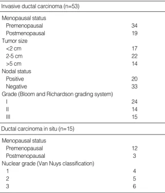
The clinical data of 68 breast carcinoma patients are listed in Table 1. There were 15 cases of pure intraductal carcino- ma (DCIS) and 53 cases of invasive ductal carcinoma (IDC).
All patients were female and the mean age was 48 yr. Twen- ty of these 53 cases had lymph node metastases and two of those 20 cases had intraductal components. Among the 33 cases of invasive carcinoma without lymph node metastasis, 14 patients had coexisting intraductal components. Tumor size was classified into three groups; 17 cases of less than 2 cm, 22 cases of 2-5 cm, 14 cases of greater than 5 cm in great- est diameter.
Microscopically, the intraductal carcinomas were grouped by Van Nuys classification into four cases of group 1, five cases of group 2, and six cases of group 3. Invasive ductal carcino- mas were also classified by Bloom and Richardson grading system into 14 cases of grade I, 24 cases of grade II, and 15 cases of grade III.
MSI at 11p15.5 in intraductal and invasive ductal carcinoma The overall incidences of MSI in the intraductal and inva-
sive carcinoma were 26.7% and 45.3% at D11S922, 13.3%
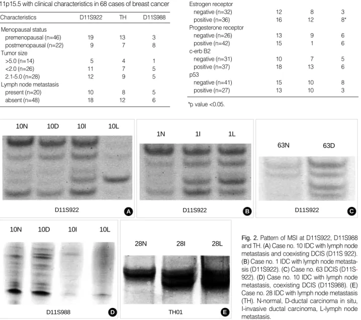
and 33.9% at TH, and 6.7% and 18.9% at D11S988 respec- tively (Table 2). Data from 41 cases that have MSI for at least one polymorphic marker are presented in Fig. 1. The patterns of MSI in 4 of 41 cases are shown in Fig. 2.
The MSI data from cases was analyzed by each tumor com- ponent. In 33 cases of IDC without metastasis, the incidence of MSI at D11S922, TH, and D11S988 were 36.4%, 27.3%, and 15.2% in invasive component and 18.7%, 18.7%, 12.5%
in 14 cases of coexisting intraductal component. In 20 cases with lymph node metastases, it was 40%, 30%, and 25% in invasive component and 35.0%, 25.0%, 20.0% in lymph node metastatic lesion, respectively. The intraductal compo- nent in IDC was only 2 cases and has no significant result.
The incidence of MSI increased through the subsequent stages of cancer progression but showed no significant difference in statistical analysis. MSI incidence in intraductal lesions coex- isting with invasive carcinoma did not significantly increase the risk of progression to invasive carcinoma.
Clinicopathological correlation
The MSI frequency at D11S922, TH, or D11S988 accord- ing to clinicopathological parameters such as the patient’s age, tumor size, and lymph node metastasis are presented in Table 3 and no significant difference was observed.
Correlation with expression of estrogen/progesterone receptor, p53, and c-erb B2 by immunohistochemistry
The positivity rates of immunohistochemical stain in 68 cases were as follows: estrogen receptor positivity was 64.7%, progesterone receptor was 61.7%, p53 was 39.7%, and c-erb B2 was 54.4%. The comparisons between MSI at D11S922, TH, or D11S988 and positivity rates of estrogen/progesterone
Invasive ductal carcinoma (n=53)
Ductal carcinoma in situ (n=15) Menopausal status
Premenopausal 34
Postmenopausal 19
Tumor size
<2 cm 17
2-5 cm 22
>5 cm 14
Nodal status
Positive 20
Negative 33
Grade (Bloom and Richardson grading system)
I 24
II 14
III 15
Menopausal status
Premenopausal 12
Postmenopausal 3
Nuclear grade (Van Nuys classification)
1 4
2 5
3 6
Table 1.Clinicopathologic characteristics of 68 cases of breast cancer patients
1 D11S922 TH D11S988
D11S922 TH D11S988
3 4 5 6 7 10111314 1617 181920 212223242526
2830 313435 36 37394244 4548 495152 54616364 66
Fig. 1.Pattern of microsatellite instability in 41 of 68 cases present- ing at least one polymorphic marker. Invasive ductal carcinoma cases are from case number 1 to 52 and ductal carcinoma in situ cases are from case number 54 to 66. microsatellite instability,
retention of heterozygosity, uninformative.
receptor, p53, c-erb B2 are listed in Table 4. The correlation between MSI of D11S988 and expression of estrogen recep- tor was statistically significant (p=0.041).
DISCUSSION
Among the theories related to the development mechanism of breast cancer presented up to now, according to the theory of Newsham, when a normal ductal epithelium progresses
to intraductal carcinoma through atypical proliferation, the locus of the genes of chromosomes 11p15.5 and 11q23 are lost and when it progresses to invasive carcinoma, chromo- some 11q13 is lost (16). However, the role of 11p15.5 is not clearly identified yet. We used D11S922, TH, and D11S988 as the microsatellite markers that correspond to chromosome 11p15.5 in intraductal and invasive carcinoma and these parts correspond to the part of 6Mb that codes the IGF-II (insulin- like growth factor-II), the p57KIP2, and the KvLQT1 gene.
The IGF-II is an important growth factor and the p57KIP2 is the cyclin-dependent-kinase inhibitor that provokes arrest of the G1-S stage (23). The KvLQT1 gene is involved in a voltage-gated potassium channel.
Characteristics D11S922 TH D11S988
Menopausal status
premenopausal (n=46) 19 13 3
postmenopausal (n=22) 9 7 8
Tumor size
>5.0 (n=14) 5 4 1
<2.0 (n=26) 11 7 5
2.1-5.0 (n=28) 12 9 5
Lymph node metastasis
present (n=20) 10 8 5
absent (n=48) 18 12 6
Table 3.The correlation of the frequency of MSI at chromosome 11p15.5 with clinical characteristics in 68 cases of breast cancer
Marker D11S922 TH D11S988
Estrogen receptor
negative (n=32) 12 8 3
positive (n=36) 16 12 8*
Progesterone receptor
negative (n=26) 13 9 6
positive (n=42) 15 1 6
c-erb B2
negative (n=31) 10 7 5
positive (n=37) 18 13 6
p53
negative (n=41) 15 10 8
positive (n=27) 13 10 3
Table 4.The correlation between the frequency of MSI at chro- mosome 11p15.5 and estrogen/progesterone receptor, c-erb B2 and p53 protein in 68 cases of breast cancer
*p value <0.05.
Fig. 2.Pattern of MSI at D11S922, D11S988 and TH. (A) Case no. 10 IDC with lymph node metastasis and coexisting DCIS (D11S 922).
(B) Case no. 1 IDC with lymph node metasta- sis (D11S922). (C) Case no. 63 DCIS (D11S- 922). (D) Case no. 10 IDC with lymph node metastasis, coexisting DCIS (D11S988). (E) Case no. 28 IDC with lymph node metastasis (TH). N-normal, D-ductal carcinoma in situ, I-invasive ductal carcinoma, L-lymph node metastasis.
A B C
E D
10N
D11S922 D11S922
D11S988 TH01
D11S922 10D
28N 28I 28L
10I 10L
10N 10D 10I 10L
1N 1I 1L
No. of case\marker D11S922 TH D11S988
Ductal carcinoma in situ (n=15) 4 (26.7%) 2 (13.3%) 1 (6.7%) Invasive ductal carcinoma (n=53) 24 (45.3%) 18 (33.9%) 10 (18.9%) Table 2.Overall incidence of microsatellite instability at 11p15.5 loci in breast ductal carcinoma (%)
63N 63D
In this research, the MSI of the locus of the gene D11S922 was relatively higher than another markers, and was 26.7%
in pure intraductal carcinoma, 36.4% in invasive ductal car- cinoma without lymph node metastasis, and 40.0% in inva- sive ductal carcinoma with lymph node metastasis. It shows a high frequency according to the progression of breast cancer, but was not statistically significant. This shows a similar result to the other research using the existing paraffin embedded tissue. MSI incidences at TH and D11S999 loci were also increased during histologic progression, but the data cannot be interpreted as representative and significant because of limited positive results.
According to the research of Lichy et al., the LOH of 11p 15.5 is 37.5% in pure intraductal carcinoma, 32% in inva- sive ductal carcinoma without lymph node metastasis, and 44% in invasive ductal carcinoma with lymph node metas- tasis (24). Their result was also different from ours because it reported that the LOH of 11p15.5 is mainly involved when the normal epithelial cell progresses to intraductal carcinoma.
In the research of Winqvist et al., it is reported at 35%
overall frequency (15). According to the report of Deng and others, the LOH of the chromosome 11p15.5 is also observed in the normal terminal duct lobular unit. This suggests that the LOH of chromosome 11p15.5 can play the role of a genet- ic shunt progressing toward the infiltrating carcinoma from normal epithelial tissues (25). That is to say, cancer may occur in another pathway and not in a series of continuous pathways of the normal epithelial cell, intraductal carcinoma, invasive carcinoma, and metastatic carcinoma. In the present research, we observed that the MSI of chromosome 11p15.5 has a lower frequency in intraductal carcinoma than the invasive carci- noma; however, the comparison of intraductal carcinoma by histology and grade has no significance probably due to the limitation of the number of cases. In the research of Fujii and others regarding the loss of genes according to the histologic grade of intraductal carcinoma, the number of lost genes in- creases significantly at a nuclear grade higher and in a lower grade, it was observed that 16q and 17p were lost (26). How- ever, the loss of 11p is related to high-grade lesion and it is observed at around 40%. It is reported that intraductal car- cinoma lesions accompanied with infiltrating cancer and pure intraductal carcinoma lesions are different from each other in the form of heterozygosity and it is considered that tumors have been developed from intraductal carcinoma lesions to infiltrating carcinoma through various genetic pathways (26).
In the present research, there is no statistical significance as a result of analyzing the correlation of the MSI of 11p15.5 with the patient’s clinical factors such as age of onset, size of tumor, grade, and presence of lymph node metastasis. As for the correlation between the factors such as p53, c-erb B2, estrogen and progesterone receptor, which are known as the prognostic factors for breast cancer, the estrogen receptor showed a significant correlation with the MSI of D11S988.
But it is necessary to evaluate the correlation to the patient’s
survival rate if the MSI of 11p15.5 could be useful as a poten- tial prognostic marker for breast cancer.
As a result, we suggest that the MSI of chromosome 11p 15.5 could play a part in the occurrence and progression of breast cancer, because the more cancer progresses, the higher its frequency increases. But its role in the continuous progres- sive stage of breast cancer could not be clearly verified. It is considered necessary to study further the worth of which MSI in the chromosome 11p15.5 is used as the method to predict the prognosis of breast cancer.
REFERENCES
1. Parker SL, Tong T, Bolden S. Wingo PA. Cancer statistics. CA Can- cer J Clin 1997; 47: 5-27.
2. Andersen TI. Genetic heterogeneity in breast cancer susceptibility.
Acta Oncol 1996; 35: 407-10.
3. Niederacher D, Schnurch HG, An HX, Ellenberger I, Dall P, Van Roeyen CR, Kuppers V, Beckmann MW. Detection of sequential genetic alterations relevant for breast cancer development. Eur J Cancer Prev 1996; 5: 497-503.
4. Lodish H, Berk A, Zipursky SL, Matsudaira P, Baltimore D, Darnell JE. Molecular cell biology. 4th ed, W.H. Freeman and company, New York, p1063-9.
5. Fearon ER, Vogelstein B. A genetic model for colorectal tumorige- nesis. Cell 1990; 61: 759-67.
6. Callahan R, Campbell G. Mutations in human breast cancer: an overview. J Natl Cancer Inst 1989; 81: 1780-6.
7. Genuardi M, Tsihira H, Anderson DE, Saunders GF. Distal deletion of chromosome 1p in ductal carcinoma of the breast. Am J Hum Genet 1989; 45: 73-82.
8. Matsumoto S, Minobe K, Utada Y, Furukawa D, Onda M, Sakamoto G, Kasumi F, Nakamura Y, Emi M. Loss of heterozygosity at 3p24- p25 as a prognostic factor in breast cancer. Cancer Letters 2000; 152:
63-9.
9. Tomlinson IP, Nicolai H, Solomon E, Bodmer WF. The frequency and mechanism of loss of heterozygosity on chromosome 11q in breast cancer. J Pathol 1996; 180: 38-43.
10. Hampton GM, Mannermaa A, Winquist R, Alavaikko M, Blanco G, Taskinen PJ, Kiviniemi H, Newsham I, Cavenee WK, Evans GA. Loss of heterozygosity in sporadic human breast carcinoma: a common region between 11q22 and 11q23.3. Cancer Res 1994; 54: 4586-9.
11. Van Den Berg J, Johannsson O, Hakansson S, Olsson H, Borg A.
Allelic loss at chromosome 13q12-q13 is associated with poor prog- nosis in familial and sporadic breast cancer. Br J Cancer 1996; 74:
1615-9.
12. Iida A, Isobe R, Yoshimoto M, Kasumi F, Nakamura Y, Emi M. Lo- calization of a breast cancer tumour-suppressor gene to a 3-cM inter- val within chromosomal region 16q22. Br J Cancer 1997; 75: 264-7.
13. Ali IU, Lidereau R, Theillet C, Callahan R. Reduction to homozygosity of genes on chromosome 11 in human breast neoplasia. Science 1987;
238: 185-8.
14. Karnik P, Plummer S, Casey G, Myles J, Tubbs R, Crowe J, Williams
BR. Microsatellite instability at a single locus (D11S988) on chro- mosome 11p15.5 as a late event in mammary tumorigenesis. Hum Mol Genet 1995; 4: 1889-94.
15. Winqvist R, Hampton GM, Mannermaa A, Blanco G, Alavaikko M, Kiviniemi H, Taskinen PJ, Evans GA, Wright FA, Newsham I. Loss of heterozygosity for chromosome 11 in primary human breast tumors is associated with poor survival after metastasis. Cancer Res 1995;
55: 2660-4.
16. Newsham IF. The long and short of chromosome 11 in breast cancer.
Am J Pathol 1998; 153: 5-9.
17. Karnik P, Paris M, Williams BR, Casey G, Crowe J, Chen P. Two distinct tumor suppressor loci within chromosome 11p15 implicated in breast cancer progression and metastasis. Hum Mol Genet 1998;
7: 895-903.
18. Phillips KK, Welch DR, Miele ME, Lee JH, Wei LL, Weissman BE.
Suppression of MDA-MB-435 breast carcinoma cell metastasis fol- lowing the introduction of human chromosome 11. Cancer Res 1996;
56: 1222-7.
19. Chen T, Dhingra K, Sahin A, Sneige N, Hortobagyi G, Aldaz CM.
Technical approach for the study of the genetic evolution of breast cancer from paraffin-embedded tissue sections. Breast Cancer Res Treat 1996; 39: 177-85.
20. Lee JY, Dong SM, Kim SY, Yoo NJ, Lee SH, Park WS. A simple, precise and economical microdissection technique for analysis of genomic DNA from archival tissue sections. Virchows Arch 1998; 433:
305-9.
21. Youngson BJ, Anelli A, Van Zee KJ, Borgen PI, Norton L, Rosen PP. Microdissection and molecular genetic analysis of HER2/neu in breast carcinoma. Am J Surg Pathol 1995; 19: 1354-8.
22. Dahiya R, Deng G. Molecular prognostic markers in breast cancer.
Breast Cancer Res Treat 1998; 52: 185-200.
23. Feinberg AP. Imprinting of a genomic domain of 11p15 and loss of imprinting in cancer: an introduction. Cancer Res 1999; 59: 1743-6.
24. Lichy JH, Zavar M, Tsai MM, O’Leary TJ, Taubenberger JK. Loss of heterozygosity on chromosome 11p15 during histological progres- sion in microdissected ductal carcinoma of the breast. Am J Pathol 1998; 153: 271-8.
25. Deng G, Lu Y, Zlotnikov G, Thor AD, Smith HS. Loss of heterozy- gosity in normal tissue adjacent to breast carcinomas. Science 1996;
274: 2057-9.
26. Fujii H, Szumel R, Marsh C, Zhou W, Gabrielson E. Genetic progres- sion, histological grade and allelic loss in ductal carcinoma in situ of the breast. Cancer Res 1996; 56: 5260-5.