INTRODUCTION
Bronchogenic cysts are derived from the embryologic bran- chial cleft and are mainly of pulmonary origin. They are rarely located in an extrathoracic site, such as subdiaphragmatic retroperitoneal area (1-17). Only a few cases of intraperitoneal area (18-26) have been documented (Table 1). To the best of our knowledge, only 22 retroperitoneal cases have been report- ed in the world literature by the year of 2001, 17 of which are English language reports (17). Cases arising from an in- traperitoneal position are more unusual. Only 8 cases have been reported by the year of 2001. We report upon the first isolated intraperitoneal bronchogenic cyst in a 48-yr-old woman, which was presented as a gallbladder mass in Korea.
CASE REPORT
A 48-yr-old female was admitted to our hospital with one- year history of dyspepsia after meals and intermittent epigas- tric pain. A physical examination demonstrated no palpable mass in the abdominal region. White blood cell (WBC) count was at 5.2×10⁹/L, and hemoglobin was at 11.9 g/dL. Blood chemistry results were normal and preoperative serum alpha- feto protein (AFP) was also within normal range (0.77 U/mL, normal 0-5 U/mL). Ultrasound sonography showed a cystic
mass adjacent to the gallbladder (Fig. 1). Abdominal CT showed a well defined and circumscribed, cystic mass 3×2.5 cm in size at the inferomedial aspect of the gallbladder (Fig.
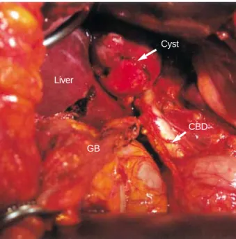
2). Radiological findings suggested a gallbladder tumor, a teratoma, bronchopulmonary sequestration, a complicated cyst or carcinoma, but the findings were insufficient for an accurate diagnosis to be made. Therefore a presumptive diag- nosis of a gallbladder tumor was made. The lesion was explor- ed because CT did not show a definite demarcation between the mass and the neighboring structures, nor did it confirm its isolation in the gallbladder area; moreover, the possibility of malignancy could not be ruled out. At laparotomy, a 3 cm- sized cystic mass was discovered adherent to the gallbladder (Fig. 3). The cyst was dissected from the liver bed, and the entire cyst and gallbladder were excised consequently. There was no connection between cyst and gallbladder. The gross appearance of the resected specimen seemed to be a benign cyst. On opening the specimen revealed one large cystic cav- ity, which contained thick brownish mucoid fluid (Fig. 4).
Microscopically, the cyst is lined by a layer of pseudostrati- fied ciliated columnar epithelial cells occasionally interspersed with goblet cells (Fig. 5). Thus, the cyst was histologically diagnosed as a bronchogenic cyst. The postoperative course was uneventful; the patient was discharged at 10th day post- operatively, and had remained asymptomatic through biweek- ly follow-ups for two months.
Kee Hwan Kim, Ji Il Kim, Chang Hyeok Ahn, Jeong Soo Kim, Young Mi Ku*, Ok Ran Shin�, Eun Jung Lee�, Keun Woo Lim
Department of Surgery, *Radiology and �Clinical Pathology, Uijeongbu St. Mary’s Hospital, College of Medicine, The Catholic University of Korea, Uijeongbu, Korea
Address for correspondence Jeong Soo Kim, M.D.
Department of Surgery, Uijeongbu St. Mary’s Hospital, College of Medicine, The Catholic University of Korea, 65-1 Gumo-dong, Uijeongbu 480-130, Korea Tel : +82.31-820-3048, Fax : +82.31-847-2717 E-mail : [email protected]
470 J Korean Med Sci 2004; 19: 470-3
ISSN 1011-8934
Copyright � The Korean Academy of Medical Sciences
The First Case of Intraperitoneal Bronchogenic Cyst in Korea Mimicking a Gallbladder Tumor
We present a case of an intraperitoneal bronchogenic cyst located at inferior surface of the liver, next to the gallbladder which clinically mimicked a gallbladder tumor.
This is the first case reported in Korea, and we offer reviews of the related literatures.
A 48-yr-old woman was admitted to our hospital because of intermittent abdominal pain in right upper quadrant. Computed tomography showed a large mass along- side the gallbladder. During laparotomy, the mass showed an ovoid cystic nature, which was attached to the normal gallbladder and liver bed. Cyst excision with chole- cystectomy was performed, and histopathological examination revealed a broncho- genic cyst. Most bronchogenic cysts have a benign nature, but malignant changes have also been reported. Therefore, if a cystic tumor in the abdomen is suspected during preoperative diagnosis, a bronchogenic cyst should be considered in the dif- ferential diagnosis.
Key Words :Bronchogenic Cyst; Gallbladder Neoplasms
Received : 27 March 2003 Accepted : 18 August 2003
Intraperitoneal Bronchogenic Cyst Mimicking a Gallbladder Tumor 471
DISCUSSION
Bronchogenic cysts are congenital abnormalities arising from the ventral foregut during the third to seventh week of fetal development. They are almost always lined, at least partially, by ciliated cuboidal to pseudostratified columnar epithelium and are often filled with mucus. Bronchial com- ponents such as cartilage, smooth muscle, elastic fibers, fibrous tissue and seromucinous glands may all be presented in the cyst wall (27). A retroperitoneal location is rarely reported.
Although the exact mechanism is unknown, Sumiyoshi et al.
(2) proposed the following theory. During early embryonic
life, the thoracic and abdominal cavities are linked via the pericardio-peritoneal canal. When the canal is later divided by the fusion of the pleuroperitoneal membranes (the future diaphragm), a portion of the tracheobronchial tree may be pinched off and migrate, resulting in a retroperitoneal bron- chogenic cyst (2). However, subdiaphragmatic bronchogenic cysts, especially in the intraperitoneal region, are extremely rare. Only 8 cases have been reported in the world literature, and all had their locations adjacent to the stomach. Our case had an unique gallbladder location. To our knowledge, no intraperitoneal cyst arising near the gallbladder had been reported in either the Korean or the English literatures. Of these retroperitoneal bronchogenic cysts, nine cases occurred in
Fig. 1.Sonographic finding showing a well-defined round cystic mass adjacent to the gallbladder, the lesion is filled with echogenic materials.
Cyst GB
Fig. 2.Post-contrast sequential axial abdominal CT scan shows a well-defined round cystic mass at the inferomedial aspect of the gallbladder. The internal density of the cystic mass appears as a subtle increase than that of the gallbladder.
Cyst GB GB Cyst
Cyst
10 cm
A B
Fig. 3.On operation, the mass is ovoid and cystic and is attached to the normal gallbladder and liver bed.
GB
CBD Liver
Cyst
Fig. 4.The cut section of the specimen shows a single large cys- tic cavity, containing a thick brownish mucoid fluid.
472 K.H. Kim, J.I. Kim, C.H. Ahn, et al.
males and eight in females. The age of the patients varied because the cases of smaller cysts were asymptomatic and the masses were incidentally discovered. In the cases of larger cysts, the patients complained of various types of pains in the
suspected region. The size of the cyst showed a increasing tendency with ages of the patients. Table 1 summarizes the eight cases of isolated intraperitoneal bronchogenic cysts that have been reported by the year of 2001. Interestingly, eight cases were located adjacent to the stomach. All of the eight cases were considered to arise in the left side of stomach. Pre- operative clinical diagnosis included the followings; benign tumor (18), leiomyoma or lipoma (19), a intestinal obstruc- tion (20), and a dermoid cyst (24). In our case, which located beside the gallbladder, apart from stomach, and no connection to stomach and gallbladder wall.
Since there are no common symptoms and specific changes in laboratory findings, CT scan has an important role in mak- ing the diagnosis. CT scan is useful for evaluating cyst con- tents so it allows a further differential diagnosis of retroperi- toneal cystic lymphangioma, hematoma, abscess, etc. (28).
Bronchogenic cysts usually show high CT values ranging from 30 to 100 HU since the cysts are filled with protein-rich fluid (12).
For histological diagnosis, they should be differentiated from bronchopulmonary sequestration and cystic teratoma.
Bronchopulmonary sequestration can be diagnosed by the fact
Author, year Ref. no. Age (yr)/sex Size (cm) Site
Retroperitoneal cysts
Miller et al., 1953 1 10 weeks/F 2 Anterior site of pancreas
Sumiyoshi et al., 1985 2 59/M 7 Superior body of pancreas
Coselli et al.,1987 3 35/F 5 Superior body of pancreas
Foerster et al.,1991 4 35/M 10.5×7.5×4.5 Superior left adrenal gland
Swanson et al.,1991 5 4/F 2 Superior left adrenal gland
Wirbel et al.,1993 6 38/M 3 Superior left adrenal gland
Fischbach et al.,1994 7 12/M 1.5×1.3 Right crus of the diaphragm
Ojika et al.,1996 8 62/M 2.2×1.5 Right crus of the diaphragm
Harvell et al., 1996 9 57/F 2.2×1.7×1.5 Superior body of the pancreas
Resl et al., 1996 10 21/M 4 Superior left adrenal gland
Tokuda et al., 1997 11 24/F 3 Superior left adrenal gland
Menke et al., 1997 12 35/M 8 Superior left adrenal gland
Doggett et al., 1997 13 44/M 10×10×6 Adherent to left adrenal gland
Cetinkursun et al., 1997 14 20 months/F 5 Superior pancreatic tail
Itoh et al., 1998 15 46/F 8×8×7 Superior left adrenal gland
Sullivan et al., 1999 16 55/F 10×8×4 Inferior left adrenal gland
Haddadin et al., 2001 17 51/M 4.0×3.5 Superior left adrenal gland I
Intraperitoneal cysts
Dewing et al., 1956 18 56/F 4×3×3 Intramural in the posterior wall of the gastric cardia
Gensler et al., 1966 19 46/F 6×8 Intramural in the gastric curvature of the stomach
Pai et al., 1971 20 67/M 9×2.5 Fused with posterior gastric wall proximally
Tanenbaum et al., 1971 21 60/M 10, 7.6 Between the spleen and stomach, intramural in the
posterior gastric wall
Benoit, 1972 22 37/F Not stated Juxtagastric
Murley and Lenz, 1979 23 17/M Not stated Attached to distal esophagus and adherent to inferior surface of left hemidiaphragm
Shireman, 1987 24 61/F 6 Intramural in gastric cardia
Braffman et al., and 25, 26 64/F 15 Communicating with the posterior wall of the stomach
Keohane et al., 1988
Present case 48/F 3×2.5 Attached to gallbladder and adherent to inferior
surface of liver
Table 1.Characteristics of the patients with subdiaphragmatic bronchogenic cysts reported in the English literature Fig. 5.Cyst lining is composed of respiratory type epithelium, under-
lying lamina propria, and smooth muscle (A, H&E, ×40). Pseudo- stratified ciliated columnar epithelial cells are interspersed occasion- ally with goblet cells (arrow head) (B, H&E, ×200).
A B
Intraperitoneal Bronchogenic Cyst Mimicking a Gallbladder Tumor 473
that it cintains has lung parenchyme and pleural tissue.
Cystic teratoma has endoderm-origin bronchial tissue and other structures from mesoderm and ectoderm. Among the cysts of foregut origin, those containing cartilage or seromu- cinous respiratory glands are designated as bronchogenic cysts;
those containing two well-developed layers of smooth muscle without cartilage are designated as esophageal cysts; and those with none of these distinguishing features are classified as foregut cysts (9). In contrast, the cysts of urogenital origin may rarely have pseudostratified ciliated epithelium, and submucosal seromucinous glands (4, 12). In our case, a ter- atoma was excluded by the absence of tissue, representing the three different germinal layers. In addition, bronchopulmonary sequestration can be diagnosed by the fact that it possesses lung parenchyma, pleural investment, and bronchial elements which were absent in our case.
Preferred treatment of intraperitoneal bronchogenic cyst is surgical removal. Although most are asymptomatic, excision is recommended to establish the diagnosis, alleviate symp- toms, and to prevent complications, such as infections and the remote, but documented risk of malignant transforma- tion (16).
Although the occurrence of bronchogenic cyst is rare, it should be considered in the differential diagnosis of an intra- abdominal mass, particularly in the case of a cystic tumor in the region adjacent to the gallbladder.
REFERENCES
1. Miller RF, Fraub M, Pashuck ET. Bronchogenic cysts: Anomalies resulting from maldevelopment of the primitive foregut and midgut.
Am J Roentgenol Radium Ther Nucl Med 1953; 70: 771-85.
2. Sumiyoshi K, Shimizu S, Enjoji M, Iwashita A, Kawakami K. Bron- chogenic cyst in the abdomen. Virchows Arch A Pathol Anat Histo- pathol 1985; 408: 93-8.
3. Coselli MP, de Ipolyi P, Bloss RS, Diaz RF, Fitzgerald JB. Broncho- genic cyst above and below the diaphragm: Report of eight cases. Ann Thorac Surg 1987; 44: 491-4.
4. Foerster HM, Sengupta EE, Montag AG, Kaplan EL. Retroperitoneal bronchogenic cyst presenting as an adrenal mass. Arch Pathol Lab Med 1991; 115: 1057-9.
5. Swanson SJ III, Skoog SJ, Garcia V, Wahl RC. Pseudoadreanl mass:
Unusual presentation of bronchogenic cyst. J Pediatr Surg 1991; 26:
1401-3.
6. Wirbel RJ, Uhlig U, Kiffner EM, Berger K. [Bronchogenic cyst as a rare differential diagnosis of retroperitoneal tumor.] Chirurg 1993;
64: 1056-9.
7. Fischbach R, Benz-Bohm G, Berthold F, Eidt S, Schmidt R. Infra- diaphragmatic bronchogenic cyst with high CT numbers in a boy with primitive neuroectodermal tumor. Pediatr Radiol 1994; 24: 504-5.
8. Ojika T, Mukouyama N, Tsuzuki T. [A case of bronchogenic cyst in
the subdiaphragmatic region.] Kyobu Geka 1996; 49: 505-7.
9. Harvell JD, Macho JR, Klein HZ. Isolated intra-abdominal esophageal cyst. Case report and review of the literature. Am J Surg Pathol 1996;
20: 476-9.
10. Resl M, Navratil P, Krajina A. Retroperitoneal bronchogenic cyst in a young adult. Respiration 1996; 63: 387-9.
11. Tokuda N, Naito S, Uozumi J, Shimura H, Takayanagi R, Kumaza- wa J. A retroperitoneal bronchogenic cyst treated with laparoscopic surgery. J Urol 1997; 157: 619.
12. Menke H, Roher HD, Gabbert H, Schweden F. Bronchogenic cyst:
A rare cause of a retroperitoneal mass. Eur J Surg 1997; 163: 311-4.
13. Doggett RS, Carty SE, Clarke MR. Retroperitoneal bronchogenic cyst masquerading clinically and radiologically as a phaeochromo- cytoma. Virchows Arch A Pathol Anat Histopathol 1997; 431: 73-6.
14. Cetinkursun S, Ozturk H, Celasun B, Sakarya MT, Surer I. Isolate abdominal bronchogenic cyst: A case report. Eur J Pediatr Surg 1997;
7: 103-5.
15. Itoh H, Shitamura T, Kataoka H, Ide H, Akiyama Y, Hamasuna R, Hasui Y, Osada Y, Koono M. Retroperitoneal bronchogenic cyst:
Report of a case and literature review. Pathol Int 1999; 49: 152-5.
16. Sullivan SM, Okada S, Kudo M, Ebihara Y. A retroperitoneal bron- chogenic cyst with malignant change. Pathol Int 1999; 49: 338-41.
17. Haddadin WJ, Reid R, Jindal RM. A retroperitoneal bronchogenic cyst: a rare cause of a mass in the adrenal region. J Clin Pathol 2001;
54: 801-2.
18. Dewing SB, Roessel CW, Olmstead EV. Enterogenous cyst of the stomach wall, a rare benign lesion: a case report. Ann Surg 1956;
143: 131-5.
19. Gensler S, Seidenberg B, Rifkin H, Rubistein BM. Ciliated lined intramural cyst of the stomach: case report and suggested embryo- genesis. Ann Surg 1966; 163: 954-6.
20. Pai SH, Cameron CT, Lev R. Accessory lung presenting as a juxta- gastric mass. Arch Pathol 1971; 91: 569-72.
21. Tanenbaum B, Levowitz BS, Ponce M, Manubay S Jr. Respiratory choristoma of stomach. N Y State Med J 1971; 71: 373-5.
22. Benoit CG. Compression de la grosse tuberosite de l’estomac par un kyste bronchogenique. Sem Hop Paris 1972; 48: 2641-4.
23. Murley GD, Lenz TR. Bronchogenic cyst, intra-abdominal. Rocky Mt Med J 1979; 76: 243-4.
24. Shireman PK. Intramural cyst of the stomach. Hum Pathol 1987; 18:
857-8.
25. Braffman B, Keller R, Gendal ES, Finkel SI. Subdiaphragmatic bron- chogenic cyst with gastric communication. Gastrointest Radiol 1988;
13: 309-11.
26. Keohane ME, Schwartz I, Freed J, Dische R. Subdiaphragmatic bronchogenic cyst with communication to the stomach: a case report.
Hum Pathol 1988; 19: 868-71.
27. Rosai J. Peritoneum, retroperitoneum, and related subjects. In: Rosai J, ed. Ackerman’s surgical pathology. New York: Mosby-Year Book, 1996: 2135-72.
28. Kajiya Y, Nakajo M, Ichinari N, Yamazumi K, Otuji T, Tanaka T.
Retroperitoneal foregut cyst. Abdom Imaging 1997; 22: 111-3.
. . . . . .
. .