INTRODUCTION
Lung cancer is a major cause of cancer deaths worldwide (1) and has become the leading cause of cancer deaths in Korea (2). Advances in cancer treatment in the past two decades have contributed to only minimal increase in survival rates of patients with non-small cell lung cancer (NSCLC) (3). Des- pite apparent complete resection of the primary tumor, recur- rence rates remain high and the overall 5-yr survival remains poor with <15% of patients surviving 5 yr from diagnosis (1). Some tumors, regardless of clinically favorable staging, are quite aggressive and progress to fatal disease. This implies that the TNM staging of NSCLC may be an acceptable, but not a satisfactory, classification system. Recent molecular stud- ies have provided increased understanding of the biology of lung cancer and have identified multiple factors responsible for the modulation of tumor growth and the prognosis (4-6).
Nevertheless, the essential genetic features, along with fac- tors for prognosis, have yet to be fully understood.
Altered regulation of the cell cycle is a hallmark of human cancers (6). The cell cycle is governed by cdks. An important mechanism for regulating the cdk activity involves the cdk
inhibitors, which are organized into two families based on structure and function: the Cip/Kip family (p21, p27, p57) and the INK4 family (p16, p18, p19). Cyclin E/cdk2 com- plex is an important regulator of entry into the S phase of the cell cycle, whereas cyclin B1/cdc2 is the classic M phase- promoting factor that drives entry into mitosis (7-10). Ki-67 proliferative index appears to be associated with survival in patients with various malignancies, but results are conflicting for NSCLC. Apoptosis or programmed cell death is a crucial mechanism of cellular homeostasis in organisms (11-14). One of the hallmark features of cancer cells is their ability to evade apoptosis. Angiogenesis is the process by which new capil- lary beds are formed from preexisting vessels, and is impor- tant in tumor growth (15). Vascular endothelial growth fac- tor (VEGF) is a potent growth factor for endothelial cells (16).
Tumors may activate angiogenic inhibitors such as angio- statin and endostatin, which control growth by suppressing endothelial cell proliferation and angiogenesis and by indi- rectly increasing apoptosis in tumor cells (17).
In the present study, we used the high-throughput tissue microarray (TMA) technology combined with immunohis- tochemistry (IHC) analysis (18, 19) to define the clinical sig-
Jinyoung Yoo, Ji Han Jung, Myung A Lee*, Kyung Jin Seo, Byoung Yong Shim*, Sung Hwan Kim�, Deog Gon Cho�, Myeong Im Ahn�, Chi Hong Kim*, Kyu Do Cho�, Seok Jin Kang, Hoon Kyo Kim*
Departments of Pathology, Internal Medicine*, Radiation Oncology�, Thoracic Surgery�, Diagnostic Radiology�, St. Vincent’s Hospital, The Catholic University of Korea, Suwon, Korea
Address for correspondence Seok Jin Kang, M.D.
Department of Pathology, St. Vincent’s Hospital, The Catholic University of Korea, 93 Ji-dong, Paldal-gu, Suwon 442-723, Korea
Tel : +82.31-249-7591, Fax : +82.31-244-6786 E-mail : [email protected]
*Supported in part by the 2006 Research Grants from St. Vincent’s Hospital and Lung Cancer Study Group at the St. Vincent’s Hospital.
318
Immunohistochemical Analysis of Non-Small Cell Lung Cancer:
Correlation with Clinical Parameters and Prognosis
Non-small cell lung cancers (NSCLC) vary in their biologic behavior. Recurrence and tumor-related mortality may be attributable to molecular abnormalities in prima- ry tumors. This study evaluated such immunophenotypes with regard to cell cycle regulation and proliferation, apoptosis, and angiogenesis, to determine their signifi- cance for patient outcome. Core biopsies from 219 patients with NSCLC were as- sembled on tissue microarrays, and the expressions of p16, p21, p27, cyclin B1, cyclin E, Ki-67, caspase-3, survivin, bcl-2, VEGF, and endostatin were evaluated by immunohistochemistry. Despite previously described prognostic relevance of some of the investigated molecules, many of those markers were not directly asso- ciated with recurrence or survival. However, there was a trend for p16 immunore- activity to be associated with a good prognosis (57% vs. 42% in 5-yr survival) (p=
0.071). bcl-2 expression was strongly correlated with a better outcome (65% vs.
45% in 5-yr survival) (p=0.029), and the hazard of death for bcl-2 positive patients was 0.42 times of that for bcl-2 negative patients (p=0.047). A multivariate analysis with Cox proportional hazards model confirmed that the lymph node status (p=0.043) and stage (p=0.003) were other independent prognostic factors. Our results sug- gest that p16 and bcl-2 provide prognostic information independent of the TNM stage in NSCLC.
Key Words : Carcinoma, Bronchogenic; Cell Cycle; Apoptosis; Angiogenesis Factor; Prognosis
Received : 8 August 2006 Accepted : 2 October 2006
nificance of altered expression of cell cycle regulatory or pro- liferation-related, apoptotic, and angiogenic factors. Immu- nophenotypes were correlated with patient outcome to deter- mine their prognostic value.
MATERIALS AND METHODS Patients
Two hundred and nineteen patients with previously un- treated NSCLC were included in this study. The study pro- tocol was reviewed and approved by the Institutional Review Board at the Catholic University St. Vincent’s Hospital. All patients underwent surgical resection at the Department of Thoracic Surgery. The patients were staged at the time of their surgery following the guidelines of the American Joint Committee on Cancer Staging (20). Clinical information was obtained through a computerized retrospective database of tumor registry. Patients who died within one month after surgery were excluded from the study to avoid bias of peri- operative mortality. Only patients with stages I to IIIA dis- ease and microscopically negative resection margins were included in this study. Patients did not receive neoadjuvant chemotherapy or radiotherapy. The survival duration was determined from the date of surgery for the primary lung cancer to date of last follow-up or date of death from lung cancer. Follow-up ranged from 1.6 to 117.8 months (medi- an 38.9 months). Eighty-seven patients (39.7%) died dur- ing follow-up, and 132 were alive at the time of this study.
Histologic examination and TMA construction
Hematoxylin and eosin stained slides were reviewed for confirmation of histopathologic diagnosis and for selection of adequate specimens for analysis. Histologic typing was performed according to the histological classification of lung cancer by the World Health Organization (21). When the tumors are composed of cells resembling the mature normal cells of the tissue of origin of the neoplasm, they were con- sidered as well differentiated, and when the tumors have prim- itive-appearing, unspecified cells, they were considered as poorly differentiated.
Areas of viable, representative tumor were identified and marked by a pathologist (JY) for the construction of microar- rays. Three to four cores, each measuring 2.0 mm in diame- ter, were sampled from different areas of the tumor with a precise instrument and were arrayed on a recipient paraffin block.
Immunohistochemistry
For immunohistochemical staining, a sensitive peroxidase- streptavidin method, as described previously, was performed
using four- m sections of these array blocks. First sections were stained for H&E to verify histology.
The antibodies for cell cycle regulation used in this study were p16 (clone 16P07, NeoMarkers, Fremont, CA, U.S.A.), p21 (clone EA10, Zymed, San Francisco, CA, U.S.A.), p27 (NeoMarkers), cyclin E (clone 13A3, NeoMarkers), and cyclin B1 (clone GNS11, NeoMarkers). Proliferative activity was determined using anti-Ki-67 (clone 7B11, Zymed). The anti- bodies to detect the apoptotic factors were caspase-3 (clone 3CSP03, NeoMarkers), survivin (clone 4F7, NeoMarkers), and bcl-2 (clone bcl-2-100, Zymed). The antibodies for stain- ing of angiogenic factors were VEGF (clone A20, Santa Cruz Biotechnology, Heidelberg, Germany), and endostatin (clone 1837-46, NeoMarkers).
Briefly, tissues cut from microarrays were deparaffinized in xylene and rehydrated in graded alcohols and water. En- dogenous peroxidase was blocked by soaking in 3% H2O2 at 45℃for 4 min. The slides were microwaved in citrate buffer (2.1 g/L, pH 6.0) at 120℃for 15 min to unmask the antigen, and treated with a protein-blocking reagent before incubation at 4℃overnight with primary antibodies at a 1:50 dilution, as recommended by the supplier. After exten- sive washing, the sections were incubated at room tempera- ture for 10 min with biotinylated anti-mouse immunoglob- ulin antibodies (Zymed, San Francisco, CA) at a 1:20 dilu- tion and subsequently with streptavidin-biotin peroxidase complexes at a 1:25 dilution. Reaction products were visu- alized by immersing slides in 3,3′-diaminobenzidine tetrahy- drochloride. Counterstaining was performed with hemato- xylin. All series included positive and negative controls. Nega- tive controls were prepared by omitting the primary antibod- ies and known positive controls were included in each run.
Three investigators independently evaluated the results from the immunohistochemical staining in a blinded fash- ion and obtained the identical results in 92% of the cases. For the discordant samples, slides were reviewed jointly and a consensus was reached. For the evaluation of immunoreactiv- ity in tumor cells, a dichotomised scoring system was used as follows: p16 positivity if >80% of tumor cells demonstrat- ed either nuclear or cytoplasmic staining; p21 positivity if
>5% tumor nuclei stained; p27 positivity if >20% of tumor nuclei stained; high Ki-67 labeling index and high cyclin E labeling index if >30% tumor cells stained; caspase-3 posi- tivity if nuclear or cytoplasmic staining was present in >25%
of tumor cells; survivin or bcl-2 positivity if >10% tumor cell stained; VEGF expression if >5% of tumor cells stained;
endostatin positivity if scores between 3 and 6 (the sum of the percentage of tumor cells [0, 1=<25%, 2=26-50%, 3=
>50%]and the staining intensity [1-3]) (9, 12, 18, 22-26).
Statistical analysis
Statistical analysis was carried out using a software pack- age (SPSS 13.0, Seoul, Korea). Survival probabilities were
estimated using the Kaplan-Meier method and then com- pared with the log rank test. The influence of various clini- copathologic variables on the survival was assessed with the Cox proportional hazards model. The level of significance was set at p<0.05.
RESULTS Clinicopathologic data
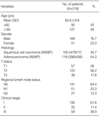
Complete clinical and histologic data were available for all patients. One hundred and sixty-eight patients (76.7%) were male and the mean age was 65.8 yr (SD 9.9; median 67; age range 19-89). Patient characteristics are shown in Table 1.
Immunohistochemistry
A significant variation was present in the expression of the individual immunohistochemical markers in tumor tissues.
The highest rate of positivity was noted for VEGF in 92.7%
of the tumors. The lowest rate of expression was observed with survivin in 2.7% of the samples. The expression profiles of all markers are presented in Table 2.
Of the cell-cycle regulation and proliferation-related pro-
W, well differentiated; M, moderately differentiated; P, poorly differenti- ated.
No. of patients (N=219)
Variables %
Age (yrs)
Mean (SD) 65.8±9.9
<65 92 42
≥65 127 58
Gender
Male 168 76.7
Female 51 23.3
Histology
Squamous cell carcinoma (W/M/P) 100 (4/79/17) 45.7 Adenocarcinoma (W/M/P) 119 (29/64/26) 54.3 T status
T1 57 26
T2 123 56.2
T3 39 17.8
Regional lymph node status
N0 141 64.4
N1 51 23.3
N2 27 12.3
Clinical stage
I 135 61.6
II 25 11.4
III 59 26.9
Table 1.Clinicopathologic charateristics
Variables No.
No. of positive cases
p16 p21 p27 Cyclin
B1
Cyclin E
Cas- pase
Sur-
vivin Bcl-2 VEGF Endo- statin Ki-67 Histology
Squamous cell carcinoma 100 22 71 22 4 43 36 3 19 93 94 11
Well differentiated 4 2 2 1 1 2 2 0 2 3 4 0
Moderately differentiated 79 14 58 17 2 33 28 2 14 74 70 9
Poorly differentiated 17 6 11 4 1 8 6 1 3 16 16 2
p-value 0.215 0.847 0.807 0.561 0.686 0.855 0.396 0.398 0.843 0.764 0.781
Adenocarcinoma 119 52 79 49 6 41 48 3 6 110 97 6
Well differentiated 29 11 17 14 0 3 9 1 0 26 22 1
Moderately differentiated 64 24 38 23 4 25 30 1 0 66 58 0
Poorly differentiated 26 9 14 8 2 13 9 1 6 18 17 5
p-value 0.408 0.313 0.297 0.13 0.001 0.366 0.805 0.002 0.803 0.017 0.909
T status
T1 57 21 36 21 2 18 22 1 10 55 53 3
T2-3 162 53 114 50 8 66 62 5 15 148 138 14
p-value 0.049 0.98 0.168 0.73 0.608 0.172 0.259 0.471 0.741 0.739 0.414
Regional lymph node status
N0 141 48 98 42 6 52 46 5 14 129 119 11
N1-3 78 26 52 29 4 32 38 1 11 74 72 6
p-value 0.535 0.037 0.733 0.948 0.378 0.049 0.964 0.523 0.469 0.699 0.454
Clinical stage
I 135 57 113 54 8 62 62 5 19 149 141 12
II-III 84 17 37 17 2 22 22 1 6 54 50 5
p-value 0.038 0.012 0.822 0.657 0.436 0.983 0.714 0.946 0.135 0.127 0.19
Total 219 74 150 71 10 84 84 6 25 203 191 17
Table 2.Molecular expression profiles
VEGF, vascular endothelial growth factor.
teins, p16 was expressed in 74 of 219 (33.8%) cases (Fig. 1A).
There were significant differences in T status and clinical stage between the p16-positive and -negative patients (p=
0.049 and 0.038, respectively); normal p16 expression was more frequent in patients with T1 than in those with T2-3 (36.8% vs. 32.7%), and in patients with stage I than in pa- tients with stage II-III (42.2% vs. 20.2%). Seventy-four tu- mors (68.5%) expressed p21. p21 expression was correlated with regional lymph node status (p=0.037) and with clini- cal stage (p=0.012). p27 immunoreactivity was identified in 71 cases (32.4%) and was more common in adenocarci- nomas than in squamous cell carcinomas (p=0.035). Cyclin- B1 immunostaining was detected in 10 tumors (4.6%) with-
out a clinically significant pattern. Cyclin E was positive in 84 samples (38.4%). In adenocarcinomas, cyclin-E staining was associated with differentiation (p=0.001); 3 of 29 well- differentiated tumors (10.3%), 25 of 64 moderately differ- entiated tumors (39.1%), and 13 of 26 poorly differentiated tumors (50%) were positive for cyclin-E (data not shown).
For Ki-67, a high Ki-67 proliferative index was observed in 17 of 219 patients (7.8%).
Of the apoptotic factors analyzed in the study, caspase-3 was expressed in 84 tumors (38.4%). Reactivity for caspase- 3 was present in 46 of 141 node-negative patients (32.6%) and 38 of 78 node-positive patients (48.7%) (p=0.049). Sur- vivin expression was rarely seen, with a mere positive stain-
Fig. 1.Immunohistochemistry for p16 (A) and bcl-2 (B), showing diffuse strong reactivity (×40).
A B
Cumulative survival
1.0
0.8
0.6
0.4
0.2
0.0
0.00 1000.00 2000.00 3000.00 4000.00
Survival (days)
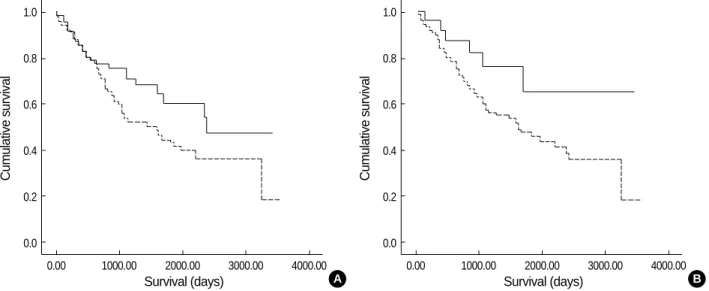
Fig. 2.Kaplan-Meier survival plots for non-small cell lung carcinoma. (A) p16 positive (solid line) vs. negative (dotted line); (B) bcl-2 posi- tive (solid line) vs. negative (dotted line). p-values by Mantel-Cox log-rank test were 0.071 (A) and 0.047 (B).
A
Cumulative survival
1.0
0.8
0.6
0.4
0.2
0.0
0.00 1000.00 2000.00 3000.00 4000.00
Survival (days) B
ing rate of 2.7%. bcl-2 immunoreactivity was observed in 25 cases (11.4%) (Fig. 1B).
Of 219 patients, 203 (92.7%) and 191 (87.2%) had posi- tive immunohistochemical staining for VEGF and endostatin, respectively. There was no significant influence of these two factors on histology, T status, regional lymph node status and clinical stage.
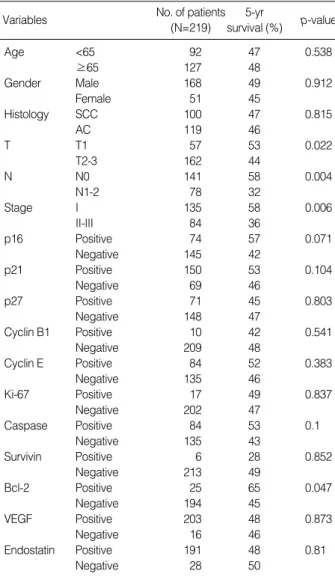
Five-year survival rates according to clinicopathologic vari- ables and immunohistochemical expression profiles are sum- marized in Table 3. T status (53% vs. 44%; p=0.022), regional lymph node status (58% vs. 32%; p=0.004) and clinical stage (58% vs. 36%; p=0.006) demonstrated a significant corre- lation with 5-yr survival, respectively, based on the log-rank test.
Of 11 markers analyzed, p16 and bcl-2 had an impact on the 5-yr survival. There was a trend for p16 immunoreac- tivity to be associated with a good prognosis (5-yr survival,
57% vs. 42%; p=0.071). Patients with negative or positive bcl-2 expression had 5-yr survival rates of 45% and 65%, respectively, which was statistically significant (p=0.047, Mantel-Cox log-rank test) (Fig. 2).
Combined analysis of bcl-2 and p16 expression identified 5-yr survival rates of 73.3% and 38.6% in patients with at least one of these factors and those with neither of these fac- tors, respectively. These molecular phenotypes had a signifi- cant influence over clinical outcome (p=0.002). In addition, Hazard ratio was used to estimate hazard of death due to NSCLC for the baseline variables (Table 4). The hazard of death for node-positive patients was 1.855 times of the haz- ard of death for node-negative patients (p=0.004). The haz- ard of death for patients with tumor stages II-III was 1.994 times of the hazard of death for patients with tumor stage I (p=0.002). For the effects on survival, bcl-2 expression was statistically significant with a p-value of 0.029. The multivari- ate analysis for overall survival revealed three independent prognostic factors: regional lymph node status, stage and bcl- 2 expression. Regional lymph node status (N1-2 vs. N0) was a significant and independent unfavorable prognostic factor (p=0.043), as was the advanced clinical stage (II-III vs. I) (p=
0.003), whereas immunohistochemical expression of bcl-2 (positive vs. negative) was a significant and independent favor- able prognostic factor (p=0.047).
DISCUSSION
Management of a patient with NSCLC could be determin- ed by the aggressiveness of the cancer, and the aggressive can- cer phenotypes are characterized by a number of molecular abnormalities. Because of the complexity of the molecular biology of NSCLC, studies on a single molecular factor have not yet been successful in creating biological risk assessment.
A few studies support the possibility that multiple markers might be more informative than any single marker for the prediction of clinical outcome of NSCLC (4, 8). This has prompted us to investigate 11 biologic markers, including those involved in cell cycle control and proliferation, apop- tosis and angiogenesis, and to construct a multivariate anal- ysis for these factors predicting the patients’ prognosis.
The major findings of our study are as follows: 1) expres- sion of p16 was associated with T status in NSCLC; 2) expres- sion of p21 or caspase-3 correlated with regional lymph node status; 3) expression of p16 or p21 correlated with stage; 4) patients with positive bcl-2 immunostaining or p16 immu- noreactivity or both had a higher 5-yr survival rate; 5) bcl-2 expression constitutes an independent prognostic parameter, along with regional lymph node status and stage.
Cyclin E is involved in the regulation of G1-S transition of the cell cycle, whereas cyclin B1 is involved in the regula- tion of G2-M transition. An important mechanism for regu- lating the CDK activity involves the CDK inhibitors, includ-
SCC, squamous cell carcinoma; AC, adenocarcinoma; VEGF, vascular endothelial growth factor.
No. of patients (N=219)
5-yr
survival (%) p-value Variables
Age <65 92 47 0.538
≥65 127 48
Gender Male 168 49 0.912
Female 51 45
Histology SCC 100 47 0.815
AC 119 46
T T1 57 53 0.022
T2-3 162 44
N N0 141 58 0.004
N1-2 78 32
Stage I 135 58 0.006
II-III 84 36
p16 Positive 74 57 0.071
Negative 145 42
p21 Positive 150 53 0.104
Negative 69 46
p27 Positive 71 45 0.803
Negative 148 47
Cyclin B1 Positive 10 42 0.541
Negative 209 48
Cyclin E Positive 84 52 0.383
Negative 135 46
Ki-67 Positive 17 49 0.837
Negative 202 47
Caspase Positive 84 53 0.1
Negative 135 43
Survivin Positive 6 28 0.852
Negative 213 49
Bcl-2 Positive 25 65 0.047
Negative 194 45
VEGF Positive 203 48 0.873
Negative 16 46
Endostatin Positive 191 48 0.81
Negative 28 50
Table 3.Five-year survival rate analysis (log-rank test) according to clinicopathologic parameters and protein expression profiles
ing p21, p27, p57, p16, p18 and p19. Some of the alterations in expression of these proteins, which lead to the failure of cell cycle arrest, have been demonstrated to be predictive for outcome and thus may serve as markers of more malignant phenotypes (27). In the present study, p16 and p21 were found to correlate with T, N or stage. Only p16 had a direct impact on the 5-yr survival in patients with NSCLC. This is in agreement with the results of the study by Esposito et al.
(8). In their multivariate analysis on cell cycle regulator pro- teins, the only immunohistochemical parameter that influ- enced overall survival was p16. It is suggested that cell cycle regulatory proteins may have different and multifunctional properties on cell proliferation and may contribute to their different effects on survival. The p16 expression may have a great value in identifying NSCLC patients with better prog- nosis.
As bcl-2 inhibits apoptosis, the bcl-2 overexpression should theoretically favor the malignant process and result in a poor outcome. However, there have been several studies describ-
ing a favorable outcome in NSCLC expressing bcl-2, although others have shown no survival advantage or even a worse prog- nosis (14, 26-29). Poleri et al. (26) described a poor outcome in patients with positive bcl-2 tumor. Cox et al. (14) inves- tigated 167 patients with resected stage I-IIIA NSCLC and found a trend for bcl-2 expression to be associated with an improved survival, which was consistent with our findings.
The reasons for this association are poorly understood. These conflicting results may have been due to the difference in the nature of tumor samples analyzed. In contrast to Poleri et al’s. study limited to the stage I NSCLC (26), we includ- ed stage I-IIIA tumors, as in Cox et al. (14). The criteria for interpreting the results as positive or negative may also have attributed to this difference. Cytoplasmic bcl-2 staining was considered either positive or negative according to the inves- tigators using different percentage criteria. While only sam- ples with at least 20% of the tumor were regarded as being positive, immunoreaction in more than 5% or even 1% of the tumor cells was also considered as being expressed by
Variables Level Hazard ratio 95% CI for HR p-value
A. Univariate analysis of potential prognostic factors
Age (yr) ≥65 vs. <65 0.874 0.570-1.341 0.539
Histology AC vs. SCC 0.951 0.623-1.452 0.815
T T2-3 vs. T1 1.604 0.987-2.605 0.056
N N1-2 vs. N0 1.855 1.214-2.834 0.004
Stage II-III vs. I 1.994 1.282-3.101 0.002
p16 Positive vs. negative 0.65 0.406-1.041 0.065
p21 Positive vs. negative 0.697 0.450-1.079 0.106
p27 Positive vs. negative 1.061 0.665-1.694 0.803
Cyclin B1 Positive vs. negative 1.325 0.536-3.270 0.542
Cyclin E Positive vs. negative 0.82 0.525-1.281 0.384
Caspase Positive vs. negative 0.686 0.437-1.075 0.095
Survivin Positive vs. negative 1.116 0.352-3.536 0.852
Bcl-2 Positive vs. negative 0.44 0.192-1.011 0.029
VEGF Positive vs. negative 0.929 0.374-2.303 0.873
Endostatin Positive vs. negative 0.925 0.491-1.744 0.811
Ki-67 Positive vs. negative 0.91 0.367-2.252 0.878
B. Multivariate analysis of prognostic factors
Age (yr) ≥65 vs. <65 0.924 0.574-1.485 0.743
Histology AC vs. SCC 0.913 0.532-1.566 0.74
T T2-3 vs. T1 1.065 0.531-2.137 0.859
N N1-2 vs. N0 1.821 1.019-3.292 0.043
Stage II-III vs. I 1.999 1.282-3.101 0.003
p16 Positive vs. negative 0.697 0.414-1.174 0.088
p21 Positive vs. negative 0.942 0.571-1.555 0.816
p27 Positive vs. negative 1.117 0.661-1.886 0.68
Cyclin B1 Positive vs. negative 1.687 0.544-5.227 0.365
Cyclin E Positive vs. negative 0.894 0.518-1.543 0.687
Caspase Positive vs. negative 0.717 0.395-1.302 0.275
Survivin Positive vs. negative 0.975 0.222-4.293 0.974
Bcl-2 Positive vs. negative 0.42 0.168-1.049 0.047
VEGF Positive vs. negative 1.063 0.365-3.096 0.911
Endostatin Positive vs. negative 1.1 0.502-2.410 0.812
Ki-67 Positive vs. negative 0.827 0.319-2.140 0.695
Table 4.Cox proportional hazards model analysis of prognostic factors in patients with NSCLC
CI, confidence interval; HR, hazard ratio; AC, adenocarcinoma; SCC, squamous cell carcinoma.
others (14, 30, 31). Further investigations using the identi- cal scoring criteria are necessary to establish a significance of bcl-2 expression in lung cancer outcome.
The bcl-2 expression has been demonstrated to be inversely related with angiogenesis, a finding that may account for the favorable prognosis. The present study, however, revealed a significant association between the bcl-2 immunoreactivi- ty and VEGF (data not shown), suggesting that other mech- anisms independent of the angiogenesis pathway may account for a better survival in patients with bcl-2 positivity. It has been suggested that loss of bcl-2 expression may represent tumor de-differentiation (14, 32).
VEGF is a potent growth factor for endothelial cells. While angiogenesis and increased VEGF have been shown to adver- sely affect NSCLC outcome by some authors, other studies showed no prognostic value of VEGF (27). We found no cor- relation between VEGF immunoreactivity and prognosis.
Endostatin, one of angiogenic inhibitors, controls growth by suppressing endothelial cell proliferation and angiogene- sis and by indirectly increasing apoptosis in tumor cells. The expression of endostatin has been demonstrated in several solid malignancies, including rectal cancer, ovarian cancer, hepatocellular carcinoma and NSCLC (17, 27, 33). Although higher expression of endostatin would be expected to improve survival, the impact of endostatin expression on clinical out- come remains unclear. Iizasa et al. (17) reported that the ex- pression of collagen XVIII, an endostatin precursor, in tumor tissue correlated with elevated levels of circulating serum endo- statin and was strongly associated with a poorer outcome in NSCLC. In this study, the majority of NSCLC cases demon- strated endostatin immunoreactivity. However, no statisti- cally significant effect on survival was found.
In conclusion, the present study demonstrated that NSCLCs have heterogeneous expression of cell cycle regulatory pro- teins, apoptotic factors, and angiogenic factors. However, the expressions of p16 and bcl-2 were associated with a trend toward a better survival in this selected group of patients.
These findings warrant additional molecular and clinicopatho- logic studies of those markers and their related pathways potentially relevant to prognosis in NSCLC.
REFERENCES
1. Cox G, Jones JL, Andi A, Waller DA, O’Byrne. A biological staging model for operable non-small cell lung cancer. Thorax 2001; 56:
561-6.
2. Korean National Statistical Office. Annual report on the cause of death statistics, 2001.
3. Jemal A, Chu KC, Tarone RE. Recent trends in lung cancer mortal- ity in the United States. J Natl Cancer Inst 2001; 93: 277-83.
4. Kwiatkowski DJ, Harpole DH Jr, Goleski J, Herndon JE II, Dar-Bin S, Richards W, Blanco R, Xu H, Strauss G, Sugarbaker DJ. Molec- ular pathologic substaging in 244 stage I non-small-cell lung cancer
patients: clinical implications. J Clin Oncol 1998; 16: 2468-77.
5. Gibbs JB. Mechanism-based target identification and drug discovery in cancer research. Science (Washington DC), 2000; 287: 1969-73.
6. Sherr CJ. Cancer cell cycle. Science (Washington DC), 1996; 274:
1672-7.
7. Cordon-Cardo C. Mutation of cell cycle regulators. Biological and clinical implications of human neoplasia. Am J Pathol 1995; 147:
858-911.
8. Esposito V, Baldi A, Tonini G, Vincenzi B, Santini M, Ambrogi V, Mineo TC, Persichetti P, Liuzzi G, Montesarchio V, Wolner E, Baldi F, Groeger AM. Analysis of cell cycle regulator proteins in non-small cell lung cancer. J Clin Pathol 2004; 57: 58-63.
9. Dosaka-Akita H, Hommura F, Mishina T, Ogura S, Shimizu M, Ka- toh H, Kawakami Y. A risk-stratification model of non-small cell lung cancers using cyclin E, Ki-67, and ras -21: Different roles of G1 cyclins in cell proliferation and prognosis. Cancer Res 2001;
61: 2500-4.
10. Volm M, Koomagi R, Mattern J, Efferth T. Expression profiles of genes in non-small cell lung carcinomas from long-term surviving patients. Clin Cancer Res 2002; 8: 1843-8.
11. Thompson CB. Apoptosis in the pathogenesis and treatment of dis- ease. Science (Washington DC), 1995; 267: 1456-62.
12. Koomagi R, Volm M. Relationship between the expression of Cas- pase-3 and the clinical outcome of patients with non-small cell lung cancer. Anticancer Res 2000; 20: 593-6.
13. Falleni M, Pellegrini C, Marchetti A, Oprandi B, Buttitta F, Barassi F, Santambrogio L, Coggi G, Bosari S. Survivin gene expression in early-stage non-small cell lung cancer. J Pathol 2003; 200: 620-6.
14. Cox G, Jones JL, Andi A, Abrams KR, O’Byrne KJ. Bcl-2 is an inde- pendent prognostic factor and adds to a biological model for predict- ing outcome in operable non-small cell lung cancer. Lung Cancer 2001; 34: 417-26.
15. Folkman J. Clinical applications of research on angiogenesis. N Engl J Med 1995; 333: 1757-63.
16. Decaussin M, Sartelet H, Robert C, Moro D, Claraz C, Brambilla C, Brambilla E. Expression of vascular endothelial growth factor (VE- GF) and its two receptors (VEGF-R1-Flt1 and VEGF-R2-Flk1.KDR) in non-small cell lung carcinomas (NSCLCs): correlation with angio- genesis and survival. J Pathol 1999; 188: 369-77.
17. Iizasa T, Chang H, Suzuki M, Otsuji M, Yokoi S, Chiyo M, Moto- hashi S, Yasufuku K, Sekine Y, Iyoda A, Shibuya K, Hiroshima K, Fujisawa T. Overexpression of collagen XVIII is associated with poor outcome and elevated levels of circulating serum endostatin in non- small cell lung cancer. Clin Cancer Res 2004; 10: 5361-6.
18. Hoos A, Nissan A, Stojadinovic A, Shia J, Hedvat CV, Leung D, Paty P, Klimstra D, Cordon-Cardo C, Wong WD. Tissue microar- ray molecular profiling of early, node-negative adenocarcinoma of the rectum: a comprehensive analysis. Clin Cancer Res 2002; 8:
3841-9.
19. Kononen J, Bubendorf L, Kallioniemi A, Barlund M, Schraml P, Leighton S, Torhorst J, Mihatsch M, Sauter G, Kallioniemi OP. Tis- sue microarrays for high-throughput molecular profiling of tumor specimens. Nature Med 1998; 4: 844-7.
20. Mountain CF. Revisions in the international system for staging lung
cancer. Chest 1997; 111: 1710-7.
21. WHO. Histological classification of tumors of the lung. Lyon, 2004;
10.
22. Oshita F, Ito H, Ikehara M, Ohgane N, Hamanaka N, Nakayama H, Saito H, Yamada K, Noda K, Mitsuda A, Kameda Y. Prognostic impact of surviving, cyclin D1, integrin beta 1, and VEGF in patients with small adenocarcinoma of stage I lung cancer. Am J Clin Oncol 2004; 27: 425-8.
23. Yoshida T, Tanaka S, Mogi A, Shitara Y, Kuwano H. The clinical significance of cyclin B1 and Wee 1 expression in non-small cell lung cancer. Ann Oncol 2004; 15: 252-6.
24. Tanaka K, Iwamoto S, Gon G, Nohara T, Iwamoto M, Tanigawa N.
Expression of surviving and its relationship to loss of apoptosis in breast carcinomas. Clin Cancer Res 2000; 6: 127-34.
25. Rodel C, Grabenbauer GG, Rodel F, Birkenhake S, Kuhn R, Martus P, Zorcher T, Fursich D, Papadopoulos T, Dunst J, Schrott KM, Sauer R.
Apoptosis, p53, bcl-2, and Ki-67 in invasive bladder carcinoma: pos- sible predictors for response to radiochemotherapy and successful bladder preservation. Int J Radiat Oncol Biol Phys 2000; 15: 1213-21.
26. Poleri C, Morero JL, Nieva B, Vazquez MF, Rodriguez C, de Titto E, Rosenberg M. Risk of recurrence in patients with surgically resect- ed stage I non-small cell lung carcinoma: Histopathologic and immu- nohistochemical analysis. Chest 2003; 123: 1858-67.
27. Singhal S, Vachani A, Antin-Ozerkis D, Kaiser LR, Albelda SM.
Prognostic implications of cell cycle, apoptosis, and angiogenesis
biomarkers in non-small cell lung cancer: a review. Clin Cancer Res 2005; 11: 3974-86.
28. Silvestrini R, Costa A, Lequaglie C, Mochen C, Veneroni S, Leutner M, Ravasi G. bcl-2 protein and prognosis in patients with potential- ly curable non-small -cell lung cancer. Virchows Arch 1998; 432:
441-4.
29. Ohsaki Y, Toyoshima E, Fujiuchi S, Matsui H, Hirata S, Miyokawa N, Kubo Y, Kikuchi K. bcl-2 and p53 protein expression in non-small cell lung cancers: correlation with survival time. Clin Cancer Res 1996; 2: 915-20.
30. Ritter JH, Dresler CM, Wick MR. Expression of bcl-2 protein in stage T1N0M0 non-small cell lung carcinoma. Human Pathol 1995; 26:
1227-32.
31. O’Neill AJ, Staunton MJ, Gaffney EF. Apoptosis occurs indepen- dently of bcl-2 and p53 over-expression in non-small cell lung car- cinoma. Histopathol 1996; 29: 45-50.
32. Koukourakis MI, Giatromanolaki A, O’Byrne KJ, Whitehouse RM, Talbot DC, Gatter KC, Harris AL. Potential role of bcl-2 as a sup- pressor of tumor angiogenesis in non-small cell lung cancer. Int J Cancer 1997; 74: 565-70.
33. Chang H, Iizasa T, Shibuya K, Iyoda A, Suzuki M, Moriya Y, Liu TL, Hiwasa T, Hiroshima K, Fujisawa T. Increased expression of collagen XVIII and its prognostic value in nonsmall cell lung carci- noma. Cancer 2004; 100: 1665-72.