Occurrence of Papaya ringspot virus Infecting Cucurbit Crops in Korea
Tae-Seong Jin, Sang-Mok Kim1, Sug-Ju Ko2, Su-Heon Lee, Hong-Soo Choi, Jin-Woo Park* and Byeong-Jin Cha3
Agricultural Microbiology Division, National Academy of Agricultural Science, Rural Development Administration, Suwon 441-857, Korea, 1Jungbu Post-Entry Quarantine Station, National Plant Quarantine Service, Suwon 443-400, Korea, 2Environmentally-Friendly Research Center, Jeollanam-do Agricultural Research and Extension Services, Naju 520-715, Korea, 3Department of Plant Medicine, Chungbuk National University, Cheongju 361-763, Korea
(Received November 13, 2009; accepted November 20, 2009)
A flexuous rod-shaped virus was isolated from Cucurbita pepo leaves showing as green mosaic and puckering symptoms at Anseong, Korea. Based on the biological analysis, electron microscopy, and reverse transcription- polymerase chain reaction (RT-PCR), the virus isolate was identified as Papaya ringspot virus type watermelon (PRSV-W). From biological analysis, the host range of PRSV-W was limited to the families Cucurbitaceae and Chenopodiaceae. Most susceptible cucurbit species, such as Cucumis lanatus, Cucumis sativus, Cucurbita pepo, and Citrullus lanatus, showed symptoms of green mosaic, malformation, puckering, and narrow laminae by infection with PRSV-W. The local lesion were showed on the inoculated leaves of both Chenopodium amaranticolor and C. quinoa. Field survey of PRSV, Watermelon mosaic virus (WMV) and Zucchini yellow mosaic virus (ZYMV), three major viruses infecting cucurbit, was done during 2001 to 2003 on 173 commercial cucurbit cultivating fields distributed over the three regions of Gyeonggi, Gyeongbuk and Jeonnam Provinces where cucurbits are grown in different environmental conditions and cropping patterns. Typical viral symptoms were observed from 107 cultivating fields, and all three kinds of potyviruses were detected from 206 samples out of the 235 samples using RT-PCR. Watermelon mosaic virus (WMV) and Zucchini yellow mosaic virus (ZYMV) are the most widely distributed viruses in outdoor and retarding-culture fields, at an infection rating of 48 and 33 percents, respectively. PRSV was detected from 12 percent of 235 samples. The nucleotide and amino acid sequences of coat proteins (CP) of eight PRSV isolates, collected from several areas including Anseong, were determined and sequenced heterogeneity among the isolates was performed. The CP gene of PRSV showed 88.6~97.3 percent homology in nucleotide sequences and 95.1~99.3 percent homology in amino acid sequences with other PRSV isolates worldwide. The phylogenetic analysis indicated that the Korean PRSV isolates belong to the southern-east Asian cluster.
Key words Cucurbits, potyvirus, Papaya ringspot virus (PRSV), Watermelon mosaic virus (WMV), Zucchini yellow mosaic virus (ZYMV)
Abstract
*연락저자 : Tel. +82-31-290-0426, Fax. +82-31-290-0406 E-mail: [email protected]
Introduction
Cucurbit is one of the most widely grown vegetables worldwide. It is grown in all provinces in Korea. The culti-
vation area and the estimated production volume were 45,436 ha and 1,847,057 metric tons in 2005, respectively (Ministry of Agriculture and Forestry, 2005). The important cucurbit species include Cucumis sativus (cucumber), C.
melo (melon), Citrullus lanatus (watermelon), Cucurbita maxima (squash), and C. pepo (courgette, pumpkin, 298
ORIGINAL ARTICLE/CONTROL
zucchini). All of these serve as primary or alternate natural hosts to several viruses. Cucurbits are easily affected by viral infection, causing significant reduction in yield. In Korea, eight viruses have been reported to infect cucurbits, such as Watermelon mosaic virus (WMV), Zucchini yellow mosaic potyvirus (ZYMV), Cucumber mosaic cucumovirus (CMV), Cucumber green mottle mosaic tobamovirus (CGMMV), and Kyuri green mottle mosaic tobamovirus (KGMMV).
Most of these viruses can infect a number of plant species belonging to different genera and families, few are restricted only to Cucurbitaceae.
Three major potyviruses cause diseases on cucurbits throughout the world: Papaya ringspot virus-W (PRSV-W) (Purcifull & Hiebert, 1979), WMV (Purcifull et al., 1984a), and ZYMV (Lisa and Lecoq, 1984). PRSV, WMV and ZYMV are now considered distinct on the basis of their serological and biological properties (Lisa & Lecoq, 1984;
Purcifull et al., 1984b). Purcifull et al. (1984b) reported that PRSV is grouped into two types: type P (PRSV-P) infects papaya and cucurbits, whereas type W (PRSV-W) infects cucurbits but not papaya. PRSV-W was previously referred to as WMV-1. Recently, it was demonstrated that WMV-1 and PRSV are serologically identical but the two strains can be distinguished by host range differences (Gonsalves & Ishii, 1980; Milne & Grogan, 1969; Purcifull
& Hiebert, 1979; Quiot-Douine et al., 1990; Webb &
Scott, 1965). In the field, PRSV-P and PRSV-W are transmitted by numerous aphid species as non-persistent manner. PRSV-P and W are widespread wherever the main hosts, papaya and cucurbits, are grown. PRSV has been recorded in Africa, India, Australia, Europe, Mexico, Middle East countries, USA and South-East Asia as early as 1975 (Abou-Jawdah et al., 2000; Grafton-Cardwell et al., 1996; Jain et al., 1998; Luis-Arteaga et al., 1998;
Wan & Conover, 1983; Yuki et al., 2000).
The study was conducted at Jeonnam, Gyeongbuk and Gyeonggi Provinces from 2001 to 2003 to identify the virus isolated in Korea and examine its general charac- teristics as well as its biological and physical properties.
The identity of the nucleotide and amino acid sequences of the PRSV-coat protein with other isolates of potyviruses including WMV and ZYMV was also discussed.
Materials and Methods
Disease survey
Samples were collected during the growing seasons from three regions of Korea (Jeonnam, Gyeongbuk and Gyeonggi Provinces) where cucurbits (squash, cucumber and melon, etc.) are widely cultivated. In these regions, cucurbit crops are transplanted from late April to early June and harvested from June to November. Sample fields were selected and monitored from May to October, so that each field was surveyed when plants are approxi- mately at the same phenological stage, i.e. the first fruit set was less than 10 cm long. Viral symptoms like mosaic, vein-clearing, mottling and lamina distortion in leaves were observed. Some fruits were distorted and showed depressed areas or abnormal colorations on their surfaces.
Each plant sample was placed separately in a plastic bag and kept at -20℃ until analyzed.
Host range
Indicator plants from 37 species and belonging to nine families were mechanically inoculated with the virus. It was done by rubbing the primary leaves with 600-mesh carborundum dusted with extracts of infected zucchini at 1:10 (w/v) dilution in 0.01 M sodium phosphate buffer, pH 7.2 and then immediately rinsed with tap water. The inoculated plants were kept in a greenhouse at 24∼26℃
and observed for 2 weeks or longer.
Physical properties
The physical properties of PRSV were determined using fresh extracted sap of young zucchini leaves. The test of thermal inactivation point was measured every 5℃ intervals by heating the sap from 45℃ to 70℃. For the dilution end point, dilutions of crude sap from 10-1 to 10-6 were made by 0.01 M sodium phosphate buffer (pH 7.2) while the determination of longevity in vitro, crude sap was left at 20℃ from 1 to 10 days (Webb & Scott, 1965).
Electron microscopy
Infected leaves were cut with a razor blade and the cut
Table 1. PRSV isolates and GenBank accession number used for comparison of homology
Isolates Host Biotypea Origin Accession No.b
PRSV- KOS1 Cucurbita moschata W Korea -
KOS2 Cucurbita moschata W Korea -
KOS3 Cucurbita moschata W Korea -
KOS4 Cucurbita moschata W Korea -
KOS5 Cucurbita moschata W Korea -
KOM1 Cucumis melo W Korea -
KOC1 Cucumis sativus W Korea -
KOC2 Cucumis sativus W Korea -
CHP Carica papaya P China AF243496
TAIP Carica.papaya P Taiwan X78557
TAIW Cucumis metaliferus W Taiwan AY027810
THAW Cucurbita pepo W Thailand AY010722
USP Carica papaya P USA D00595
USW Cucurbit W USA D00594
AUP Carica.papaya P Australia U14736
AUW Cucurbita pepo W Australia S89893
HA Carica papaya P USA Hawaii X67672
INW Cucurbita moschata W India AF063221
WMV-AUS Cucurbit Australia D00535
ZYMV-TW Cucurbit Taiwan NC-003224
a W : watermelon strain, P : papaya strain
b GenBank accession number
edge was dipped into 1.5% PTA on carbon coated formvar grids, and drained with a piece of filter paper. Ultra thin sections were prepared as described by Choi et al. (2005).
Data were taken from the leaf sections and virus particles viewed under electron microscope LEO 912AB (Carl Zeiss, Germany) at 80 kV.
DAS-ELISA and RT-PCR
Plant samples were placed separately in a plastic bag and kept at 4℃ until analyzed. The presence of PRSV, WMV, ZYMV, and CMV were determined using the double antibody sandwich enzyme-linked immuno-sorbent assay (DAS-ELISA). Commercial ELISA kits were obtained from Agdia (Elkhart, Indiana, USA). ELISA reactions were measured spectrophotometically at 405 nm using a Micro- plate Reader (Molecular Devices, USA). A sample was considered virus-positive if the A405 nm value was twice greater than that of a healthy plant or buffer controls,
whichever is higher. Complementary DNA (cDNA) was synthesized as described earlier (Choi et al, 2005). PCR reactions were performed by denaturation of the samples at 95℃ for 1 min, followed by 39 cycles at 94℃ for 45 sec, 58℃ for 1 min, and 72℃ for 80 sec, and a final elongation step at 72℃ for 10 min (Peltier thermal cycler - MJ, DNA engine DYADTM). Amplified products were electrophoresed in 1.0% agarose gel containing 0.5 μg/㎖
ethidium bromide and then photographed.
Sequence analyses of the CP gene of PRSV The DNA samples used for cycle sequencing were pre- pared using the Applied Biosystems Big Dye Terminator kits. Nucleotide sequences were determined by an automated DNA sequencer (ABI Prism 310) with POP-6TM polymer (PE Applied Biosystems, Co.) and 47 cm, 50 μm (i.d.) of capillary as followed: laser power 10 mW / 15 kV / 50℃. Nucleotide and amino acid sequences presenting
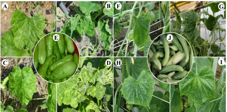
Fig. 1. Viral symptoms of squash in naturally infected fields. A: Vein-banding and systemic mosaic induced by WMV. B: Severe mosaic and malformation induced by ZYMV. C, I: Mildly green mosaic infected by PRSV. D: Severe malformation and stunting caused by multiple infection (CMV+WMV+ZYMV+PRSV). F: Vein-banding and leaf curling caused by co-infection (PRSV+CMV).
G: Saw-edged and/or aster-leaf induced by triple infection (PRSV+WMV+ZYMV). H: Systemic green mosaic and vein-banding induced by co-infection (ZYMV+WMV). E, J: fruits of field-infected squash and cucumber are often prominently affected with malformation, such as knobby overgrown accompanied by color break.
CP-coding region of PRSV isolate were compared with other isolates by using the DNAMAN software package (version 4.02; Lynnon Biosoft, Quebec, Canada). Phy- logenetic tree was constructed by the neighbor-joining algorithm based on the calculations from pairwise nucleo- tide and amino acid sequence distances derived from the multiple alignment formats. The data set was subjected to 1000 bootstrap replicates. The sequence of eight Korean PRSV isolates were compared with ten isolates of PRSV and two isolates each of WMV and ZYMV chosen from the Blast output obtained from the National Center of Biotechnology Information (Table 1).
Results
Incidence of cucurbit-Infecting viruses
From a total of 173 commercial cucurbit cultivating fields distributed over three regions, typical viral symptoms were shown from samples collected in 107 fields. In retarding-culture fields, incidence of symptomatic plants
ranged from 0.3 to 99%. Virus-like symptoms were observed in outdoor-cultured fields as mottle-mosaic, vein banding or clearing and malformation of leaves and fruits (Fig. 1). However, symptomatic plants were not found in forcing-culture fields. Four viruses were detected from 206 out of 235 symptomatic samples. The relative fre- quencies of four viruses detected from cucurbits were described in Table 2 and 3. WMV and ZYMV were the most wide spread viruses in outdoor-cultured fields and retarding-culture fields, reaching 48 and 33%, respectively.
PRSV was detected from 36 samples, representing 12%
of the total samples. Incidence of PRSV in cucurbita- ceous plants was lower than that of WMV and ZYMV.
Single infections accounted to 62.1% for the total virus- infected plants, while double and mixed infection was occurred in 37.9% of them.
Host range and symptoms
The reactions of indicator plants to PRSV and other two potyviruses were summarized in Table 4. The Korean
Table 2. Detection of viral diseases on cucurbits in different cultivating regions during 2001 to 2003
Locality Fieldsb
No. of plants (detected
/tested)
No. of plants detected witha P
R S V
W M V
Z Y M V
C M V
P + W
P + Z
P + C
Z + W
W + C
Z + C
P+
Z+
W Z+
W+
C P+
C+
W P+
Z+
W+
C N
/ Dc
Gyeonggi 43/70 80/93 3 15 23 2 3 2 2 18 1 1 3 5 1 1 13
Gyeongbuk 18/32 55/60 0 29 12 1 3 2 0 7 0 0 1 0 0 0 5
Jeonnam 46/71 71/82 8 33 5 3 2 2 2 13 0 0 1 2 0 0 11
Total 107/173 206/235 11 77 40 6 8 6 4 38 1 1 5 7 1 1 29
a P+W ; mixed infections with PRSV and WMV, P+Z ; mixed infections with PRSV and ZYMV, P+C; mixed infections with PRSV and CMV, W+Z ; mixed infections with WMV and ZYMV2, Z+C; mixed infections with ZYMV+CMV, P+W+Z ; mixed infections with PRSV, WMV, and ZYMV, Z+W+C; mixed infections with ZYMV, WMV, and CMV, P+C+W; mixed infections with PRSV, CMV, and WMV, P+Z+W+C; mixed infections with PRSV, ZYMV, WMV, and CMV,
b Fields with symptomatic plants/fields surveyed
c N/D; not determined
Table 3. Detection of viruses from cucurbit cultivated under different cropping pattern by RT-PCR and ELISA during 2001 to 2003
Cropping pattern No. of samples
No. of plants infected with Single infections
(%)
Mixed Infection
PRSV WMV ZYMV CMV (%)
Forcing Culture 8 0 0 0 0 0 0
Outdoor Culture 139 13 88 65 5 72.8 27.2
Retarding Culture 88 23 55 33 16 51.4 48.6
Total
(%)a 235 36
(12.1)
143 (48.0)
98 (32.9)
21 (7.0)
62.1 37.9
a Percentage over the total number infected with viruses.
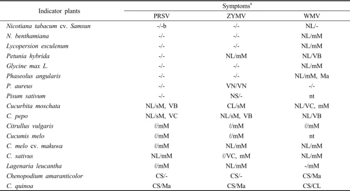
Table 4. Reactions of indicator plants to mechanical inoculation of PRSV and other potyviruses
Indicator plants Symptomsa
PRSV ZYMV WMV
Nicotiana tabacum cv. Samsun -/-b -/- NL/-
N. benthamiana -/- -/- NL/mM
Lycopersion esculenum -/- -/- NL/mM
Petunia hybrida -/- NL/mM NL/VB
Glycine max L. -/- -/- NL/mM
Phaseolus angularis -/- -/- NL/mM, Ma
P. aureus -/- VN/VN -/-
Pisum sativum -/- NS/- nt
Cucurbita moschata NL/sM, VB CL/sM NL/VC, mM
C. pepo NL/sM, VC NL/sM, VB NL/VB
Citrullus vulgaris ℓ/mM ℓ/mM ℓ/mM
Cucumis melo ℓ/mM ℓ/mM nt
C. melo cv. makuwa ℓ/mM NL/mM NL/mM
C. sativus NL/mM ℓ/VC, mM NL/mM
Lagenaria leucantha ℓ/mM NL/mM -/mM
Chenopodium amaranticolor CS/- CS/- CS/Ma
C. quinoa CS/Ma CS/Ma CS/CL
a The abbreviations of the symptoms are as follows: NL ; necrotic lesion, NS ; necrotic spot, CL ; chlorotic lesion, CS ; Chlorotic spot, mM ; mild mosaic, Ma ; malformation, VN; vein necrosis, VC ; vein clearing, VB ; vein banding, ℓ ; latent, nt ; not tested
b Inoculated leaves / upper leaves
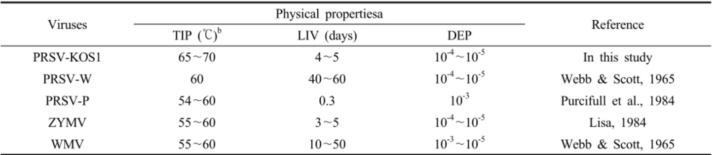
Table 5. Physical properties of PRSV-KOS1 and other potyviruses infecting cucurbitsa
Viruses Physical propertiesa
Reference
TIP (℃)b LIV (days) DEP
PRSV-KOS1 65~70 4~5 10-4~10-5 In this study
PRSV-W 60 40~60 10-4~10-5 Webb & Scott, 1965
PRSV-P 54~60 0.3 10-3 Purcifull et al., 1984
ZYMV 55~60 3~5 10-4~10-5 Lisa, 1984
WMV 55~60 10~50 10-3~10-5 Webb & Scott, 1965
a The properties of PRSV-KOS1 were determined in zucchini (Cucurbita pepo).
TIP and DEP were determined by methods described by Webb & Scott, 1965.
b TIP ; thermal inactivation point, LIV ; longevity in vitro, DEP ; dilution end point PRSV isolates infected all seven species of Cucurbitaceae
with manual inoculations. The 28 other species representing 13 non-cucurbitaceous plant families inoculated with PRSV did not show any systemic infection (data not shown). All isolates of PRSV, ZYMV and WMV produced local lesions on Chenopodium quinoa and C. amaranticolor and induced systemic symptoms on most cucurbitaceous plants. All symptoms developed within 14 days after inoculation. The Korean PRSV isolate induced severe systemic mosaic, vein clearing and vein banding symptoms both in Cucurbita moschata and C. pepo and systemic mild-mosaic symptom in Citrullus vulgaris, Cucumis melo, C. sativus and Lagenaria leucantha. Vein banding and clearing of true leaves were observed from three indicator plants of squash, Cucurbita moschata and C. pepo 10 days after inoculation with PRSV followed by severe mosaic with malformation and distortion noted on the upper leaves.
Physical properties of PRSV
Zucchini (C. pepo) was used as the source and test plants to determine the physical properties of the Korean PRSV isolate. Sodium phosphate buffer (0.01 M, pH 7.2) was used to dilute infectious plant sap. It was observed that the thermal inactivation point (TIP) of PRSV was between 65 and 70℃/10 minutes. Tolerance to dilution (dilution end point; DEP) was 10-4 to 10-5 and longevity in vitro (LIV) was less than five days at room tempera- ture. Table 5 summarized the physical properties of PRSV and other potyviruses.
Morphology of PRSV particles
The viral particles of 235 cucurbitaceae crop samples collected were observed using transmission electron micro- scope. Two hundred six samples out of these 235 specimens, showed filamentous viral-particles. Electron microscopy showed that PRSV-KOS1 was flexuous, approximately 780 nm in length and 12 nm in width, which are typical features of potyviruses (photograph not shown).
Ultrastructure of PRSV-infected squash leaf tissue All three types of inclusion bodies were observed from PRSV-infected squash leaves. Crystalline inclusions were observed in the nuclei, while cylindrical inclusions (pin- wheels and scrolls) in the cytoplasm (Fig. 2). Another type of inclusion, which in some sections appeared as two layers or bands alternating with a layer of circles, was seen mainly in the cytoplasm, and rarely in the nuclei.
Sequence diversity in PRSV worldwide
The open reading frame corresponding to the CP cistron of PRSV was composed of 864 nucleotides, which encode 287 amino acid residues (Fig. 3). The base composition of this viral RNA indicated high adenine content at 35.4%, followed by uracil (23.7%), guanine (22.6%) and cytosine (18.3%). This structure was similar to those of the other PRSV isolates reported by Yeh et al. (1992). The complete nucleotide and amino acid sequences of the PRSV CP were compared with other PRSV isolates resisterd in GenBank.
The homology percent of the nucleotide sequences showed
Fig. 2. Ultrastructure of the leaf tissues of Cucurbit pepo (Zucchini) infected with PRSV-KOS1. Pinwheel and cylindrical inclusions are shown. Chl: chloroplast, Va: vacuole, LI: laminated inclusion, PW: pinwheel, CI: cylindrical inclusion (scroll shape)
Fig. 3. Nucleotide sequences of the coat protein gene of PRSV-KOS1 isolate. The predicted amino acid sequence is presented below the nucleotide sequence. Asterisk (*) indicates the stop triplet codon.
that of PRSV-KOS1 was the closest to PRSV-TAIW, showing 97.2% homology. The nucleotide sequence homo- logy of the CP gene among PRSV isolates was ranged from 87.6 to 97.5% range, however they showed 51.9 to
52.6% homology with ZYMV and WMV. The multiple amino acid sequence alignment of the CP of 18 PRSV isolates including 8 Korean isolates showed that C-terminal three quarters of the CP was highly conserved.
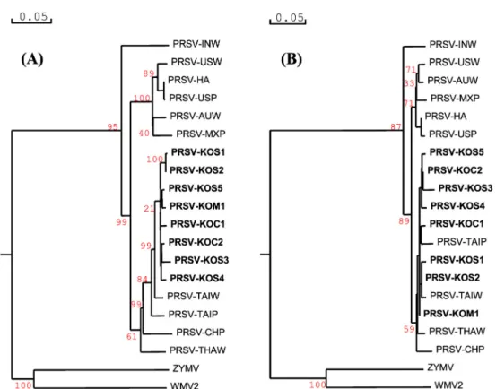
The genetic distances of nucleotide and amino acid sequences for all PRSV isolates were used to generate phylogenetic tree with two related potyvirus, ZYMV and WMV, as the out-group. As shown in Fig. 4, the Korean PRSV isolate, clustered with Taiwan isolate (PRSV-TAIW) into PRSV watermelon strain (PRSV-W biotype) at nucleo- tide level, and clustered with Taiwan, Thailand, and China isolates (PRSV-TAIP, TAIW, THAW, CHP) at amino acid level. The Korean PRSV isolates also formed a sister cluster with the American and Australian isolates, PRSV-USW, USP, HA, AUW, and MXP but not with the Indian isolate, PRSV-INW which is distantly apart.
The phylogenetic tree showed that PRSV and other poty- viruses (ZYMV and WMV) clusters were distinct group but genetic relationships among the PRSV isolates was high on the basis of their nucleotide and amino acid sequences.
Discussion
The study revealed that WMV and ZYMV are the most
Fig. 4. Phylogenetic analyses of CP gene present and other worldwide isolates of PRSV at nucleotide level (A) and amino acid level (B) by Neighbor-joining algorithm.
widely spread viruses in cucurbit cultivated under outdoor culture and retarding culture cropping type in Korea.
PRSV was also detected in the fields, but its presence on cucurbit plants was less than the other potyviruses WMV and ZYMV. Single infection of PRSV, WMV, ZYMV and CMV manifested a viral incidence of 12.1, 48.0, 32.9 and 7.0 percents, respectively. In the case of co-infection, PRSV+WMV, PRSV+ZYMV, PRSV+CMV, and ZYMV+
WMV manifested the infection rate of 3.4, 2.5, 1.7 and 16.0 percents, respectively. The incidence of single and multiple infections in outdoor culture were 72.8 and 27.2%, while single and mixed infections in retarding culture were 51.4 and 48.6%, respectively. Towards the end of the growing season, virus incidence became high in most fields. Various levels of infection in the plants at the early growth stage caused by a particular virus type among the different regions could be attributed to the factors that triggered the onset of infection (Luis-Arteaga et al., 1998;
Abou-Jawdah et al., 2000; Grafton-Cardwell et al., 1996;
Yuki et al., 2000).
In other countries, PRSV-W was reported to infect cucumber, melon, squash, watermelon and other cultivated cucurbits at economic level. In Korea, only ZYMV and WMV infected squash, cucumber and watermelon but not PRSV (Ko et al., 2006; Lee, 1981). The biological, physical, cytological and molecular properties of PRSV isolated from cucurbits in Korea proved that PRSV is distinct from the other two potyviruses, ZYMV and WMV commonly occurring on cucurbit crops worldwide. In the biological tests, PRSV-KOS1 caused systemic mosaic and veinal symptoms in cucurbit species and produced local lesion on Chenopodium quinoa and C. amaranticolar as described in previous studies (Purcifull & Hiebert, 1979; Pucifull et al., 1998; Yeh et al., 1984; Milne & Grogen, 1969; Grogen et al., 1959; Webb & Scott, 1965; Anderson, 1953). Based on host reactions, PRSV-KOS1 only infected Cucurbitaceae and Chenopodiaceae, while WMV formerly classified as PRSV can systemically infect Nicotiana benthamiana of the Solanaceae family and shows several indicator plants in Legumiosae family (Jin et al., 2003). These results
indicate that the host ranges of PRSV-W are Cucurbitaceae and Chenopodiaceae families while N. benthamiana can be used to distinguish PRSV-W from WMV (Yeh et al., 1984; Purcifull & Hiebert, 1979).
Majority of the physical properties of Korean PRSV isolates were closely related to those of PRSV as defined by Webb & Scott (1965). The viral particle of PRSV was flexuous filament-type, which is typical of potyvirus. The three types of inclusions in zucchini tissues as revealed by electron microscopy conformed to those described by Mejia et al. (1985), Purcifull et al. (1998) and Zettler et al. (1968). These are the crystalline inclusions in the nuclei, the cylindrical inclusions (pinwheels and scrolls) in the cytoplasm and the alternating bands with circles layers in the cytoplasm.
The RT-PCR procedure was able to identify biotype W of PRSV with leaves and fruits infected squash samples collected in the field. Amplification of a 648 bp product of the CP gene using PRS-C20 primer complementary to the UTR for reverse transcription and PRS-N60 primer provided a convenient method for differentiating PRSV from other viruses that infect cucurbits. The optimum annealing temperature for this PRSV specific primer sets was 58℃ and the temperature is beyond the range of 57 to 59℃, reduced detection sensitively and included negative results. The RT-PCR assay has been effectively used in the study to diagnose viral diseases as reported by Henson
& French (1993), Thomson et al. (1995). Langeveld et al.
(1991) also demonstrated that RT-PCR using degenerated oligo-nucleotide primers designed from conserved sequences was useful in identifying potyviruses. So far, ELISA test that is based on serological relationship has been employed to detect viruses, but often fail because of low titer and non-specific reaction. In this study, RT-PCR assays showed higher sensitivity than other diagnostic tools such as ELISA and symptomatic detection methods.
Shukla & Ward (1988) reported homology in amino acid sequence of CP as a basis for identification and classification of the potyvirus group. According to their hypothesis, 38 to 71% sequence homology between two viruses indicates that they are distinct species, while 90 to 99% sequence homology indicates that they are related
strains. The study had demonstrated that the CP gene of PRSV-KOS1 showed 88.6~97.3% nucleotide homologies and 95.1~99.3% amino acid homologies with other PRSV isolates worldwide. However, PRSV-KOS1 was differen- tiated from ZYMV and WMV at a range of 51.9 to 52.6% homologies in amino acid levels. Moreover, the phylogenetic tree indicated that the Korean PRSV isolates belongs to South-East Asian PRSV group as well as the TAIP and CHP isolates. These isolates belong to the American and Australian PRSV subgroup. PRSV isolates showed low relationships with the ZYMV and WMV.
Based on the biological tests, electron microscopy, RT-PCR and sequence analyses of the CP cistron, the Korean PRSV isolates are clearly identified as PRSV W type of the potyvirus family as shown by its homology to PRSV isolates worldwide based on the partial biochemical and molecular characterizations done. The study is the first attempt to investigate and characterize the incidence of PRSV in Korea. Further study is needed to assess the extent of viral infection on commercially important cucurbit crops as squash, cucumber, melon and watermelon. The molecular characterization and ecological distribution of PRSV is also worth pursuing.
Literature Cited
Abou-Jawdah, Y., Sobh, H., El-Zammar, S., Fayyad, A. and Lecoq, H. 2000. Incidence and management of virus disease of cucurbits in Lebanon. Crop Protection 19:217~224.
Anderson, C. W. 1953. Two watermelon mosaic virus strains from Central Florida. Phytopathology 44:198~202.
Choi, H. S., Ko, S. J., Kim, M. K., Park, J. W., Lee, S. H., Kim, K. H., Hassan, K. W., Choi, J. K. and Takanami, Y. 2005.
Characteristics of Potato virus Y isolated from paprika in korea. Plant Pathol. J. 21:349~354.
Gonsalves, D. and M. Ishii. 1980. Purification and serology of papaya ringspot virus. Phytopathology 70:1028~1032.
Grafton-Cardwell, E. E., Perring, T. M., Smith, R. F., Valencia, J. and Farrar, C. A. 1996. Occurrence of mosaic viruses in melons in the Central Valley of California. Plant Dis.
80:1092~1097.
Henson, J. M. and French, R. 1993. The polymerase chain reaction and plant disease diagnosis. Annu. Rev. Phytopathol.
31:81~109.
Jain, RK., Pappu, HR., Varma, A. and Ram, RD. 1998. Molecular characterisation of papaya ringspot potyvirus isolates from India. Annals of Applied Biology 132(2):413~425.
Jin, T. S., Lee, S. H., Park, J. W., Choi, H. S., Kim, S. M., Shin, D. B., Cheon, J. U. and Cha, B. J. 2003. Identification of Papaya ringspot potyvirus type W infecting squash in Korea. Plant Pathol. J. 19:339 (abstract)
Ko, S. J., Lee, Y. H., Cha, K. H., Lee, S. H., Choi, H. S., Choi, Y. S., Lim, G. C. and Kim, K. H. 2006. Incidence and distribution of virus diseases on cucumber in jeonnam province during 1999-2002. Plant Pathol. J. 22(2):147~
151.
Langeveld, S. A., Dore, J. M., Memelink, J., Derks, A. F., van der Vlugt, C. I., Asjes, C. J. and Bol, J. F. 1991. Identifi- cation of potyviruses using the polymerase chain reaction with degenerate primers. J. Gen. Virol. 72:1531~1541.
Lee, S. H. and Lee, K. W. 1981. Incidence of watermelon mosaic virus in cucurbits. Korean J. Plant Prot. 20(4):191~
195.
Lisa, V. and Lecoq, H. 1984. Zucchini yellow mosaic virus.
CMI/AAB Descriptions of Plant Viruses No. 282. Walling- ford, UK: CAB International.
Luis-Arteaga, M., Alvarez, J. M., Alonso-Prados, J. L., Garcia- Arenal, F., Lavina, A., Batlle, A. and Moriones, E. 1998.
Occurrence, distribution, and relative incidence of mosaic viruses infecting field-grown melon in Spain. Plant Dis.
82:979~982.
Mejia M. V. G., Hiebert E., Purcifull, D. E, Thornbury, D. W.
and Pirone, T. P. 1985. Identification of potyviral amorphous inclusion as a nonstructural, virus-specific protein related to helper component. Virology 142:34~43.
Milne, K. S. and Grogan, R. G. 1969. Characterization of water- melon mosaic strains by serology and other properties.
Phytopathology 59:809~818.
Purcifull, D. E. and E. Hiebert. 1979. Serological distinction of watermelon mosaic virus. Phytopathology 69:112~116.
Purcifull, D. E., Hiebert, E., Petersen, M. A., Simone, G. W., Kucharek, T. A., Gooh, M. D., Crawford, E. E, Beckham, K. A. and De Sa, P. B. 1998. Partial characterization of a
distinct potyvirus isolated from watermelon in Florida. Plant Dis. 82:1386~1390.
Purcifull, D., Edwardson, J. and Hiebert, E. 1984. Watermelon mosaic virus 2. CMI/AAB Descriptions of Plant Viruses No. 293. Wallingford, UK: CAB International.
Purcifull, D., Edwardson, J., Hiebert, E. and Gonsalves, D.
1984. Papaya ringspot virus. CMI/AAB Descriptions of Plant Viruses No. 292. Wallingford, UK: CAB International.
Quiot-Douine, L., Lecoq, H., Quiot, J. B., Pitrat, M. and Labonne, G. 1990. Serological and biological variability of virus isolates related to strains of papaya ringspot virus.
Phytopathology 80:256~263.
Shukla, D. D. and Ward, C. W. 1988. Amino acid sequence homology of coat proteins as a basis for identification and classification of the potyvirus group. J. Gen. Virol. 69:2703~
2710.
Thomson, K. G., Dietzgen, R. G., Gibbs, A. J., Tang, Y. C., Liesak, W. Teakle, D. S. and Stackebrand, E. 1995. Identifi- cation of zucchini yellow mosaic potyvirus by RT-PCR and analysis of sequence variability. J. Virol. Methods 55:83~
96.
Wan, S. -H. and Conover, R. A. 1983. Incidence and distribution of papaya viruses in southern Florida. Plant Dis. 67:353~
356.
Webb, R. E. and Scott, H. A. 1965. Isolation and identification of watermelon mosaic virus 1 and 2. Phytopathology 55:
895~900.
Yeh, S. -D., Jan, FJ., Chiang CH., Doong, TJ., Chen, MC., Chung PH. and Bau HJ. 1992. Complete nucleotide sequence and genetic organization of papaya ringspot virus RNA. J.
Gen. Virol. 73:2531~2541.
Yuki, VA., Rezende, JAM., Kitajima, EW., Barroso, PAV., Kuniyuki, H., Groppo, GA. and Pavan, MA. 2000. Occurrence, distribution, and relative incidence of five viruses infecting cucurbits in the state of Sao Paulo, Brazil. Plant Dis.
84(5):516~520.
Zettler, F. W., Edwardson, J. R. and Purcifull, D. E. 1968.
Ultramicroscopic differences in inclusions of papaya mosaic virus and papaya ringspot virus correlated with differential aphid transmission. Phytopathology 58:332~335.
박과작물에 발생하는 파파야원형반점바이러스의 발생 보고
진태성 ・ 김상목1・ 고석주2・ 이수헌 ・ 최홍수 ・ 박진우 ・ 차병진3
국립농업과학원 농업미생물과, 1국립식물검역원 중부격리재배관리소, 2전남농업기술원 친환경연구소, 3충북대학교 식물의학과
요 약 안성에서 모자이크와 주름증상을 보이는 호박으로부터 사상형 바이러스가 분리되었으며, 생물학적 특성과 전자현 미경 검정, RT-PCR 검정에 의해 파파야원형반점바이러스(Papaya ringspot virus) 수박계통(PRSV-W)으로 동정되었다.
PRSV-W의 기주범위는 박과작물과 명아주과작물에 한정되었고 감수성 기주인 오이, 호박, 수박 등에는 녹색모자이크, 기형, 주름 등의 증상을 나타냈지만 명아주과의 기주에는 국부병징만을 나타냈다. 2001년에서 2003년에 걸쳐, 경기, 경북, 전남에 서 다양한 작형의 박과작물이 재배되는 173지역에서 박과작물의 주요 바이러스인 PRSV, 수박모자이크바이러스(WMV), 쥬키니황화모자이크바이러스(ZYMV)와 오이모자이크바이러스(CMV) 등 4종 바이러스의 발생상황을 조사하였다. 173지역 중 107지역에서 수집한 시료로부터 바이러스 병징이 관찰되었으며 RT-PCR 검정 결과, 235점의 시료 중 206시료에서 3종 의 바이러스가 검출되었다. 검출빈도는 WMV가 48%, ZYMV가 33%였으며, PRSV는 12%였다. 8종 PRSV 시료의 핵산 과 외피단백질 아미노산을 분석하였다. 분석된 결과를 전세계의 다른 PRSV 계통과 유사도를 비교한 결과, 핵산은 88.6~
97.3%, 아미노산은 95.1~99.3%로 조사되었다. 이들 분리주의 유연관계를 분석한 결과, PRSV-W는 남동 아시아 계통과 근연종으로 판명되었다.
색인어 박과작물, 포티바이러스, 파파야원형반점바이러스, 수박모자이크바이러스, 쥬키니황화모자이크바이러스