Machilus Thunbergii Water Extract Induces Cytotoxic Effect against Human Acute Jurkat T Lymphoma
Min Hwan Kim3 and Jong-Hwan Lee1,2,3*
1Biomedical Engineering and Biotechnology Major, Division of Applied Bioengineering, College of Engineering, Dong-eui University, Busan 614-714, Korea
2Department of Biotechnology and Bioengineering, College of Engineering, Dong-eui University, Busan 614-714, Korea
3Department of Smart Bio-Health, Dong-eui University, Busan 614-714, Korea Received April 24, 2017 /Revised June 2, 2017 /Accepted June 8, 2017
To understand the cytotoxic activity of Machilus thunbergii, which has been used as a traditional ori- ental medicine, the mechanism underlying the cytotoxic effect of its extract on human acute Jurkat T cells was investigated. The methanol extract of roots (3 kg) of M. thunbergii was evaporated, dis- solved in, and then extracted by water. The water-extracted active substance was designated MTWE.
When Jurkat T cells were treated with MTWE at concentrations of 0, 25, 50, and 100 μg/ml, the apop- totic phenomenon of cells accompanying several subsequent biochemical reactions, such as mitochon- drial cytochrome c release, activation of caspase-3, and ICAD degradation, was detected in the Jurkat T cells. Moverover. the expression of Bcl-xL, which is a suppressor for mitochondrial cytochrome c release pathway, was reduced in the Jurkat T cells. As DUSP6, a growth suppressor of cancer cells, ranged from 0, 25, 50, 100 μg/ml of MTWE, the expression level was elevated in the Jurkat T cells.
The apoptotic morphological change of the nuclei was observed by DAPI staining. Although the po- tential involvement of the other factors and DUSP6 is currently being investigated in more detail, these findings support the notion that MTWE is able to achieve the apoptosis of Jurkat T cells, and it seems that MTWE is useful as a method of evaluating a chemotherapeutic agent or tonic materials for human acute leukemia.
Key words : Apoptosis, cytotoxicity, Jurkat T cells, Machilus thunbergii
*Corresponding author
*Tel : +82-51-890-2280, Fax : +82-505-182-6897
*E-mail : [email protected]
This is an Open-Access article distributed under the terms of the Creative Commons Attribution Non-Commercial License (http://creativecommons.org/licenses/by-nc/3.0) which permits unrestricted non-commercial use, distribution, and reproduction in any medium, provided the original work is properly cited.
Journal of Life Science 2017 Vol. 27. No. 8. 951~957 DOI : https://doi.org/10.5352/JLS.2017.27.8.951
Introduction
Cancer is a diverse collection of life-threatening diseases that is caused by abnormal and invasive cell proliferation.
For a normal cell to evolve into a disease-causing cancer is a lengthy process. Most emerging cancers are likely to be eliminated by the immune system before they are detectable or cause any symptoms. Only a minority of cancers defeat the immune system and progress to cause the diseases that are so feared. Cancers of immune system cells are known as leukemias when they involve circulating cells. T cell acute lymphoblastic leukemia (T-ALL) is an aggressive hemato- logical cancer that is mainly diagnosed in children and arises from the malignant transformation of T cell progenitors [17].
In treating leukemias, physicians resort to surgery, radiation, bone marrow transplantation and cytotoxic drugs, some- times referred to slash, burn, and poison. Although these treatments give remission of cure to some patients, more of- ten they are limited by the incomplete elimination of cancer cells and the deleterious side-effects such as reduced in- tellectual capacity, osteonecrosis and growth deficiencies, in- fertility, and an increased risk of secondary tumor develop- ment later in life. Therefore, integration of novel targeted therapies into contemporary T-ALL treatment protocols will be required to increase the quality of life for pediatric T-ALL survivors [2]. Natural products have played a significant role in drug discovery and specifically in the development of new tonic agents; more than 79.8% of the anticancer drugs introduced from 1981 to 2008 were natural products, semi- synthetic analogs, or synthetic compounds based on natu- ral-product pharmacophores [3].
Machilus thunbergii belongs to the Lauraceae family and is one of the most commonly distributed in Asia. The bark of M. thunbergii SIEB. et ZUCC. (Lauraceae) has been used as a folk medicine for the treatment of leg edema, abdominal - Note -
pain and abdominal in Korea [6]. It has been reported that compounds such lignans, alkaloids, flavonoids, butanolides, essential oils, and machilin derived from M. thunbergii have marked anti-oxidative activity with hepatoprotective, an- ti-bacterial acivities [5, 19, 21], anti-inflammatory activities in RAW 264.7 cells [18] and neuroprotective activity against glutamate-induced neurotoxicity [13], in vitro osteoblast dif- ferentiation [11]. Magnolol and honokiol, a phenolic compo- nent of water extracts of the stem bark of Magnolia officinalis has multiple pharmacological effects [8, 10]. Moreover, an ingredients of this medicinal herb has been reported to have anti-oncogenic properties against A549 cells, HeLa, B16F10 metastatic melanoma cells, F9 mice teratocarcinoma cells and human promyelocytic leukemia HL-60 cells [6, 16]. Targeting specific molecules is a promising cancer treatment because certain types of cancer cells are dependent on specific oncogenes. Although there have been studies on the an- ti-cancer activity of M. thunbergii, however, its activity in T acute lymphoblastic leukemia (T-ALL) is still unclear.
Therefore, in the present study, we investigated a role for the anti-cancer activity of M. thunbergii extract using bio- chemical assay in the human acute leukemia, Jurkat T cells of T-ALL cell lines.
Materials and Methods
Sample preparation from M. thunbergii
M. thunbergii was purchased from Hyun-dae Pharmaco- logical Company (Busan, Korea). A sample has been de- posited in the author’s laboratory. The dried roots (3 kg) from M. thunbergii were extracted with 100% methanol. The methanol extract was evaporated (295.2 g), resuspended in water, and then freeze-dryed by freezer. The cytotoxic com- pound was designated as MTWE.
Cell culture
The human T cell lymphoma line, Jurkat T E6.1 cells were obtained from Albert A Nordin (Gerontology Research Center, NIA/NIH, Baltimore, MD, USA). The Jurkat T E6.1 cells were grown in RPMI1640 medium (Sigma, USA) sup- plemented with 10% fetal bovine serum (FBS, Invitrogen, USA), 100 μg/ml streptomycin, and 100 U/ml penicillin.
Male Balb/c mice were purchased from Samtako, Inc. (Osan, Korea). Mice used in all experiments were 12 weeks old.
These mice were housed in a specific pathogen-free facility with appropriate temperature and humidity and allowed
free access to food and water. The mice for this study (DEU-R2013-002) were approved by the Institutional Animal Care and Use Committee at Dong-eui University. Mice were killed by cervical dislocation, and spleens were aseptically removed and stripped of fat. Single-cell suspensions were obtained by grinding the spleens with a syringe plunger against a fine steel mesh. Erythrocytes were lysed with am- monium chloride haemolysis buffer (0.8% NH4Cl with 0.1 mM EDTA) and then washed twice in complete RPMI-1640 Medium. Splenocytes (5×10⁵ cells/well) were plated in tripli- cate in 96-well culture plates and cultured in RPMI-1640 Medium supplemented with 5% foetal bovine serum (FBS) at 37°C in a humidified 5% CO2 incubator.
Cytotoxicity assay with Jurkat E6.1 T cell
Cytotoxicity of MTWE against Jurkat T cell was de- termined by Cell Titer 96 AQueous One Solution Cell Proliferation Assay (MTS) (Promega, USA). Briefly, the cells (5×10⁵) were added to a serial dilution of the MTWE in 96-well plates. After 24 hr, 5% MTS was added in a 96-well plate and incubated for 2 hr before reading at a wavelength of 490 nm by a microplate reader (Bio-Rad, USA). Jurkat T cells (5×10⁵ cells/well) were treated with phytohemag- glutinin (PHA, 12.5 μg/ml, Sigma Aldrich, St. Louis, MO, USA) to stimulate T cells cytotoxicity in the absence or pres- ence of MTWE and incubated in 96-well culture plates (Corning, NY, USA) for 24 hr.
Immunoblotting
Cells pretreated with or without MTWE were lysed with lysis buffer [137 mM NaCl, 15 mM EDTA, 1 mM sodium orthovanadate, 15 mM MgCl2 , 0.1% Triton X-100, 25 mM 3-N-morpholino-propanesulfonic acid (MOPS, Sigma, USA), and 2.5 μg/ml proteinase inhibitor E-64, pH 7.2]. Protein concentration was measured by BCA (Peirce, USA). After 12% SDS-PAGE, the samples were transferred onto im- mobilon-P membranes. The membranes were soaked in a blocking solution (5% skim milk and 0.2% Tween 20-PBS) for 1 hr, and then incubated with anti-cytochrome c (Phar- mingen), anti Bcl-xL, anti-caspase-3, anti-DUSP6, anti-ICAD and anti-β-actin (Santa Cruz Biotechnology, Inc., USA). After being washed with Tween 20-PBS, membranes were in- cubated with appropriate HRP-conjugated secondary anti- bodies for 1 hr. Specific bands were visualized by an ECL method (ECL+Amersham Biosciences, Arlington Heights, IL, USA). The relative amount of cytochrome c, caspase-3,
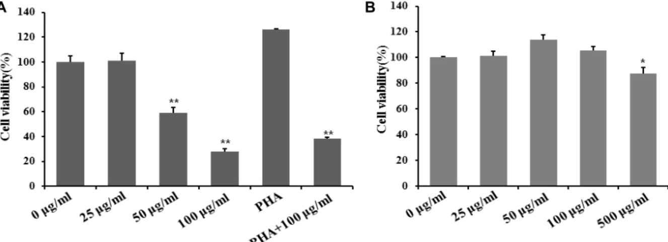
A B
Fig. 1. Effect of MTWE on cell viability in Jurkat T cells and splenocytes. MTWE shows cytotoxicity dose-dependently in Jurkat T cells. Continuously growing Jurkat T cells (5×10⁵ cells/well) (A) and splenocytes (5×10⁵ cells/well) (B) were incubated with indicated concentrations of MTWE in a 96-well plate for 24 hr and further incubated with MTS for 4 hr. Jurkat T cells (5×10⁵ cells/well) were treated with PHA (12.5 ug/ml) in the absence or presence of MTWE and incubated in 96-well culture plates for 24 hr. The cells were sequentially processed to assess the colored formazan crystal produced from MTS as an index of cell viability. Data were given as means of values from three independent assays. Level of significance was identified statistically (*, p<0.05; **, p<0.01) using ANOVA test.
Bcl-xL, DUSP6 and ICAD compared with that in the control was calculated by measuring the band density of cyto- chrome c, caspase-3, Bcl-xL, DUSP6 and ICAD and normal- ized to β-actin density.
DAPI staining
Cells were washed with PBS and were treated with 95%
ethanol for 1hr at 4℃ as a fixative. After washing the sample, RNase (12.5 μg) in 1.12% sodium citrate buffer (pH 8.45) was added at 37°C for 30 min. MTWE-treated jurkat T cells were stained with DAPI (4 μg/ml) in 100% methanol for 15 min at 37°C and observed under the microscope with ultraviolet (UV) excitation at 300-500 nm (Microphot-FX, Nikon, Tokyo, Japan). Cells with nuclei that contained clear condensed chromatin or cells with fragmented nuclei were scored as apoptosis.
Statistical analysis
For statistical analysis, the ex vivo and in vitro assays were performed independently at least three times. The P values were determined through one-way ANOVA with less than 0.05 considered to be statistically significant. The P-val- ues are represented as an asterisk (*) or (**).
Results and Discussion
MTWE has the cytotoxicity against Jurkat T cells
We tested cell apoptosis in Jurkat T cell stimulated with MTWE. Cells (5×10⁵) treated at concentration ranging 0, 25, 50, 100 μg/ml MTWE were incubated for 24 hr and carried out with MTS assay. The cells (5×10⁵) were incubated with MTWE solution containing 0, 25, 50, 100 μg/ml concen- tration in 96-well plate for 24 hr. The MTWE (25.0 μg/ml) treated-cells showed the cell viability about 95% and the MTWE (100.0 μg/ml) treated-cells showed the cell viability less than 50% (Fig. 1A). To see whether MTWE suppresses T cell proliferation, Jurkat T cells were treated with PHA in the presence or absence of MTWE. As shown in Fig. 1A, MTWE inhibited T cell proliferation. Splenocytes (5×10⁵) were incubated with MTWE containing 0, 25, 50, 100, 500 μg/ml concentration for normal cell cytotoxicity for 24 hr.
The MTWE (25, 50, 100 μg/ml) treated-splenocytes showed the cell viability similar to control and the MTWE (500 μg/
ml) treated-cells showed the cell viability less than 87%(Fig.
1B). This result suggested that no cytotoxicity showed at concentration ranged from 0, 25, 50, 100 μg/ml MTWE. This means that MTWE induces the apoptosis of Jurkat T cells.
Although it has been reported that M. thunbergii has been used for tonic materials or fork remedies in several countries including Korea [6], but little is known that biochemicals in M. thunbergii shows potential cytotoxicity against leuke- mic cells, and especially signaling pathways during apopto- sis is still unknown. In this research, we have first demon- strated that a partial extracted phytochemical ingredient des-
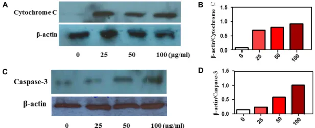
A B
C D
Fig. 2. Effect of MTWE on cytochrome C and caspase-3 expression in Jurkat T cells. MTWE induced-cell death is through mitochondrial cytochrome c release. (A, C) The cells (5×10⁵) were incubated with indicated concentrations of MTWE for 24 hr and prepared for the cell lysates. Equivalent amounts of cell lysates were electrophoresed on 12% SDS polyacrylamide gels and electro- transferred to Immobilon-P membrane. Western blot analysis was performed as described in materials and methods using the ECL Western blot detection system. Cytochrome c (A), caspase-3 activation (C), and β-actin. (B, D) The relative intensity compared with the control level was normalized by the amount of β-actin (histograms).
ignated as MTWE from M. thunbergii induces human acute Jurkat T cell apoptosis.
Apoptosis pathway triggered by MTWE is involved in mitochondrial cytochrome c release and caspase- 3 activation
We investigated the apoptosis mechanism by utilizing dose-dependent treated Jurkat T cells stimulated by MTWE to find molecular mediators. Significant differences from an- tibody reaction related to mitochondrial cytochrome c re- lease pathway [14] were detected in control cell compared to MTWE-treated cells. Release level of mitochondrial cyto- chrome c to cytosol was increased in cells incubated with 0, 25, 50, 100 μg/ml of MTWE (Fig. 2A, Fig. 2B). Subsequently, released cytochrome c is related to switching caspase 3, one of downstream partners of cytochrome c related to apoptotic pathway. The amount of caspase 3 was increased in the range of 0, 25, 50, 100 μg/ml of MTWE in Jurkat T cells (Fig. 2C, Fig. 2D). In the present results, in Jurkat T cells exposed to MTWE, mitochondrial cytochrome c release and activation of caspase-3 were detected. Several reports sug- gest that caspase-3 can be activated through active caspase-8 in turn can cleave Bid, leading to cytochrome c release from mitochondria [12] and setting up a self-amplification loop to amplify caspase-9. Anyway, these findings support the notion that apoptotic signaling of MTWE in Jurkat T cell is regulated by mitochondrial cytochrome c release pathway.
MTWE decreases protein levels of B-cell lymphoma- extra large (Bcl-xL)
Anti-apoptotic Bcl-2 protein family including Bcl-2 itself and Bcl-xL acts as key regulators in the intrinsic or mi- tochondrial apoptosis pathway [22]. To determine the mech- anism involved, we examined the effect of the MTWE on Bcl-2 family anti-apoptotic signaling molecule. MTWE re- duced levels of the pro-survival protein Bcl-xL under dose-dependent concentration (Fig. 3). Major function of Bcl-xL is known to suppress mitochondrial cytochrome c re- lease [7, 20]. Therefore, these experiments suggest that MTWE is involved in facilitation the activation of mitochon- dria-dependent death-signaling pathway in Jurkat T cells, and subsequent cascade events.
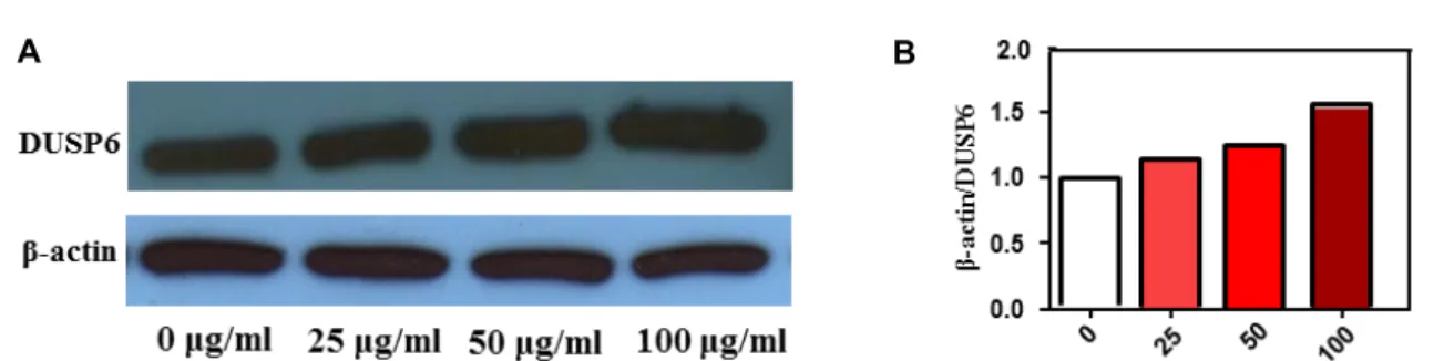
DUSP6 is involved in apoptosis pathway triggered by MTWE
Ectopic expression of DUSP6/MKP-3, a pivotal negative feedback regulator of the RAS-ERK pathway, in either pan- creatic or lung cancer cells resulted in the suppression of cell growth and apoptosis related to restrain oncogenic ERK signalling [4, 15]. Thus, we performed western blotting for DUSP6. As DUSP6 ranged from 0, 25, 50, 100 μg/ml of MTWE, the expression level was elevated in Jurkat T cells (Fig. 4). DUSP6 expression levels were found to be weaker in most lung cancer cell lines, and DUSP6 restoration sup- pressed cellular growth [15]. However, the potential involve- ment between the other factors and DUSP6 is currently be-
A B
Fig. 3. Effect of MTWE on Bcl-xL expression in Jurkat T cells. MTWE facilitates the attenuation of Bcl-xL expression in Jurkat T cells. (A) Jurkat T cells (5×10⁵ cells) were incubated with indicated concentrations of MTWE in microplate for 20 hr. The cells were further incubated with DMSO for 4 hr to solve the colored formazan crystal produced from MTS as an index of cell viability. (B) The relative intensity compared with the control level was normalized by the amount of β-actin (histograms).
A B
Fig. 4. Effect of MTWE on DUSP 6 expression in Jurkat T cells. MTWE facilitates the enhancement of DUSP6 expression in Jurkat T cells. (A) The cells (5×10⁵) were incubated with indicated concentrations of MTWE for 24 hr and prepared for the cell lysates. Equivalent amounts of cell lysates were electrophoresed on 12% SDS polyacrylamide gels and electrotransferred to Immobilon-P membrane. Western blot analysis was performed as described in materials and methods using the ECL Western blot detection system. (B) The relative intensity compared with the control level was normalized by the amount of β-actin (histograms).
ing investigated in detail. Anyway, these findings support the notion that MTWE is able to carry out apoptosis of Jurkat T cells and it seems that MTWE is used as the evaluation for chemotherapeutic agent. Collectively, these results dem- onstrate that MTWE, a partially purified constituent from M. thunbergii, induces apoptosis of Jurkat T cells via mi- tochondria dependent pathway by release of mitochondria cytochrome c, and activation of caspase-3. These things will be helpful and useful for evaluation of its chemotherapeutic potency and tonic material.
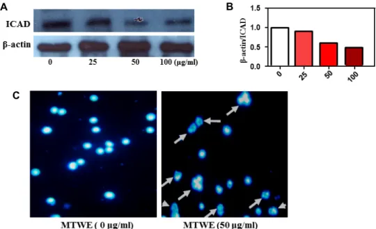
MTWE induces apoptosis of jurkat T cells
Caspases, a family of cysteine proteases, contribute to a diverse range of functions in cell including apoptosis.
Caspase 3 in particular is a key factor in canonical apoptotic signaling promoting both the cytosolic and nuclear alter- ations required for cellular disassembly [9]. Caspase 3 acti- vates caspase-activated DNase (CAD) by proteolytic in- activation of the inhibitor of CAD (ICAD) to promote DNA
fragmentation [9]. So, we performed Western blotting for ICAD, which is one of the downstream targets of active-cas- pase 3. Protein level of ICAD ranged from 0, 25, 50, 100 ug/ml of MTWE was reduced (Fig. 5A, Fig. 5B). To address the apoptotic characteristics including chromatin crescent formation, DNA fragmentation and apoptotic body for- mation [1], we stained jurkat T cells with DAPI. In DAPI staining, nuclear morphology of cells treated with 50 ug/ml of MTWE showed fragmented morphology and apoptotic body compared to untreated-control cells (Fig. 5C, arrow).
The cells undergoing apoptosis evidently may increase when the concentration of MTWE is elevated and reaction time is prolonged. In order to elucidate the mechanisms of in- hibitory effects of MTWE on jurkat T lymphoma growth, we determined the incidence of MTWE-mediated apoptosis in jurkat T lymphomas by cell cytotoxic assay, biochemical assay and DAPI staining on the basis of molecular and mor- phological level. The results demonstrated that MTWE-treat- ed jurkat T cells were characteristic by typical apoptotic al-
A B
C
Fig. 5. Effect of MTWE on ICAD expression and apoptotic body formation (arrow) in Jurkat T cells with DAPI staining. (A) MTWE facilitates the enhancement of ICAD expression and the formation of apoptotic body in Jurkat T cells. (B) The relative intensity compared with the control level was normalized by the amount of β-actin (histograms). (C) The cells (1×10⁴) were incubated with indicated concentration of MTWE for 24 hr and stained with DAPI.
terations including morphological changes by DAPI staining.
Thus, the data above implicated that MTWE inhibits human acute T lymphoma growth by inducing apoptosis.
References
1. Bártová, E., Jirsová, P., Fojtová, M., Soucek, K. and Kozubek, S. 2003. Chromosomal territory segmentation in apoptotic cells. Cell Mol. Life Sci. 60, 979-990.
2. Chiaretti, S. and Foà, R. 2009. T-cell acute lymphoblastic leukemia. Haematologica 94, 160-162.
3. Cragg, G. M., Grothaus, P. G. and Newman, D. J. 2009.
Impact of natural products on developing new anti-cancer agents. Chem. Rev. 109, 3012-3043.
4. Furukawa, T., Sunamura, M., Motoi, F., Matsuno, S. and Horii, A. 2003. Potential tumor suppressive pathway involv- ing DUSP6/MKP-3 in pancreatic cancer. Am. J. Pathol. 162, 1807-1815.
5. Kim, N. Y. and Ryu, J. H. 2003. Butanolides from Machilus thunbergii and their inhibitory activity on nitric oxide syn- thesis in activated macrophages. Phytother. Res. 17, 372-375.
6. Km, W., Lyu, H. N., Kwon, H. S., Kim, Y. S., Lee, K., H., Kim, D. Y., Chakraborty, G., Choi, K. Y., Yoon, H. S. and Kim, K. T. 2013. Obtusilactone B from Machilus Thunbergii targets barrier-to-autointegration factor to treat cancer. Mol.
Pharmacol. 83, 367-376.
7. Kluck, R. M., Bossy-Wetzel, E., Green, D. R. and Newmeyer, D. D. 1997. The release of cytochrome c from mitochondria:
a primary site for Bcl-2 regulation of apoptosis. Science 275, 1132-1136.
8. Kuribara, H., Kishi, E., Hattori, N., Yuzurihara, M. and Maruyama, Y. 1999. Application of the elevated plus-maze test in mice for evaluation of the content of honokiol in wa- ter extracts of magnolia. Phytother. Res. 13, 593-596.
9. Larsen, B. D. and Sørensen, C. S. 2017. The caspase-activated DNase: apoptosis and beyond. FEBS J. 284, 1160-1170.
10. Lee, B. C., Doo, H. K., Lee, H. J., Jin, S. Y., Jung, J. H., Hong, S. J., Lee, S. H., Kim, S. D., Park, J. K., Leem, K. H. and Ahn, S. Y. 2004. The inhibitory effects of aqueous extract of Magnolia officinalis on human mesangial cell pro- liferation by regulation of platelet-derived growth factor-BB and transforming growth factor-beta1 expression. J. Pharma- col. Sci. 94, 81-85.
11. Lee, M. K., Yang, H., Ma, C. J. and Kim, Y. C. 2007.
Stimulatory activity of lignans from Machilus thunbergii on osteoblast differentiation. Biol. Pharm. Bull. 30, 814-817 12. Luo, X., Budihardjo, J., Zou, H., Slaughter, C. and Wang,
X. 1998. BID, a Bcl-2 is an inner mitochondrial membrane protein that blocks programmed cell death. Nature 348, 334-336.
13. Ma, C. J., Sung, S. H. and Kim, Y. C. 2004. Neuroprotective lignans from the bark of Machilus thunbergii. Planta Med.
70, 79-80.
14. McDonnell, J. M., Fushman, D., Milliman, C. L., Korsmeyer, S. J. and Cowburn, D. 1999. Solution structure of the proa- poptotic molecule BID: a structural basis for apoptotic ago- nists and antagonists. Cell 96, 625-634.
15. Okudela, K., Yazawa, T., Woo, T., Sakaeda, M., Ishii, J. and Mitsui, H. 2009. Down-regulation of DUSP6 expression in lung cancer: its mechanism and potential role in carcino- genesis. Am. J. Pathol. 175, 867-881.
초록:후박 열수 추출물의 Jurkat T 세포에서 세포사멸 효과
김민환3․이종환1,2,3*
(1동의대학교 바이오응용공학부 의생명공학전공, 2동의대학교 생명공학과, 3동의대학교 스마트바이오헬스학과)
후박은 전통적으로 동양의학에서 사용되어왔는데 인간 급성 백혈병 세포주인 Jurkat T 세포를 사용하여 후박의 세포독성 관련 기작을 알아보았다. 후박 뿌리(3 kg)를 메탄올로 추출, 증류한 후 내용물을 물에 녹여 동결 건조 후 사용 하였다. 그 활성물질을 MTWE이라 명명하였다. MTWE을 0, 25, 50, 100 μg/ml의 농도로 처리하고 세포사 멸 과정을 보았다. 즉, mitochondria cytochrome c 방출, caspase-3의 활성화 및 ICAD 분해를 관찰하였다. 더욱이, mitochondria cytochrome c 방출 억제자인 Bcl-xL이 발현이 감소되는 것을 Jurkat T 세포에서는 확인하였다. 이러 한 결과는 MTWE가 mitochondria 신호전달 과정을 통해서 세포사멸을 유도 한다고 할 수 있다. 또한, MTWE를 0, 25, 50, 100 μg/ml 처리에 대한 암세포 성장억제인자인 DUSP6가 증가되는 것을 확인하였고 핵의 apoptotic morphology 변화를 DAPI를 통해 관찰할 수 있었다. 비록 DUSP6와 다른 관련인자들간의 관련성을 찾아야 하지 만, 이상의 결과는 MTWE가 T세포에 의한 급성 백혈병을 조절하는데 이용 될 수 있다는 것의 의미한다.
16. Park, B. Y., Min, B. S., Kwon, O. K., Oh, S. R., Ahn, K. S., Kim, T. J., Kim, D. Y., Bae, K. and Lee, H. K. 2004. Increase of caspase-3 activity by lignans from Machilus thunbergii in HL-60 cells. Biol. Pharm. Bull. 27, 1305-1307.
17. Pui, C. H., Relling, M. V. and Downing, J. R. 2004. Acute lymphoblastic leukemia. New Engl. J. Med. 350, 1535-1548.
18. Ryu, J. H., Ahn, H., Kim, J. Y. and Kim, Y. K. 2003. Inhibitory activity of plant extracts on nitric oxide synthesis in LPS-ac- tivated macrophages. Phytother. Res. 17, 485-489.
19. Su, Y. C., Hsu, K. P., Li, S. C. and Ho, C. L. 2015. Composi- tion, in vitro Cytotoxicity, and anti-mildew activities of the leaf essential oil of Machilus thunbergii from Taiwan. Nat.
Prod. Commun. 10, 2013-2016.
20. Yang, J., Liu, X., Bhalla, K., Kim, C. N., Ibrado, A. M., Cai, J., Peng, T. I., Jones, D. P. and Wang, X. 1997. Prevention of apoptosis by Bcl-2: release of cytochrome c from mi- tochondria blocked. Science 275, 1129-1132.
21. Yu, Y. U., Kang, S. Y., Park, H. Y., Sung, S. H., Lee, E. J., Kim, S. Y. and Kim, Y. C. 2000. Antioxidant lignans from Machilus thunbergii protect CCl4-injured primary cultures of rat hepatocytes. J. Pharm. Pharmacol. 52, 1163-1169.
22. Zhai, Z., Liu, Y., Wu, L., Senchina, D. S., Wurtele, E. S., Murphy, P. A., Kohut, M. L. and Cunnick, J. E. 2007.
Enhancement of innate and adaptive immune functions by multiple Echinacea species. J. Med. Food 10, 423-434.