Copyrightⓒ 2009, The Korean Academy of Oral Biology
61
Journal of Oral Biology
Mechanism underlying Chios gum mastic-induced apoptosis on SCC25 human tongue squamous cell carcinoma cell line
Seung-Eun Lee, Young-Joo, Hur, In-Ryoung Kim,Hyun-Ho Kwak, Gyoo-Cheon Kim, Sang-Hun Shin1, Chul-Hoon Kim2, and Bong-Soo Park*
Department of Oral Anatomy, 1Department of Oral and Maxillofacial Surgery, School of Dentistry, Pusan National University
2Department of Dentistry, College of Medicine, Dong-A University (received April 06, 2009 ; revised May 11, 2009 ; accepted May 15, 2009)
Chios gum mastic (CGM) is a resin produced from the stem and leaves of Pistiacia lentiscus L var chia, a plant which grows only on Chios Island in Greece. CGM has been used for many centuries as a dietary supplement and folk medicine for stomach and duodenal ulcers in many Mediterranean countries and is known also to induce cell cycle arrest and apoptosis in some cancer cells. In this study, we further investigated the induction and mechanisms underlying the apoptotic response to CGM treatment in the SCC25 human tongue squamous cell carcinoma cell line.
The viability of SCC25 cells, human normal keratinocytes (HaCaT cells) and human gingival fibroblasts (HGF-1 cells), and the growth inhibition of SCC25 cells were assessed by MTT assay and clonogenic assay, respectively.
Staining with Hoechst and hemacolor dyes and TUNEL assays were employed to detect SCC25 cells undergoing apoptosis. SCC25 cells were treated with CGM, and this was followed by western blotting, immunocytochemistry, confocal microscopy, FACScan flow cytometry, MMP activity and proteasome activity analyses. CGM treatment of SCC25 cells was found to result in a time- and dose- dependent decrease in cell viability, a dose-dependent inhibition of cell growth, and apoptotic cell death. Intere- stingly, CGM showed a remarkable level of cytotoxicity in SCC25 cells but not in normal cells. Tested SCC25 cells also showed several lines of apoptotic manifestation. Taken together, our present findings demonstrate that CGM strongly
inhibits cell proliferation by modulating the expression of G1 cell cycle-related proteins and induces apoptosis via the proteasome, mitochondria and caspase cascades in SCC25 cells.
Key words : Chios gum mastic, apoptosis, SCC25 human tongue squamous cell carcinoma cells, cell cycle-related proteins
Introduction
The plant Pistiacia lentiscus L. var. Chia. grows particularly and almost exculsively in the South region of Chios Island, Greece, and produces a resin, known as Chios gum mastic (CGM). It is obtained from the stem and leaves of Pistacia lentiscus trees and has been extensively used for centuries in Mediterranean and Middle Eastern countries, both as a dietary supplement and herbal remedy (Balan et al., 2007;
He et al., 2007).
Apoptosis is an essential physiological process required for embryonic development, regulation of immune responses and maintenance of tissue homeostasis. However, apoptosis is also implicated in a wide range of pathological conditions, including immunological diseases, allergy and cancer (Carson and Ribeiro, 1993; Ohta and Yamashita, 1999). The induction of apoptosis leads to specific morphological and biochemical changes, including cell blebbing, exposure of cell surface phosphatidylserine, cell size reduction including cell shrin- kage, chromatin condensation and internucleosomal cleavage of genomic DNA (Wyllie et al., 1980; Williams, 1991).
Carcinoma of the oral cavity, especially oral squamous cell carcinoma (OSCC), are one of the most leading causes
*Corresponding author: Bong-Soo Park, Department of Oral Anatomy, School of Dentistry, Pusan National University, Yangsan, 626-814, Korea. Tel.: +82-51-510-8242, Fax.: +82-51-510-8241, E-mail : [email protected]
of cancer related death and affect nearly 500,000 patients annually world-wide. And OSCC is one of the most malignancies that remain incurable with current therapies (Shen et al., 2007).
Recently, it has been reported that some natural medicinal products extracted from herbal plants may enhance apoptosis induction and reduce mitosis in a variety of cancer cells (Kang et al., 2005; Park et al., 2005; Lian et al., 2006;
Yoon et al., 2006; Jun et al., 2007; Kumagai et al., 2007; Wu and Lou, 2007). A number of studies have been pursued on the targeted induction of apoptosis to control the unlimited cell growth and proliferation. In addition, induction of apoptosis in the activated cancer cells may be an effective strategic approach for cancer therapy.
Although few studies elicited the apoptosis-inducing efficacy of CGM on cancer cells in vitro, there is no report about the apoptotic effect of CGM on human tongue sqaumous carcinoma cell lines. The present study was conducted in order to examine the effects of cytotoxicity and cell growth inhibition, and the molecular mechanism underlying the expression alterations of cell cycle-related proteins and apoptosis induction in SCC25 human tongue squamous cell carcinoma cell line treated with CGM in vitro.
Materials and Methods
Reagents
Chios gum mastic resin was obtained from Mastic Korea (Seoul, Korea). The following reagents were obtained com- mercially: TUNEL reaction mixture was from Boehringer Mannheim (Mannheim, Germany); Suc-LLVY-AMC was from Calbiochem (EMD Biosciences, Germany); 5,5',6,6'- tetrachloro-1,1',3,3-tetraethyl- benzimidazol carbocyanine iodide (JC-1) was from Molecular Probes (Eugene, OR, USA); Dulbecco's modified Eagle's medium (DMEM) and fetal bovine serum (FBS) were from Gibco (Gaithersburg, MD, USA); Dimethyl sulfoxide (DMSO), Hoechst 33342, RNase A, proteinase K, aprotinin, leupeptin, phenylmethyl- sulfonyl fluoride (PMSF), thiazolyl blue tetrazolium bromide, crystal violet, collagenase and propidium iodide (PI) were from Sigma (St. Louis, MO, USA); SuperSignal West Pico enhanced chemiluminescence Western blotting detection reagent was from Pierce (Rockford, IL, USA).
Antibodies
Rabbit polyclonal anti-human AIF antibody was from Upstate (NY, USA); mouse monoclonal anti-human p27KIP1, caspase-9, caspase-3, Bax, Bcl-2, cytochrome c, lamin A/C DFF45 (ICAD), cyclin D1, Cdk2, Cdk4, poly(ADP-ribose) polymerase (PARP) antibodies, and rabbit polyclonal anti- human β-actin antibody, and FITC-conjugated goat anti- mouse and anti-rabbit IgGs were from Santa Cruz Biotechnology (Santa Cruz, CA, USA); Mouse monoclonal
anti-human cyclin D3 was from Cell Signaling (Danver, MA, USA); rabbit polyclonal anti-human DFF40 (CAD) antibody was from Stressgen (San Diego, USA); HRP- conjugated sheep anti-mouse and anti-rabbit IgGs were from Amersham GE Healthcare (Little Chalfont, UK).
Cell culture and treatment of CGM
SCC25 human tongue squamous cell carcinoma cell line, HaCat cells human keratinocyte and HGF-1 human gingival fibroblast were purchased from the ATCC (Rockville, MD, USA). These cells were maintained at 37oC with 5% CO2 in air atmosphere in Dulbecco's modified Eagle's medium (DMEM) with 4 mM L-glutamine, 1.5µg/L sodium bicar- bonate, 4.5 g/L glucose and 1.0 mM sodium pyruvate supplemented with 10% fetal bovine serum (FBS). Cells were cultured on culture dishes and/or several type of wells for 24 h. The original medium was removed and that washed with phosphate-buffered saline (PBS). It was changed that the fresh medium on the plates. CGM (100 mg/ml) stock solution was added to the medium to obtain 10, 20, 30, 40, 50, 60, 70, 80, 90, 100µg/ml concentrations of the drug. CGM was dissolved in DMSO and it was kept frozen at -20oC until use. The concentrations of DMSO, 0.01-0.1% (vol/vol) used in this study, both as a vehicle for CGM and as a control, had no effect on cells proliferation in my preliminary studies.
MTT assay
The cells were cultured in a 96-well plate and incubated for 24 h. The cells treated with various concentrations and time points of CGM. And then cells were treated with 500µg/ml of MTT stock solution. After the cells were incubated at 37oC with 5% CO2 for 4 h. The medium was aspirated and formed formazan crystals were dissolved in the mixture solution of DMSO and absolute ethanol (1 : 1).
Cell viability was monitored on a ELISA reader (Sunrise Remote Control, Tecan, Austria) at 570 nm excitatory emission wavelength. Since viability assays demonstrated evident induction of SCC25 cell death at 50µg/ml CGM for 48 h, this concentration was utilized for further assessment of apoptosis induced by CGM.
Clonogenic (Colony-forming) assay
Cells were seeded at 2.5× 10²per well (6-well culture plate) and incubated overnight. The cells were next treated with CGM 0, 5, 10, 15, 20 and 25µg/ml and allowed to grow (7 days). The colonies were then fixed 100% methanol and stained with a filtrated solution of 0.5% (w/v) crystal violet for 10 min. The wells were then washed with tap water and dried at room temperature. The colonies, defined as groups of≥ 50 cells, were scored manually and photo- graphed under an IMT-2 inverted microscope (Olympus, Japan). Clonogenic survival was expressed as the percentage of colonies formed in CGM-treated cells with respect to control cells. Three independent experiments were conducted.
Hoechst staining
After CGM treatment, cells were harvested and cytocentri- fuged onto a clean, fat-free glass slide with a cytocentrifuge.
Cells were stained in 4µg/ml Hoechst 33342 for 10 min at 37oC in the dark and washed twice in PBS. The slides were mounted with glycerol. The samples were observed and photographed under an epifluorescence microscope (Carl Zeiss, German). The number of cells that showed condensed or fragmented nuclei was determined by a blinded observer from a random sampling of 3× 10²cells per experiment.
Three independent experiments were conducted.
TUNEL technique
To identify apoptotic cells by terminal deoxynucleotidyl transferase (TDT) - mediated dUTP nick and labelling (TUNEL), An In Situ Cell Death Detection Kit was used as recommended by the manufacturer. Cells were harvested after treatment of CGM on 60 mm dishes. The cell suspension was centrifuged onto a clean fat-free glass slide with a cytocentrifuge. After fixing with 4% paraformaldehyde for 1 h, washing with PBS and permeabilizing with 0.1%
Triton X-100 solution for 2 min on ice, cells were added with reaction mixture for 1 h at 37oC. Total cell number, at least 300 cells from each group, was counted under DIC optics and the percentage of TUNEL positive cells were calculated and photographed under epifluorescence micros- cope (Carl Zeiss, German).
Proteasome activity assay
1× 10⁶cells were lysed in proteasome buffer [10 mM Tris-HCl, pH 7.5, 1 mM EDTA, 2 mM ATP, 20% glycerol, and 4 mM dithiothreitol (DTT)], sonicated, and then centrifuged at 13,000 g at 4oC for 10 min. The supernatant (20µg of protein) were incubated with proteasome activity buffer [0.05 M Tris-HCl, pH 8.0, 0.5 mM EDTA, 50µM Suc-LLVY-AMC] for 1 h 37oC. The intensity of fluores- cence of each solution was measured by a modular fluorimetric system (Spex Edison, NJ, USA) at 380 nm excitatory and 460 nm emission wavelengths. All readings were standardized using the fluorescence intensity of an equal volume of free AMC solution (50µM).
Western blot analysis
Cells were plated at a density of 2× 10⁶cells in 100 mm culture dishes. Cells treated with CGM were washed twice with ice-cold PBS and centrifuged at 2,000 rpm for 10 min.
Total cell proteins were lysed with a RIPA buffer [300 mM NaCl, 50 mM Tris-HCl (pH 7.6), 0.5% TritonX-100, 2 mM PMSF, 2µl/ml aprotinin and 2 µl/ml leupeptin] and incubated at 4oC for 1 h. The lysates were centrifuged at 14,000 revolutions per min for 15 min at 4oC, and sodium dodecyl sulfate (SDS) and sodium deoxycholic acid (0.2% final concentration) were added. Protein concentrations of cell lysates were determined with Bradford protein assay (Bio- Rad, Richmond, CA, USA) and BSA was used as a protein
standard. A sample of 50µg protein in each well was separated and it was loaded onto 7.5-15% SDS/PAGE. The gels were transferred to Nitrocellulose membrane (Amersham Pharmacia Biotech, Piscataway, UK) and reacted with each antibody. Immunostaining with antibodies was performed using SuperSignal West Pico enhanced chemiluminescence substrate and detected with Alpha Imager HP (Alpha Innotech, San Leandro, USA). Equivalent protein loading was confirmed by Ponceau S staining.
Measurement of mitochondrial membrane potential (MMP)
JC-1 was added directly to the cell culture medium (1µM final concentration) and incubated for 15 min. Flow cytometry to measure MMP was performed on a CYTOMICS FC500 flow cytometry system (Beckman Coulter, FL, CA, USA).
Data were acquired and analyzed using CXP software version 2.2. The analyzer threshold was adjusted on the FSC channel to exclude noise and most of the subcellular debris.
Immunofluorescent staining
Cells were placed on slides by cytocentrifuge and fixed for 10 min in 4% paraformaldehyde. After blockingnonspecific binding with 3% bovine serum albumin, the cells were incubated with a primary antibody at a dilution of 1 : 100 for 1 h.
After the incubation, the cells were washed 3 each for 5 min, and then incubated with FITC-conjugated secondary antibody at a dilution of 1 : 100 for 1 h at room temperature. Fluorescent images were observed and analyzed under Zeiss LSM 510 laser-scanning confocal microscope (Gettingen, Germany).
Flow cytometry analysis
Cells were seeded into a 6-well plate at 1× 10⁶cells/ml and incubated overnight. Cells treated with CGM were incubated for various time points. In each time point, the harvested cells were washed with PBS containing 1%
bovine serum albumin and centrifuged at 2,000 rpm for 10 min. The cells were resuspended ice-cold 95% ethanol with 0.5% Tween 20 to a final concentration of 70% ethanol.
Fixed cells were pelleted, and washed in 1% BSA-PBS solution. Cells were resuspended in 1 ml PBS containing 20µg/ml RNase A, incubated at 4oC for 30 min, washed once with BSA-PBS, and resuspended in PI solution (10µg/
ml). After cells were incubated at 4oC for 5 min in the dark, DNA content were measured on a CYTOMICS FC500 flow cytometry system (Beckman Coulter, FL, CA, USA) and data was analyzed using the Multicycle software which allowed a simultaneous estimation of cell-cycle parameters and apoptosis.
Results
Effects of CGM on the viability and proliferation of the SCC25 cells
The cytotoxic effect of CGM was performed to measure
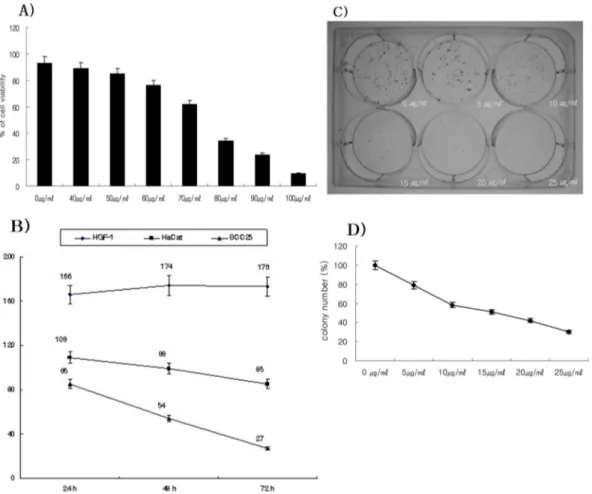
the viability of SCC25 cells by MTT assay. After CGM treatment on SCC25 cells (0 to 100µg/ml) at 24 h, the cell viability was reduced the concentrations of 50µg/ml (84.8%) to 100µg/ml (9.5%) of CGM (Fig. 1A). Also, after treatment of 50µg/ml CGM to the cells, the decreasing viability was undertaken at 24, 48 and 72 h. At 50µg/ml CGM, the cell viability was showed in a time-dependent manner (24 h, 85.0%; 48 h, 54.0%; 72 h, 27.0%) (Fig. 1B).
Furthermore, the cytotoxic effect of CGM was performed to measure the viability of HaCat cells and HGF-1 cells compared with that of SCC25 cells by MTT assay. The viability of SCC25 cells treated with 50µM CGM over a period of 72 h remarkably reduced. But the viability of HGF-1 cells treated with CGM at 50µg/ml over a period of 72 h slightly increased. In addition, the viability of HaCaT cells slightly reduced (Fig. 1B). Hence, the half maximal inhibitory concentration (IC50) of CGM was at the 50µg/ml for 48 h. This concentration was utilized for further assess-
ment of apoptosis and alternation of the cell cycle-related proteins.
To investigate whether CGM inhibited the growth of SCC25 cells, clonogenic assay was performed. After exposure to low level CGM concentrations (0 to 25µg/ml) on SCC25 cells for 7 days, the inhibition of colony formation was determined and was shown in Fig. 1C and 1D. The growth of CGM treated group was determined by percentage of control. The values on colony formation were 79.2% (5µg/ml CGM treated cells), 58.3% (10µg/ml CGM treated cells), 51.0% (15µg/ml CGM treated cells), 42.1% (20 µg/ml CGM treated cells), 30.0% (25µg/ml CGM treated cells).
Morphological and biochemical changes in CGM treated SCC25 cells
SCC25 cells treated with CGM at 50µg/ml resulted in morphological and biochemical changes associated with apoptosis. Hoechst stain demonstrated that CGM induced a
Fig. 1. Effects of cytotoxicity and growth inhibition in CGM-treated SCC25 cells as determined by MTT assay (A and B) and clonogenic assay (C and D). (A) SCC25 cells were treated with CGM (40~100µg/ml) for 24 h. The viability of SCC25 cells were decreased in a does- dependent manner. (B) MTT assay of SCC25 cells, HaCat cells and HGF-1 cells treated with 50µg/ml CGM at various time points. SCC25 cells showed the remarkable reduction of viability in a time-dependent manner. But HGF-1 cells showed the slight increase of viability. And HaCat cells showed the slight reduction of viability. Result is expressed as percentage of the control± SD of three separate experiments (C and D). The effect of growth inhibition on SCC25 cells was examined by clonogenic assay. SCC25 cells were cultured in the presence of the indicated concentrations (0 to 25µg/ml) of CGM for 7 days. (C) The photograph showing colony formation in SCC25 cells. (D) The growth of CGM treated groups is expressed as percentage of the control. Note that CGM at low concentrations significantly inhibited the growth of SCC25 cells. Values are means± SD of triplicates of each experiment.
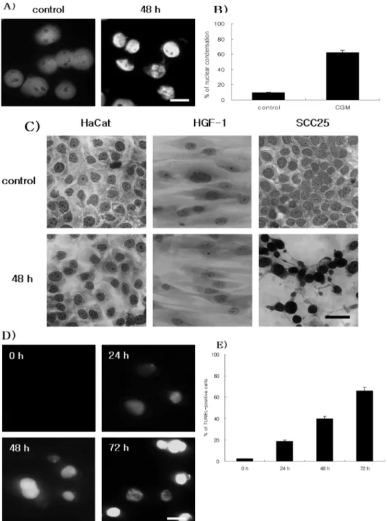
change in nuclear morphology. Compared with the typical round nuclei of the control cells, SCC25 cells treated with CGM at 50µg/ml for 48 h displayed condensed and fragmented nuclei (Fig. 2A & 2B). In addition, to investigate
whether CGM was displayed morphological changes on SCC25 cells and normal cells (HaCat cells and HGF-1 cells) or not, hemacolor staining was conducted. SCC25 cells treated with CGM at 50µg/ml for 48 h displayed condensed
Fig. 2. Demonstration of apoptosis in SCC25 cells treated with 50 µg/ml CGM. (A) Immunofluorescent micrographs after Hoechst staining.
Control cells showing round-shape nuclei (left panel). Cells treated with CGM for 48 h show the production of nuclear condensation (right panel). (B) The values are the mean± SD of the mean of apoptotic cells as determined by Hoechst staining. The results presented are repre- sentatives of three independent experiments. (C) Hemacolor staining of SCC25 cells, HaCat cells and HGF-1 cells treated with CGM at 50µg/ml for 48 h. In SCC25 cells treated with CGM, numerous apoptotic cells show but in HaCat and HGF-1 cells treated with CGM, cells show normal morphology. (D and E) TUNEL assay. (D) SCC25 cells were treated with 50µg/ml CGM at various time point. TUNEL posi- tive cells in control group (0 h) were not shown. Numerous TUNEL positive cells increased in a time-dependent manner. Scale bar, 10µm.
(E). The values are the mean± SD of the means of TUNEL positive cells as determined by TUNEL method. The each result was obtained by three times experiments.
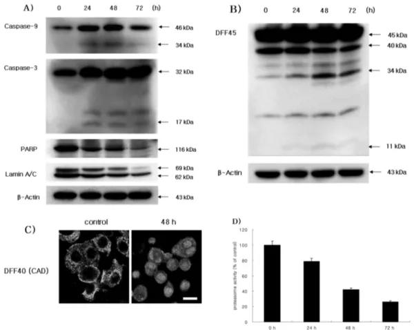
and dark stained nuclei whereas HaCat cells and HGF-1 cells showed morphology of normal cells (Fig. 2C). DNA fragmentation which is the biochemical hallmark of apoptosis, was demonstrated by TUNEL technique. The TUNEL positive SCC25 cells in the control cells did not show but the number of TUNEL positive SCC25 cells treated with 50µg/ml CGM was increased in a time- dependent manner (Fig. 2D & 2E). The Western blot assay showed that CGM treatment at various time points induced degradations of PARP and lamin A/C, and produced caspase-9 34 kDa, caspase-3 17 kDa and DFF45 34 kDa cleaved products (Fig. 3A & 3B). And confocal microscopy showed that CGM led to the translocation of DFF40 (CAD) from cytosol onto nuclei (Fig. 3C).
Proteasome activity in SCC25 cells treated with CGM In order to investigate the inhibition effect of proteasome activity at 50µg/ml CGM, proteasome activity assay was employed. In this assay, CGM remarkably abolished proteasome activity in a time-dependent manner (Fig. 3D).
Mitochondrial events were closely associated with CGM-induced apoptosis of SCC25 cells
Induction of apoptosis is regulated by Bcl-2 family members. Bcl-2 has a function of antiapoptosis, whereas Bax promotes apoptosis. And also, proapoptotic Bcl-2 family such as Bax, Bad and Bid induces loss of mitochondrial membrane potential (∆Ψm) and released cytochrome c and AIF. To examine the role of Bcl-2 family proteins in CGM-induced apoptosis, it was performed by western blot assay. The up-regulation of Bax and the down- regulation of Bcl-2 were shown in a time-dependent manner (Fig. 4A). The mitochondria were stained with JC-1 dye, and the mitochondrial membrane potential (∆Ψm) was measured by flow cytometry. SCC25 cells treated with 50µg/ml CGM at various time points showed the loss of mitochondrial membrane potential (∆Ψm) in a time- dependent manner (Fig. 4B). The confocal microscopy was conducted to examine whether AIF and cytochrome c are released in the mitochondria or not, AIF was translocated to
Fig. 3. Western blot analyses (A and B) of caspase-9, caspase-3, PARP, Lamin A/C and DFF45, confocal microscopy (C), and proteasome activity assay in SCC25 cells treated with 50µg/ml CGM. (A) CGM treatment induced PARP and Lamin A/C degradation, and produced the processed caspase-9 34 kDa and caspase-3 17 kDa cleaved products. The levels of β-actin were used as an internal standard for quantifying caspase-9, caspase-3, PARP and Lamin A/C expression. (B) CGM treatment induced the activation of DFF45 (ICAD). DFF45 (ICAD) induced cleaved products (34 kDa and 11 kDa). The levels of β-actin were used as an internal standard for quantifying DFF45 (ICAD) expression. (C) Confocal microscopy showing that DFF40 (CAD) translocated from cytosol into nuclei. Scale bar, 10µm. (D) The protea- some activity on SCC25 cells was measured by fluoro-count. Proteasome activity remarkably decreased in a time-dependent manner. Three independent assays were performed.
from mitochondria to nuclei and cytochrome c was released from mitochondria into the cytosol in SCC25 cells treated with 50µg/ml CGM (Fig. 4C & 4D).
Quantification of DNA hypoplpoidy in SCC25 cells treated with CGM
The evaluation of apoptotic percentages were confirmed Fig. 4. Demonstration of mitochondrial events in SCC25 cells after 50 µg/ml CGM treatment. (A) Western blot analysis of Bax and Bcl-2.
Pro-apoptotic factor, Bax significantly up-regulated in a time-dependent manner whereas anti-apoptotic factor, Bcl-2 down-regulated. The levels of β-actin were used as an internal standard for quantifying Bax and Bcl-2 expression. (B) Mitochondrial membrane potential (∆Ψm) at various time points. ∆Ψm was reduced in SCC25 cells in a time-dependent manner. ∆Ψm was measured by flow cytometry. Three indepen- dent assays were performed. (C) Confocal microscopy showing that cytochrome c was released from mitochondria into the cytosol of SCC25 cells. Scale bar, 10µm. (D) Confocal microscopy showing that AIF was released from mitochondria into the cytosol, and that translocation onto nuclei was evident in SCC25 cells. Scale bar, 10µm.
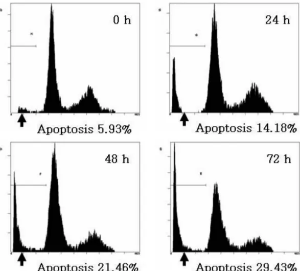
with flow cytometry analysis. A flow cytometry showed that the treatment of 50µg/ml CGM significantly increased apoptotic cells with DNA hypoploidy compared to the control group (Fig. 5).
The alteration of the cell cycle-related proteins in SCC25 cells treated with CGM
To investigate the alteration of cell cycle-related proteins, Western blot assay was conducted. Western blotting data showed that expression level of cyclin D1, cyclin D3, Cdk2 and Cdk4 regulating G0/G1 phase decreased in a time- dependent manner. The expression level of Cdk inhibitor, p27KIP1 remarkably increased over a period of 72 h (Fig. 6).
Discussion
Public attention on natural products as herbal remedies continues to grow. Moreover, the revelation of the pharmacological mechanisms of herbal plant compounds is contributing to their acceptance by healthcare professionals
and the public. A number of studies has elucidated that individual herbal medicines extracted from herbal plants have a number of pharmacological activities, e.g. anti-allergic, anti-pyretic, analgesic, anti-inflammatory and anticancer effects (Kim et al., 1998; Kim et al., 1999; Park et al., 2001;
Na et al., 2002; Kim et al., 2003). In spite of numerous in vitro and in vivo studies, the mode of action of most herbal medicines remains elusive.
Chios gum mastic (CGM) is a resinous exudate obtained from the stem and the main leaves of Pistacia lenticulus tree native to Mediterranean areas. Previous studies have demonstrated that CGM is effective in the treatment of benign gastric and duodenal ulcers and it have definite antibacterial activity against Helicobacter pylori (Al- Habbal et al., 1984; Al-Said et al., 1986; Huwez and Al- Habbal, 1986; Hawez et al., 1998). It has also been shown to have antimicrobial properties (Aksoy et al., 2006). Recently, He et al.(2007) have demonstrated that CGM treatment inhibited the proliferation of androgen-independent prostate cancer cells apparently through modulation of the NF-κB target gene. In addition, CGM has also been shown to
Fig. 5. The kinetic analysis of the effect of 50 µg/ml CGM treatment on SCC25 cell cycle progression and induction of apoptosis using flow cytometry. Representative DNA histograms are shown. CGM treatment significantly showed the increase of apoptotic cells with DNA hypoploidy in a time-dependent manner. Data shown are representatives of three independent experiments.
contain compounds that can induce in vitro apoptosis of human colon cancer cells through caspase-dependent pathways (Balan et al., 2007). Thus, the present study investigating the effects of CGM on cell viability in SCC25 human tongue squamous cell carcinoma cells revealed that CGM produced a dose- and time-dependent reduction on viability of SCC25 cells in MTT assay. In addition, the clonogenic assay (colony forming assay) confirmed that CGM at 5 to 25µg/ml remarkably inhibited the growth of SCC25 cells. Interestingly, CGM remarkably revealed cytotoxicity in SCC25 cells treated with 50µg/ml CGM over a period of 72 h but not normal cells. These data indicate that CGM exerts a specific cytotoxic effect on SCC25 cells compared to normal cells. Based on these data, CGM could have an anti-cancer effect for human oral squamous cell carcinoma.
Apoptosis and necrosis are conceptually distinct forms of cell death and can be distinguished by their specific morphological changes. During apoptosis, apoptotic cells undergo specific morphological changes such as the cell blebbing, reduction of cell size, cell shrinkage, chromatin condensation and DNA fragmentation (Wyllie et al., 1980;
Williams, 1991). In the results of Hoechst stain and TUNEL assay, SCC25 cells treated with CGM showed apoptotic hallmarks such as the formation of apoptotic bodies and the increase of TUNEL-positive cells. These results indicate that CGM induced SCC25 cell death via activating the apoptosis.
Apoptotic stimuli may induce apoptosis by inhibiting the proteasome activity of the target cells (Meng et al., 1999).
However, other studies have reported that a proteasome
inhibitor itself can induce apoptosis in certain cells (Drexler et al., 2000). Generally, the proteasome-mediated step(s) in apoptosis are located upstream of mitochondrial changes and caspase activation, and can involve different systems, including various cyclins, p53, NF-κB, Bax and Bcl-2 (Grimm et al., 1996; Orlowski, 1999; Li and Dou, 2000).
Thus, the possibility existed that CGM may have affected proteasome activity in SCC25 cells and induced the mitochondrial pathway of apoptosis. In this study, the proteasome activity showed the remarkable reduction in SCC25 cells treated with CGM time-dependently. This data suggests that CGM induced apoptosis via proteasome pathway.
Because mitochondria play a crucial role in apoptosis, the induction of the mitochondrial permeability transition plays a key part in the regulation of apoptosis (Kroemer et al., 1997; Green and Reed, 1998; Susin et al., 1999; Orrenius, 2004). The mitochondrial pathway can also be triggered by various intracellular and extracellular stress signals, which result in activation of pro-apoptotic proteins, including Bax and Bak, or inactivation of anti-apoptotic Bcl-2 family members, such as Bcl-2 and Bcl-xL (Orrenius, 2004). As a result of activation/inactivation of Bcl-2 family proteins, changes in mitochondrial membrane lead to the dissipation of inner membrane potential and permeabilization of the outer mitochondrial membrane (OMM). This in turn induces the release of various proapoptotic proteins, such as cytochrome c, Smac/Diablo, endonuclease G and AIF (Hengartner, 2000; Barczyk et al., 2005). The present study showed a significant shift of the ratio of Bax to Bcl-2 in SCC25 cells treated with CGM. This result indicates that a shift of the ratio of Bax to Bcl-2 may be the molecular mechanisms by which CGM induces apoptosis of SCC25 cells. It has been reported that the pro-apoptotic Bcl-2 family in isolated mitochondria induces cytochrome c release, the loss of mitochondrial membrane potential and results in AIF release (Hunter and Parslow, 1996; Narita et al., 1998). Cytochrome c release and disruption of mitochondrial membrane potential (MMP) are known to contribute to apoptosis triggered by proteasome inhibition (Wagenknecht et al., 2000; Marshanskya et al., 2001).
Generally, cytochrome c is released into the cytosol during apoptosis, where it binds to Apaf-1. This cytochrome c/
Apaf-1 complex (apoptosome) promotes the autoactivation of procaspase-9 to caspase-9. Caspase-9 then acts on procaspase-3 to initiate a caspase activation cascade (Li et al., 1997; Zou et al., 1999). Released AIF through pro- apoptotic Bcl-2 family activation induces its translocation to the nucleus, resulting in chromatin condensation and large- scale DNA fragmentation (Douglas et al., 2000). In the present study, CGM treatment also induced translocation of AIF from mitochondria into nuclei, cytochrome c release from mitochondria into the cytosol, a significant loss of MMP and the prodcution of caspase-9 cleavage. These data have clearly demonstrated that the CGM-induced apoptosis Fig. 6. Western blot analysis of cell cycle-related proteins in
SCC25 cells treated with 50µg/ml CGM. Cdk2, Cdk4, Cyclin D1, and Cyclin D3 were down-regulated in a time dependent manner.
Cdk inhibitors, p27KIP1 was up-regulated in a time-dependent man- ner. The levels of β-actin were used as an internal standard for quantifying p27KIP1, Cdk2, Cdk4, Cyclin D1 and Cyclin D3 expres- sion.
in SCC25 cells was involved with mitochondrial events as mentioned above.
A common final event of apoptosis is nuclear condensation, which is controlled by caspases, DFF (DNA fragmentation factor), and PARP. Caspases, the aspartate- specific intracellular cysteine protease, play an essential role during apoptotic death (Acehan et al., 2002). Once activated, the effector caspases (caspase-3, caspase-6 or caspase-7) are responsible for the proteolytic cleavage of a broad spectrum of cellular targets, leading ultimately to cell death. The known cellular substrates include structural components (such as actin and nuclear lamin), inhibitors of deoxyribonuclease (such as DFF45 or ICAD) and DNA repair proteins (such as PARP) (Gross et al., 1999; Porter, 1999). In apoptotic cells, activation of DFF40 (CAD), also a substrate of caspase-3, occurs with the cleavage of DFF45 (ICAD). Once DFF40 is activated and released from the complex of DFF45 and DFF40, it can translocate to the nucleus and then degrade chromosomal DNA and produce DNA fragmentation (Cheng AC, 2007). Furthermore, in apoptotic cells, the degradation of the lamin A/C, a substrate of caspase-6 was sometimes occurred. In this study, cleavage of caspase-3 and DFF45, and degradation of PARP and lamin A/C were shown in CGM-treated SCC25 cells.
Furthermore, confocal microscopy showed that CGM led to the translocation of DFF40 (CAD) from cytosol onto nuclei in SCC25 cells. Therefore, these data have demonstrated that CGM-induced apoptosis in SCC25 cells is associated with caspase-3 activation, and activated caspase-3 leads to the activation of PARP and DFF45, and the translocation of DFF40 from cytosol into nucleus which degrades the chromosomes into fragments.
Various studies on the molecular analyses of cancers have revealed that cell cycle regulators are frequently mutated in most common malignancies. Therefore, the control of cell cycle progression in tumor cells is considered to be a potentially effective strategy for the control of tumor growth. In the case of Cdks, Cyclins and Cdk inhibitors, these play critical roles in the regulation of cell cycle progression (Pavletich, 1999). Cdk inhibitors inhibit the active Cdk-Cyclin complex (el-Deiry et al., 1994). p21WAF1/
CIP1
and p27KIP1 have been demonstrated to play an important role in regulating progression from the G1/S phase, by binding to and preventing premature activation of Cdk4/
cyclin D and Cdk2/cyclin E complexes (Polyak et al., 1994;
Coats et al., 1996). It is known that the cell cycle G1 arrest may be related to activation of the p53 tumor suppressor protein, which acts as a transcription factor and regulates expression of several components, implicated in pathways that regulate cell cycle progression and apoptosis induction (Teyssier et al., 1999; Colman et al., 2000). In this study, Cdk2, Cdk4, Cyclin D1 and Cyclin D3 were remarkably down-regulated whereas p27KIP1 were remarkably up- regulated. However, in SCC25 cell (p53 deficient cancer cell), p53 was not detected over a experimental period (data
not shown). These data have demonstrated that the CGM- induced apoptosis in SCC25 cells was involved with the expression alterations of the G1 cell cycle-related proteins.
Moreover, we suggest that p27KIP1 may play a key role in CGM-induced SCC25 cell death.
Taken collectively, this study demonstrate that CGM strongly inhibits cell proliferation via the expression modulations of the G1 cell cycle-related proteins and induces apoptosis via proteasome, mitochondria and caspase cascade in SCC25 cells. Therefore, our data provide the possibility that a natural product, CGM could be considered as a novel therapeutic strategy for human oral squamous cell carcinoma.
Acknowledgement
This work was supported by for two years Pusan National University research grant.
References
Acehan D, Jiang X, Morgan DG, Heuser JE, Wang X, Akey CW. Three-dimensional structure of the apoptosome: Impli cations for assembly, procaspase-9 binding, and activation.
Mol Cell. 2002;9:423-32.
Aksoy A, Duran N, Koksai F. In vitro and in vivo antimic- robial effects of mastic chewing gum against Streptococcus mutans and mutans streptococci. Arch Oral Biol. 2006;
51:476-81.
Al-Habbal MJ, Al-Habbal Z, Huwez FU. A double-blind controlled clinical trial of mastic and placebo in the treatment of duodenal ulcer. Clin Exp Pharmacol Physiol.
1984;11:541-4.
Al-Said MS, Ageel AM, Parmar NS, Tariq M. Evaluation of mastic, a crude drug obtained from Pistacia lentiscus for gastric and duodenal anti-ulcer activity. J Ethnopharmacol.
1986;15:271-8.
Balan KV, Prince J, Han Z, Dimas K, Cladaras M, Wyche JH, Sitaras NM, Pantazis P. Antiproliferative activity and induction of apoptosis in human colon cancer cells treated in vitro with constituents of a product derived from Pistacia lentiscus L. var. chia. Phytomedicine. 2007;14:263-72.
Barczyk K, Kreuter M, Pryjma J, Booy EP, Maddika S, Ghavami S, Berdel WE, Roth J, Los M. Serum cytochrome c indicates in vivo apoptosis and can serve as a prognostic marker during cancer therapy. Int J Cancer. 2005;116:167-73.
Carson DA, Ribeiro JM. Apoptosis and disease. Lancet.
1993;341:1251-4.
Cheng AC, Jian CB, Huang YT, Lai CS, Hsu PC, Pan MH.
Induction of apoptosis by Uncaria tomentosa through reactive oxygen species production, cytochrome c release, and caspases activation in human leukemia cells. Food Chem Toxicol. 2007;45:2206-18.
Coats S, Flanagan WM, Nourse J, Roberts JM. Requirement of p27Kip1 for restriction point control of the fibroblast cell
cycle. Science. 1996;272:877-80.
Colman MS, Afshari CA, Barret JC. Regulation of p53 stability and activity in response to genotoxic stress. Mutat Res. 2000;462:179-88.
Douglas E, Susin SA, Zamzami N, Ferri KF, Irinopoulou T, Larochette N, Prevost MC, Leber B, Andrews D, Penninger J, Kroemer G. Mitochondrio-nuclear translocation of AIF in apoptosis and necrosis. FASEB J. 2000;14:729-39.
Drexler HC, Risau W, Konerding MA. Inhibition of proteasome function induces programmed cell death in proliferating endothelial cells. FASEB J. 2000;14:65-77.
el-Deiry WS, Harper JW, O'Connor PM, Velculescu VE, Canman CE, Jackman J, Pietenpol JA, Burrell M, Hill DE, Wang Y, et al. WAF1/CIP1 is induced in p53-mediated G1 arrest and apoptosis. Cancer Res. 1994;54:1169-74.
Green DR, Reed JC. Mitochondria and apoptosis. Science.
1998;281:1309-12.
Grimm LM, Goldberg AL, Poirier GG, Schwartz LM, Osborne BA. Proteasome play an essential role in thymocyte apoptosis. EMBO J. 1996;15:3845-52.
Gross A, McDonnell JM, Korsmeyer SJ. BCL-2 family members and the mitochondria in apoptosis. Genes Dev.
1999;13:1899-911.
Hawez FU, Thirlwell D, Cockayne A, Ala’Aldeen PA. Mastic gum kills Helicobacter pylori. N Engl J Med. 1998;339 :1946.
He M, Li A, Xu CS, Wang SL, Zhang MJ, Gu H, Yang YQ, Tao HH. Mechanisms of antiprostate cancer by gum mastic:
NF-kappaB signal as target. Acta Pharmacol Sin. 2007;28:
446-52.
Hengartner MO. The biochemistry of apoptosis. Nature. 2000;
407:770-6.
Hunter JJ, Parslow TG. A peptide sequence from Bax that converts Bcl-2 into an activator of apoptosis. J Biol Chem.
1996;271:8521-4.
Huwez FU, Al-Habbal MJ. Mastic in treatment of benign gastric ulcers. Gastroenterol Jpn. 1986;21:273-4.
Jun DY, Kim JS, Park HS, Han CR, Fang Z, Woo MH, Rhee IK, Kim YH. Apoptogenic activity of auraptene of Zanthoxylum schinifolium toward human acute leukemia Jurkat T cells is associated with ER stress-mediated caspase-8 activation that stimulates mitochondria-dependent or -independent caspase cascade. Carcinogenesis. 2007;28:1303-13.
Kang JX, Liu J, Wnag J, He C, Li FP. The extract of huanglian, a medicinal herb, induces cell growth arrest and apoptosis by upregulation of interferon-beta and TNF-alpha in human breast cancer cells. Carcinogenesis. 2005;26:1934-9.
Kim HM, Lee EH, Hong SH, Song HJ, Shin MK, Kim SH, Shin TY. Effect of Syzygium aromaticum extract on immediate hypersensitivity in rats. J Ethnopharmacol. 1998;60:125-31.
Kim HM, Yi JM, Lim KS. Magnoliae flos inhibits mast cell- dependent immediate-type allergic reactions. Pharmacol Res. 1999;39:107-11.
Kim JH, Bae HR, Park BS, Lee JM, Ahn HB, Rho JH, Yoo KW, Park WC, Rho SH, Yoon HS, Yoo YH. Early mito- chondrial hyperpolarization and intracellular alkalinization in lactacystin-induced apoptosis of retinal pigment epithelial cells. J Pharmacol Exp Ther. 2003;305:474-81.
Kroemer G, Zamzami N, Susin SA. Mitochondrial control of apoptosis. Immunol Today. 1997;18:44-51.
Kumagai T, Müller CI, Desmond JC, Imai Y, Heber D, Koeffler HP. Scutellaria baicalensis, a herbal medicine: anti- proliferative and apoptotic activity against acute lym- phocytic leukemia, lymphoma and myeloma cell lines. Leuk Res. 2007;31:523-30.
Lian Z, Niwa k, Onogi K, Mori H, Harrigan RC, Tamaya T.
Anti-tumor effects of herbal medicines on endometrial carcinomas via estrogen receptor-alpha-related mechanism.
Oncol Rep. 2006;15:1133-6.
Li B, Dou QP. Bax degradation by the ubiquitin/proteasome- dependent pathway: Involvement in tumor survival and progression. Pro Natl Acad Sci USA. 2000;97:3850-5.
Li P, Nijhawan D, Budihardjo I, Srinivascula SM, Ahmad M, Alnemri ES, Wang X. Cytochrome c and dATP-dependent formation of Apaf-1/caspase-9 complex initiates an apoptotic protease cascade. Cell. 1997;91:479-89.
Marshansky V, Wang X, Bertrand R, Luo H, Duguid W, Chinnadurai G, Kanaan N, Vu MD, Wu J. Proteasomes modulate balance among proapoptotic and antiapoptotic Bcl-2 family members and compromise functioning of the electron transport chain in leukemic cells. J Immunol.
2001;166:3130-42.
Meng L, Kwok BH, Sin N, Crews CM. Eponemycin exerts its antitumor effect through the inhibition of proteasome function. Cancer Res. 1999;59:2798-801.
Na HJ, Jeong HJ, Bae H, Kim YB, Park ST, Yun YG, Kim HM.
Tongkyutang inhibits mast cell-dependent allergic reactions and inflammatory cytokines secretion. Clin Chim Acta.
2002;319:35-41.
Narita M, Shimizu S, Ito T, Chittenden T, Lutz RJ, Matsuda H, Tsujimoto Y. Bax interacts with the permeability transition pore to induce permeability transition and cytochrome c release in isolated mitochondria. Proc Natl Acad Sci U S A.
1998;95:14681-6.
Ohta K, Yamashita N. Apoptosis of eosinophils and lym- phocytes in allergic inflammation. J Allergy Clin Immunol.
1999;104:14-21.
Orlowski RZ. The role of the ubiquitin-proteasome pathway in apoptosis. Cell Death Differ. 1999;6:303-13.
Orrenius S. Mitochondrial regulation of apoptotic cell death.
Toxicol Lett. 2004;149:19-23.
Park BS, Song YS, Yee SB, Lee BJ, Seo SY, Park YC, Kim JY, Kim HM, Yoo YH. Phospho-ser 15-p53 translocate into mitochondria and interacts with Bcl-2 and Bcl-xL in eugenol-induced apoptosis. Apoptosis. 2005;10:193-200.
Park HI, Jeong MH, Lim YJ, Park BS, Kim GC, Lee YM, Kim HM, Yoo KS, Yoo YH. Szygium aromaticum (L.) Merr. Et Perry (Myrtaceae) flower bud induces apoptosis of p815 mastocytoma cell line. Life Sci. 2001;69:553-66.
Pavletich NP. echanisms of cyclin-dependent kinase regulation:
structures of Cdks, their cyclin activators, and Cip and INK4 inhibitors. J Mol Biol. 1999;87:821-8.
Polyak K, Lee MH, Erdjument-Bromage H, Koff A, Roberts JM, Tempst P, MassaguJ. Cloning of p27Kip1, a cyclin- dependent kinase inhibitor and a potential mediator of extracellular antimitogenic signals. Cell. 1994;78:59-66.
Porter AG. Protein translocation in apoptosis. Trends Cell Biol.
1999;9:394-401.
Shen J, Huang C, Jiang L, Gao F, Wang Z, Zhang Y, Bai J, Zhou H, Chen O. Enhancement of cisplatin induced apoptosis by suberoylanilide hydroxamic acid in human oral squamous cell carcinoma cell lines. Biochem Pharmacol. 2007;73:1901- 9.
Susin SA, Lorenzo HK, Zamzami N, Marzo I, Snow BE, Brothers GM, Mangion J, Jacotot E, Costantini P, Loeffler M, Larochette N, Goodlett DR, Aebersold R, Siderovski DP, Penninger JM, Kroemer G. Molecular characterization of mitochondrial apoptosis-inducing factor. Nature. 1999;397:
441-6.
Teyssier F, Bay JO, Dionet C, Verrelle P. Cell cycle regulation after exposure to ionizing radiation. Bull Cancer.
1999;86:345-57.
Wagenknecht B, Hermission M, Groscurth P, Liston P, Krammer PH, Weller M. Proteasome inhibitor-induced
apoptosis of glioma cells involves the processing of multiple caspases and cytochrome c release. J Neurochem. 2000;75:
2288-97.
Williams GT. Programmed cell death: apoptosis and onco- genesis. Cell. 1991;65:1097-8.
Wu YD, Lou YJ. A steroid fraction of chloroform extract from bee pollen of Brassica campestris induces apoptosis in human prostate cancer PC-3 cells. Phytother Res. 2007;21:
1087-91.
Wyllie AH, Kerr JF, Currie AR. Cell death: the significance of apoptosis. Int Rev Cytol. 1980;68:251-306.
Yoon JS, Seo JC, Han SW. Pinelliae Rhizoma herbal- acupuncture solution induced apoptosis in human cervical cancer cells, SNU-17. Am J Chin Med. 2006;34:401-8.
Zou H, Li Y, Liu X, Wang X. An APAF-1, cytochrome c multimeric complex is a functional apoptosome that activates procaspase-9. J Biol Chem. 1999;274:11549-56.