• 검색 결과가 없습니다.

Trans-differentiation Induction of Human-mesenchymal Stem Cells Derived from Different Tissue Origin and Evaluation of their Potential for Differentiation into Corneal Epithelial-like Cells

N/A
N/A
Protected

Academic year: 2021

Share "Trans-differentiation Induction of Human-mesenchymal Stem Cells Derived from Different Tissue Origin and Evaluation of their Potential for Differentiation into Corneal Epithelial-like Cells"

Copied!
13
0
0

로드 중.... (전체 텍스트 보기)

전체 글

(1)

Trans-differentiation Induction of Human-mesenchymal Stem Cells Derived from Different Tissue Origin and Evaluation of their Potential for Differentiation into Corneal Epithelial-like Cells

Sun-Woung Moon

1,*

, Hyeon-Jeong Lee

1,*

, Won-Jae Lee

2

, Sun-A Ock

3

and Sung-Lim Lee

1,4,†

1

College of Veterinary Medicine, Gyeongsang National University, Jinju 52828, Gyeongnam, Republic of Korea

2

College of Veterinary Medicine, Kyungpook National University, Daegu, 41566 Republic of Korea

3

AnimalBiotechnology Division, National InstituteofAnimalScience, Rural Development Administration, Wanju-gun, Jeollabuk-do,Republic of Korea

4

Research Institute of Life Sciences, Gyeongsang National University, Jinju 52828, Gyeongnam, Republic of Korea

Abstract

The trans-differentiation potential of mesenchymal stem cells (MSCs) is employed, but there is little understanding of the cell source-dependent trans-differentiation potential of MSCs into corneal epithelial cells. In the present study, we induced trans-differentiation of MSCs derived from umbilical cord matrix (UCM-MSCs) and from dental tissue (D-MSCs), and we comparatively evaluated the in vitro trans-differentiation properties of both MSCs into corneal epithelial-like cells.

Specific cell surface markers of MSC (CD44, CD73, CD90, and CD105) were detected in both UCM-MSCs and D-MSCs, but MHCII and CD119 were significantly lower (P < 0.05) in UCM-MSCs than in D-MSCs. In UCM-MSCs, not only expression levels of Oct3/4 and Nanog but also proliferation ability were significantly higher (P < 0.05) than in D-MSCs.

In vitro differentiation abilities into adipocytes and osteocytes were confirmed for both MSCs.

UCM-MSCs and D-MSCs were successfully trans-differentiated into corneal epithelial cells, and expression of lineage-specific markers (Cytokeratin-3, -8, and -12) were confirmed in both MSCs using immunofluorescence staining and qRT-PCR analysis. In particular, the differentiation capacity of UCM-MSCs into corneal epithelial cells was significantly higher (P < 0.05) than that of D-MSCs.

In conclusion, UCM-MSCs have higher differentiation potential into corneal epithelial-like cells and have lower expression of CD119 and MHC class II than D-MSCs, which makes them a better source for the treatment of corneal opacity.

Received :31 March 2018 Revised :19 June 2018 Accepted:21 June 2018

Key Words : Mesenchymal stem cells, Corneal epithelial cells, Trans-differentiation, Umbilical cord matrix-MSC, Dental tissue-MSC

* These authors equally contributed to this work.

(2)

INTRODUCTION

The cornea consists mainly of three layers: a non-keratinized stratified epithelium, a collagen type I-rich stroma layer interspersed with fibroblast, and an inner layer of endothelial cells which retain optimal hydration (Katikireddy et al., 2013; Secker et al., 2008; Ouyang et al., 2014). The cornea protects the eyeball from injury and infection, and its sensitivity to damage including chemical, mechanical, and thermal injury can lead to severe inflammation and neovascularization, which results in loss of transparency and opacity of the eye (Rohaina et al., 2014). The visual function has to restore the corneal epithelium that is maintained by limbal epithelial stem cells located in the stem cell niche called the limbus (West et al., 2015). Fundamentally, the main pathological factor is deficiency in limbal epithelium and corneal epithelium, which is converted into keratinized skin-like epithelium (Ouyang et al., 2014).

Replacement of the stem cell population has been achieved by transplanting limbal tissue by means of autografts or allografts (Lavker et al., 2003), but risks remain, such as damage to a healthy eye by removal of autologous tissue for transplantation or immune reaction to allogenic tissue (Shahdadfar et al., 2012; Ramaesh et al., 2003). Most stem cell research into corneal epithelial regeneration is based on the use of progenitor cells isolated from corneal tissue, but a growing body of research suggests that corneal regeneration with MSCs isolated from non-corneal tissues is more likely to be considered. Therefore, other stem cells, including embryonic stem cells, hair follicle stem cells, and MSC derived from bone marrow (BM-MSCs), have been reported as sources for induction into corneal epithelial lineage (Rohaina et al., 2014; Ahmad et al., 2007; Blazejewska et al., 2009). In this way, MSC trans-differentiation into corneal epithelial cells has emerged, and therefore cell source-dependent differentiation ability into corneal epithelial cells requires comparative study to find an effective cell source of MSC for corneal regeneration.

We identified MSCs derived from dental pulp tissue (D-MSCs) and umbilical cord matrix (UCM-MSCs) as having good potential for trans-differentiation into corneal epithelial cells. D-MSCs can be another source for stem cell therapy because of their multi-potent potential and their immunomodulatory function that regulates T and B lymphocytes, dendritic cells, and natural killer cells (Park et al., 2014; Kang et al., 2015). Umbilical cord matrix-derived MSCs (UCM-MSCs) can also be a source of regenerative medicine because of their multi-lineage differentiation potential and self-renewal capacity (Troyer et al., 2008). They also have immunological features that do not provoke an immune reaction because they lack major histocompatibility complex (MHC) II and co-stimulatory molecule expression (Anzalone et al., 2010; Krampera et al., 2013).

Therefore, the present study comparatively evaluated the characterization and trans-differentiation ability of UCM-MSCs and D-MSCs in order to understand the cell source-dependent efficiency of corneal epithelial regeneration.

MATERIALS AND METHODS

Chemicals and media

All media and chemicals for the experiments were purchased from Gibco (Life Technologies, Grand Island, NY, USA) and Sigma-Aldrich Chemical Company (St. Louis, MO, USA) unless otherwise specified.

Cell isolation and culture of UCM- and D-MSCs

The study was performed in approval by Institutional Review Board of Gyeongsang National University hospital (GNUH-

2012-09-004). Umbilical cord matrix tissues were obtained from term deliveries and dental tissues were obtained from orthodontic

treatment patients. The isolation of cells and the establishment of UCM- and D-MSCs were performed using the protocols

described in previous studies (Kang et al., 2013; Park et al., 2012). Briefly, two umbilical arteries and one vein were removed

in the umbilical cord matrix tissues and the sliced small fragments were attached to 35 mm dish (Nunc, Denmark) for cell

explantation. For isolation of D-MSCs, the sliced dental tissues into small piece to isolate MSCs by digestion with 0.1%

(3)

Dulbecco’s Modified Eagle Medium (A-DMEM), 10% fetal bovine serum (FBS), 1% GlutaMax™ and 1% penicillin-streptomycin at 38.5 ° C in a humidified incubator at 5% CO

2

. Once confluent, the cells were trypsinized (0.25% trypsin-EDTA solution) and sub-cultured.

Analysis of cell surface markers

D-Cells were analyzed for the expression of cell surface antigens of MSC (CD44, CD73, CD90 and CD105), hematopoietic cell (CD34 and CD45) and immune related markers (MHCII and CD119) using a flow cytometry analysis (BD FACS Calibur;

Becton Dickinson, NJ, USA) as previously described (Kang et al., 2010). Briefly, cells at 70~80% confluence were harvested and fixed with 4% paraformaldehyde (PFA). They were labeled with FITC-conjugated isotype IgG antibodies (control, BD Pharmingen

™, 0.5 mg/ml); anti-CD34 (BD Pharmingen™, 0.5 mg/ml), anti-CD44 (BD Pharmingen™, 0.5 mg/ml), anti-CD45 (BD Pharmingen

™, 0.5 mg/ml) and anti-CD90 (BD Pharmingen™, 0.5 mg/ml) antibodies; and anti-CD105 (Santa Cruz biotechnology, 200 ug/ml), anti-CD119 (Santa Cruz biotechnology, 0.5 mg/ml) and anti-MHC class II (Santa Cruz biotechnology, 200 ug/ml) primary antibodies with FITC-conjugated secondary antibodies. All antibodies were diluted 1:100 with DPBS supplemented with 1% bovine serum albumin.

Analysis of cell proliferation

The cell proliferation was evaluated by using MTT Cell Proliferation Assay, following the manufacturer’s protocol. Briefly, ~ 1 × 10

3

/ml cells at passage 3 were suspended in 500 μL of ADMEM, supplemented with 10% FBS and cultured for 192 hours, and the culture medium was changed every 3 days. The cell proliferation was quantified by using an MTT colorimetric assay according to a VersaMaxTM microplate reader (Molecular Devices, Sunnyvale, California, USA), at an absorbance of 405-nm wavelength.

Induction and confirmation of in vitro differentiation

In vitro differentiation of the cells into adipocytes and osteocytes as mesenchymal lineages were performed as previously described (Lee et al., 2015; Ock et al., 2010). Briefly, the cells were cultured for 3 weeks in Dulbecco’s modified Eagle medium (DMEM) containing 10% FBS, 100 uM indomethacin, 10 uM insulin and 1 uM dexamethasone, and differentiation induced cells were determined by staining intracellular lipid vacuoles using 0.5% Oil red O solution for adipogenic differentiation. For osteogenesis, cells were induced in DMEM contained 10% FBS, 200 uM ascorbic acid, 10 mM β-glycerophosphate and 0.1 uM dexamethasone, and osteogenic differentiation was identified by the accumulation of calcium deposits and proteoglycan following staining with 2% Alizarin Red S solution and 5% silver nitrate.

Induction of trans-differentiation into corneal epithelial cells

Cells were induced trans-differentiation into corneal epithelial cells with the protocol described in a previous study, with minor modifications (Ahmad et al., 2007). For the preparation of collagen type-I and-IV (Col-1 and 4, Gibco) coated culture dishes, 5 ug/cm

2

Col-1 solution was treated into the 35 mm culture dishes for 1 hour incubation at room temperature, after then the coated dishes was rinsed three times with DPBS. Subsequently, 0.5 mg/ml Col-4 solution was added on the Col-1 coated dishes and kept at 4℃ overnight, followed by the Col-4 coated dishes were washed with DPBS.

Briefly, cells were treated with induction medium for 4 days following that the inducted cells were cultured in the differentiation medium for 18 days to induce trans-differentiation into corneal epithelial cells (Fig 4. A). Induction medium was comprised of no glucose DMEM without pyruvate, 1.0 g/L D-glucose, 25 ng/ml human recombinant bone morphogenetic protein-4 (BMP-4;

R&D Systems), 1 uM all-trans retinoic acid (RA) and 10 ng/ml recombinant human epidermal growth factor (EGF). Differentiation medium was comprised of three parts low-glucose DMEM with pyruvate and one part Ham’s F-12 medium, 5% FBS, 1%

penicillin-streptomycine, 500 ng/ml hydrocortisone, 5 ug/ml insulin, 2 nM tri-iodothyronine, adenine and 10 ng/ml EGF.

(4)

Immunofluorescence staining

Expression of specific markers of corneal epithelial cells was determined by immunofluorescence staining. After being fixed in 4% PFA cells were stained with primary antibodies (1:100X dilution) against to cytokeratin 3 (CK3), cytokeratin 8 (CK8) and cytokeratin 12 (CK12) at 4℃ overnight, and followed by incubation with FITC-conjugated secondary antibody (Jackson ImmunoResearch, PA, USA; 1:200 dilution) for 1 hour at 37° C. Nuclei of the cells were then counterstained with 10 ug/ml DAPI for 5 min and images were observed under a confocal laser scanning microscope. For the measurement of fluorescence intensities of the specific markers in the immunofluorescence stained MSCs, the integrated optical density (IOD) was analyzed by image analysis system (Image J software).

Analysis of quantitative reverse-transcription polymerase chain reaction (qRT-PCR)

The qRT-PCR analysis based on the multiplex method was conducted to quantify the mRNA levels of the genes involved in early transcription factors (Oct3/4 and Nanog) and corneal specific markers (CK3, CK8 and CK12) in UCM- and D-MSCs. Total RNAs were extracted from the cells before and after differentiation into corneal epithelial like cells, using RNeasy mini Kit (Qiagen, Valencia, CA, USA) on the basis of the manufacturer’s instruction and measured by OPTIZEN 3220 UV BIO Spectrophotometer (Mecasys Co, Ltd, Korea). Each cDNA of 100 ng was synthesized at 37℃ for 1 hour using Omniscript

TM

Reverse Transcription Kit (Qiagen, Valencia, CA, USA). qRT-PCR was performed using Roter gene Q (Qiagen) qRT-PCR machine with Rotor-Gene™ 2X SYBR® Green mix (Qiagen) including 1 uM forward and reverse primers with 0.01 ug cDNA per one reaction. The qRT-PCR program was consist of pre-denaturation at 95℃ for 5 minutes followed by 40 PCR cycles at 95℃ for 5 seconds, annealing at 60℃ for 10 seconds and melting curve from 60℃ to 95℃ by 1℃ per 1 second; cooling at 40℃ for 30 seconds on the basis of qRT-PCR program with minor modification in manufacturer’s protocol. Amplification curves, melting curves and Ct values were analyzed by using Rotor-Gene Q Series Software (Qiagen) and all products were confirmed by 1.5%

agarose gel electrophoresis for nonspecific product in targeted gene product and negative control after qRT-PCR. All primers used for qRT-PCR were summarized in Table 1.

Statistical analysis

For the analysis of the differences between the cells, this study used T-test analysis with SPSS software (via IBM SPSS Statistics, ver. 21). Threshold cycle (Ct) values and integrated optical density (IOD) were presented as means ± SEM and the differences were considered significant when P-values were less than 0.05.

RESULTS

Cellular- and immune-phenotype differences between UCM- and D-MSCs

The two expanded types of MSC isolated from umbilical cord matrix and dental tissue were both observed to have fibroblastic spindle-like morphology and the feature of adhering to culture dishes (Fig. 1A). CD44, CD73, CD90, and CD105 as the specific cell surface markers of MSC were highly expressed (≥ 96.9∽100% of positive cells) in UCM- and D-MSCs, whereas the expression of CD34 and CD45 was low in both cells (≤ 2% of positive cells; Fig. 1B).

Confirmation of CD119 and MHC II expression levels for immune phenotyping was performed in both types of cells. As shown

in Fig. 1C and D, the expression levels of MHCII and CD119 in UCM-MSCs (0.69% and 11.4% of positive cells, respectively)

were lower than those in D-MSCs (2.53% and 39.4% of positive cells). In particular, the expression of CD119 in UCM-MSCs

was significantly lower (P < 0.05) than in D-MSCs.

(5)

Expression of early transcription factors in UCM- and D-MSCs

Expression of early transcription factors (Oct3/4 and Nanog) was determined in MSCs derived from both types of cell (umbilical cord matrix and dental tissue) using qRT-PCR (Fig. 2A). Both MSCs expressed Oct3/4 and Nanog, but the level of expression of each early transcription factor varied. The expression levels of Oct3/4 and Nanog were significantly higher (P < 0.01) in UCM-MSCs compared to D-MSCs.

Figure 1. Characterization of UCM- and D-MSCs. (A) Morphology of UCM- and D-MSCs. Both cells at passage 4 showed a characteristic of adherence to the plastic flask with spindle-like morphology. (Original magnification ×40. Bars = 100 μm). (B, C, and D) Analysis of specific cell surface markers of MSCs by flow cytometry. Gates which were based on isotype controls are shown in white, and each antigen expression as MSC specific markers is shown in grey. A total of 10,000 MSCs at passage 4 were counted. Bars with * indicate a significant difference (P < 0.05)

Cell proliferation capacity of UCM- and D-MSCs

As shown in Fig. 2B, the population of UCM-MSCs was significantly higher (P < 0.05) than the population of D-MSCs after

144 hours and until 192 hours of cultivation.

(6)

Figure 2. Expression of transcription factors and proliferation ability in UCM- and D-MSCs. (A) Expression of early transcription factors in MSCs. Products of early transcription factors (Oct3/4 and Nanog) were performed by qRT-PCR. Both MSCs were obtained at passage 4. β‑actin was used as a reference gene. (B) The proliferation capacities of both MSCs were evaluated for 192 hours. * indicates a significant difference (P < 0.05).

In vitro differentiation into adipocytes and osteoblasts

After differentiation into adipocytes and osteoblasts of the UCM- and D-MSCs, cytochemical staining was performed to confirm

differentiation ability. The accumulation of lipid vacuoles was observed in adipocytes using Oil red O staining, and the deposition

of calcified extracellular matrix was observed in osteoblasts using Alizarin Red S and von Kossa staining in both differentiation

induced MSCs (Fig. 3).

(7)

Figure 3. In vitro differentiation induced into adipocytes and osteocytes. At passage 5, both MSCs were induced and differentiated into adipocytes and osteocytes and cytochemical staining with Oil red O (adipocytes), von Kossa and Alizarin Red S (osteocytes). (Magnification

×40. Bars = 100 μm)

Morphological changes and expression of lineage-specific markers of trans-differentiation induced MSCs into corneal epithelial cells

UCM- and D-MSCs were trans-differentiated into corneal epithelial cells for four days in induction medium (Fig. 4A (a)) followed by 18 days of differentiation medium (Fig. 4A (b)). Morphological changes were observed in both MSCs following induction of differentiation (Fig. 4B). Both undifferentiated MSCs were observed to have a fibroblastic morphology with a spindle shape. However, the UCM-MSCs trans-differentiated into corneal epithelial cells for a total of 22 days were morphologically changed into a round shape, whereas there were no distinct morphological changes in the trans-differentiated D-MSCs, which maintained their spindle-like morphology (Fig. 4B).

Figure 4. Induction of trans-differentiation into corneal epithelial cells of UCM- and D-MSCs. (A and B) Trans-differentiation induction

and morphological changes of MSCs into corneal epithelial cells. Both MSCs at passage 5 were induced and differentiated into corneal

epithelial cells. (Original magnification ×40. Bars = 100 μm).

(8)

Expression of lineage-specific markers of corneal epithelial differentiated MSCs

Expression of specific markers (CK3, CK8, and CK12) of corneal epithelial cells was observed using immunofluorescence staining after trans-differentiation induction (Fig. 5). Two types of MSCs were both positive for corneal epithelial specific proteins CK3, CK8, and CK12 (Fig. 5A-2, B-2, and C-2, respectively) after trans-differentiation for 22 days. We had confirmed that they were negative for CK3, CK8, and CK12 before trans-differentiation induction (Fig. 5A-1, B-1, and C-1, respectively). We observed that the fluorescence intensities of specific markers of UCM-MSCs were higher than in D-MSCs, and we measured integrated optical density (IOD) in both MSCs (Fig. 6). We found that the IOD of CK3, CK8, and CK12 immunofluorescence stained proteins of UCM-MSCs were significantly higher (P < 0.05) than in D-MSC (Fig. 6A).

For the analysis of corneal epithelial cell related mRNAs, we measured the mRNA levels of CK3, CK8, and CK12 in UCM-MSCs and D-MSCs using qRT-PCR before and after trans-differentiation into corneal epithelial cells. These three corneal epithelial cell related genes were shown in both types of MSCs after trans-differentiation to have significantly increased expression (P < 0.05) compared to undifferentiated MSCs, and the expression level of CK3 was significantly higher (P < 0.05) in both trans-differentiated MSCs than in undifferentiated MSCs. However, the expression levels of CK8 and CK12 were significantly increased (P < 0.05) only in trans-differentiated UCM-MSCs, not in D-MSCs (Fig. 6B).

Figure 5. Evaluation of corneal specific markers in UCM- and D-MSCs. Expression of immunofluorescence of corneal specific markers

(CK3, CK8, and CK12) in both MSCs. (A-, B-, and C-1). Expression of DAPI (blue) and specific proteins (red or green)

(9)

Figure 6. (A) Integrated optical density (IOD) of trans-differentiated MSCs. Immuno photos were analyzed using ImageJ. * indicates a significant difference (P < 0.05). (B) Expression of corneal epithelial specific genes CK3, CK8, and CK12 in trans-differentiated MSCs using qRT-PCR. The normalized gene expression to β-actin ratio is represented on the y-axis. * indicates a significant difference (P < 0.05).

DISCUSSION

MSCs derived from various adult tissue sources have different pluripotency and regeneration capacity. Moreover, differentiation of corneal epithelial cells is known to be the most effective and valuable method to overcome blindness due to limbal corneal epithelial stem cell deficiency without the need for corneal tissue transplantation. The trans-differentiation process in MSCs should bring about the regeneration of corneal epithelial cells, but there are limitations in inducing trans-differentiation into ectodermic corneal epithelial cells. The aim of the present study was to investigate the trans-differentiation efficiency of cell source-dependent MSCs into ectodermic corneal epithelial cells by comparing the differentiation characteristics of the corneal epithelial cells of dental pulp tissues and umbilical cord matrix-derived MSCs.

It has been found that MSCs derived from umbilical cord matrix and dental tissue have similar characteristics to BM-MSCs

in terms of self-renewal, differentiation capacity, and expression of early transcriptional factors (Potdar et al., 2015). Furthermore,

MSCs are multi-potent to differentiate into mesoderm (adipocytes, osteoblasts), ectoderm (neuronal, epithelial cells), and endoderm

(cardiomyocytes, hepatocytes); they possess the ability to proliferate and commit to different cell types based on the environmental

conditions (Ullah et al., 2015). In addition, MSCs are hypo-immunogenic stem cells that lack MHC II and CD119 as co-stimulatory

molecule (Anzalone et al., 2010; Krampera et al., 2013). On the basis of these findings, MSCs are considered as promising sources

(10)

of stem cell therapy (Picinich et al., 2007; Ozawa et al., 2008).

Research into cell therapy for treating blindness has continued, and previous studies have reported that BM-MSCs can be used to regenerate corneal epithelium (Gu et al., 2009; Hou et al., 2010) that can be trans-differentiated into corneal epithelial cells (Katikireddy et al., 2014). However, there is no reported comparative analysis of the trans-differentiation capacity of MSCs isolated from various tissues into corneal epithelial cells. This study therefore investigated the suitability of autologous sources for corneal cell therapy, namely UCM-MSCs and D-MSCs.

There have been numerous reports of UCM-MSCs and D-MSCs regarding their cellular and molecular aspects (Mennan et al., 2013; Huang et al., 2009; Shivakumar et al., 2015; Ullah et al., 2015). Upon isolation, both types of MSC (i.e. those from umbilical cord matrix and those from dental tissue) showed plastic adherence and the spindle-shaped morphology that is one of the basic characteristics of MSCs (Fig. 1). The expression of specific cell surface markers was similar in both MSCs, but CD119 was significantly higher (P < 0.05) in UCM-MSCs than in D-MSCs (Fig. 1). CD119 is the surface marker of endothelial or epithelial cells, T lymphocytes, B lymphocytes, NK cells, and granulocytes used for characterization of MSC (Rojewski et al., 2008). These molecules occasionally caused immune rejection when allogenic cells were transplanted in recipients. In this study, there were no significant differences in MHC II expression between UCM-MSCs and D-MSCs, but UCM-MSCs were significantly lower than D-MSCs in expression of CD119 (Fig. 5). These results indicate that UCM-MSCs are less likely to evoke immune rejection in recipients. Thus, UCM-MSCs might be more suitable cell sources than D-MSCs because of the usefulness of MSCs as a tolerance inducer when they are transplanted in allogenic tissues. Furthermore, MSCs were capable of multi-lineage differentiation, including adipogenic and osteogenic differentiation, under the specific conditions (Pittenger et al., 1999). After specific induction into adipogenic and osteogenic lineages, both MSCs were successfully differentiated into adipocytes and osteoblasts, as confirmed using intra-cytochemical staining with Oil red O, von Kossa, and Alizarin Red S staining, respectively (Fig. 3), and there was no difference in the differentiation potential of the MSCs.

Moreover, as previous studies have described, early transcription factors Oct3/4 and Nanog were essential in maintaining undifferentiated status and pluripotent capacity in MSCs (Tsai and Hung, 2012; Carlin et al., 2006). We found product bands of early transcription factors such as Oct3/4 and Nanog in both UCM-MSCs and D-MSCs using qRT-PCR (Fig. 4). For the induction of trans-differentiation into corneal epithelial cells, UCM-MSCs and D-MSCs were differentiation induced for 22 days under the specific conditions. Previous studies have reported that trans-differentiated MSCs possessed round shapes (a cobblestone morphology) and expressed specific markers of corneal epithelial cells (Katikireddy et al., 2013; Ouyang et al., 2014; Kim et al., 2004). In the present study, the trans-differentiation induced UCM-MSCs acquired a round morphology, but the D-MSCs did not change morphologically and maintained their spindle-like fibroblastic morphology (Fig. 4B). Protein expression of corneal specific markers CK3, CK8, and CK12 was positive in both MSCs as evaluated using immunofluorescence staining (Fig. 5). However, when we digitized immunofluorescence photos using the ImageJ program to compare trans-differentiated UCM-MSCs and D-MSCs, the intensity of fluorescence in trans-differentiated UCM-MSCs was significantly higher (P < 0.05) than that of D-MSCs (Fig. 6A).

The relative mRNA levels of corneal specific markers CK3, CK8, and CK12 were also observed in undifferentiated MSCs and MSCs trans-differentiated into corneal epithelial cells. In line with the findings of previous studies (Schermer et al., 1986; Wilson et al., 1994), the present study confirmed that the mRNA expression of corneal specific markers in trans-differentiated UCM-MSCs and D-MSCs displays an increasing tendency compared to undifferentiated MSCs. The trans-differentiated D-MSCs did not show any significant difference in the mRNA expression of corneal specific markers, with the exception of CK3. In contrast, the trans-differentiated UCM-MSCs showed significantly higher difference in all the mRNA expressions of corneal specific markers (Fig. 6B). These results, confirmed using immunofluorescence staining, IOD, and qRT-PCR, showed that UCM-MSCs had higher trans-differentiation potential into corneal epithelial cells than D-MSCs.

It has previously been reported that BM-MSCs and adipose tissue derived MSCs are capable of being trans-differentiated into

corneal epithelial-like cells (Katikireddy et al., 2014; Nieto-Miguel et al., 2013; Jiang et al., 2010). Similarly, the present study

demonstrates that UCM-MSCs and D-MSCs could be trans-differentiated into corneal epithelial-like cells. In addition, this study

investigated the comparative potential of UCM-MSCs and D-MSCs for trans-differentiation into corneal epithelial cells and for

(11)

as MHC II and co-stimulatory molecule, had lower expression in UCM-MSCs than in D-MSCs. Therefore, UCM-MSCs were found to be hypo-immunogeneic stem cells. Moreover, we confirmed that UCM-MSCs had a higher capacity for trans-differentiation into corneal epithelial cells at the mRNA level and protein level than D-MSCs. These results suggest that UCM-MSCs are a more suitable source than D-MSCs of cell therapy for corneal opacity.

To study the pre-clinical application of MSCs for corneal opacity in animal model, UCM-MSCs could be a good cell source;

they not only express immune tolerance related molecules but are also capable of trans-differentiation into corneal epithelial cells.

The present study has considered only collagen types I and IV, which are the main components of corneal stroma and basement membrane, respectively, as the microenvironment is similar to the cornea. However, it is important to construct a microenvironment similar to the cornea in terms of extracellular matrix, supporting cells, adhesion, and signaling receptors. On the basis of this study, we conclude that UCM-MSCs have greater potential for trans-differentiation into corneal epithelial cells, but before this finding can be used for corneal opacity, it is essential to construct a microenvironment with similar characteristics and components to those of the cornea.

DISCLOSURE STATEMENT

The authors declare that they have no conflict of interest.

ACKNOWLEDGMENTS

This work was supported by a grant from the National Research Foundation (NRF) of Korea, funded by the government of the Republic of Korea (grant no. NRF-2015R1D1A1A01056639).

REFERENCES

Ahmad S, Stewart R, Yung S, Kolli S, Armstrong L, Stojkovic M, Figueiredo F and Lako M. 2007. Differentiation of human embryonic stem cells into corneal epithelial-like cells by in vitro replication of the corneal epithelial stem cell niche. Stem Cells 25: 1145-1155.

Anzalone R, Lo Iacono M, Corrao S, Magno F, Loria T, Cappello F, Zummo G, Farina F and La Rocca G. 2010. New emerging potentials for human Wharton's jelly mesenchymal stem cells: immunological features and hepatocyte-like differentiative capacity. Stem Cells Dev. 19: 423-438.

Blazejewska EA, Schlotzer-Schrehardt U, Zenkel M, Bachmann B, Chankiewitz E, Jacobi C and Kruse FE. 2009. Corneal limbal microenvironment can induce transdifferentiation of hair follicle stem cells into corneal epithelial-like cells. Stem Cells 27:

642-652.

Carlin R, Davis D, Weiss M, Schultz B and Troyer D. 2006. Expression of early transcription factors Oct-4, Sox-2 and Nanog by porcine umbilical cord (PUC) matrix cells. Reprod. Biol. Endocrinol. 6; 4: 8.

Griffin MD, Ritter T and Mahon BP. 2010. Immunological aspects of allogeneic mesenchymal stem cell therapies. Hum. Gene.

Ther. 21: 1641-1655.

Gu S, Xing C, Han J, Tso MO and Hong J. 2009. Differentiation of rabbit bone marrow mesenchymal stem cells into corneal epithelial cells in vivo and ex vivo. Mol. Vis. 15: 99-107.

Hou GH, Ye N, Wu J, Xu JT, Shi WJ, Chen Y, Liu XH and Zhang ZH. 2010. Preliminary study on human bone marrow mesenchymal stem cells differentiation into epithelial-like cells. Zhonghua Yan Ke Za Zhi. 46: 719-724.

Huang GT, Gronthos S and Shi S. 2009. Mesenchymal stem cells derived from dental tissues vs. those from other sources: their

(12)

biology and role in regenerative medicine. J. Dent. Res. 88: 792-806.

Jiang TS, Cai L, Ji WY, Hui YN, Wang YS, Hu D and Zhu J. 2010. Reconstruction of the corneal epithelium with induced marrow mesenchymal stem cells in rats. Mol. Vis. 16: 1304-1316.

Kang EJ, Byun JH, Choi YJ, Maeng GH, Lee SL, Kang DH, Lee JS, Rho GJ and Park BW. 2010. In vitro and in vivo osteogenesis of porcine skin-derived mesenchymal stem cell-like cells with a demineralized bone and fibrin glue scaffold. Tissue Eng. Part A. 16: 815-827.

Kang EJ, Lee YH, Kim MJ, Lee YM, Kumar BM, Jeon BG, Ock SA, Kim HJ, Rho GJ. 2013. Transplantation of porcine umbilical cord matrix mesenchymal stem cells in a mouse model of Parkinson's disease. J. Tissue Eng. Regen. Med. 7: 169-182.

Kang YH, Lee HJ, Jang SJ, Byun JH, Lee JS, Lee HC, Park WU, Lee JH, Rho GJ and Park BW. 2015. Immunomodulatory properties and in vivo osteogenesis of human dental stem cells from fresh and cryopreserved dental follicles. Differentiation 90: 48-58.

Katikireddy KR, Dana R and Jurkunas UV. 2013. Differentiation potential of limbal fibroblasts and bone marrow mesenchymal stem cells to corneal epithelial cells. Stem Cells. 32: 717-729.

Kim HS, Jun Song X, de Paiva CS, Chen Z, Pflugfelder SC and Li DQ. 2004. Phenotypic characterization of human corneal epithelial cells expanded ex vivo from limbal explant and single cell cultures. Exp. Eye Res. 79: 41-49.

Krampera M, Galipeau J, Shi Y, Tarte K and Sensebe L. 2013. Immunological characterization of multipotent mesenchymal stromal cells. The International Society for Cellular Therapy (ISCT) working proposal. Cytotherapy 15: 1054-1061.

Ock SA, Jeon BG and Rho GJ. 2010. Comparative characterization of porcine mesenchymal stem cells derived from bone marrow extract and skin tissues. Tissue Eng Part C Methods. 16: 1481-1491.

Ouyang H, Xue Y, Lin Y, Zhang X, Xi L, Patel S, Cai H, Luo J, Zhang M, Zhang M, Yang Y, Li G, Li H, Jiang W, Yeh E, Lin J, Pei M, Zhu J, Cao G, Zhang L, Yu B, Chen S, Fu XD, Liu Y and Zhang K. 2015. WNT7A and PAX6 define corneal epithelium homeostasis and pathogenesis. Nature 511: 358-361.

Ozawa K, Sato K, Oh I, Ozaki K, Uchibori R, Obara Y, Kikuchi Y, Ito T, Okada T, Urabe M, Mizukami H and Kume A. 2008.

Cell and gene therapy using mesenchymal stem cells (MSCs). J. Autoimmun. 30: 121-127.

Lavker RM, Tseng SC and Sun TT. 2003. Corneal epithelial stem cells at the limbus: looking at some old problems from a new angle. Exp. Eye Res. 78: 433-446.

Lee WJ, Hah YS, Ock SA, Lee JH, Jeon RH, Park JS, Lee SI, Rho NY, Rho GJ and Lee SL. 2015. Cell source-dependent in vivo immunosuppressive properties of mesenchymal stem cells derived from the bone marrow and synovial fluid of minipigs.

Exp. Cell Res. 333: 273-288.

Mennan C, Wright K, Bhattacharjee A, Balain B, Richardson J and Roberts S. 2013. Isolation and characterisation of mesenchymal stem cells from different regions of the human umbilical cord. Biomed. Res. Int. 916136: 8.

Nieto-Miguel T, Galindo S, Reinoso R, Corell A, Martino M, Pérez-Simón JA and Calonge M. 2013. In vitro simulation of corneal epithelium microenvironment induces a corneal epithelial-like cell phenotype from human adipose tissue mesenchymal stem cells. Curr. Eye Res. 38: 933-344.

Park BW, Kang EJ, Byun JH, Son MG, Kim HJ, Hah YS, Kim TH, Mohana Kumar B, Ock SA and Rho GJ. 2012. In vitro and in vivo osteogenesis of human mesenchymal stem cells derived from skin, bone marrow and dental follicle tissues.

Differentiation 83: 249-259.

Park BW, Jang SJ, Byun JH, Kang YH, Choi MJ, Park WU, Lee WJ and Rho GJ. 2017. Cryopreservation of human dental follicle tissue for use as a resource of autologous mesenchymal stem cells. J. Tissue Eng. Regen. Med. 11: 489-500.

Picinich SC, Mishra PJ, Mishra PJ, Glod J and Banerjee D. 2007. The therapeutic potential of mesenchymal stem cells. Cell- &

tissue-based therapy. Expert Opin. Biol. Ther. 7: 965-973.

Pittenger MF, Mackay AM, Beck SC, Jaiswal RK, Douglas R, Mosca JD, Moorman MA, Simonetti DW, Craig S and Marshak DR. 1999. Multilineage potential of adult human mesenchymal stem cells. Science 284: 143-147.

Potdar PD and Jethmalani YD. 2015. Human dental pulp stem cells: Applications in future regenerative medicine. World J. Stem

(13)

Ramaesh K and Dhillon B. 2003. Ex vivo expansion of corneal limbal epithelial/stem cells for corneal surface reconstruction. Eur.

J. Ophthalmol. 13: 515-524.

Rohaina CM, Then KY, Ng AM, Wan Abdul Halim WH, Zahidin AZ, Saim A and Idrus RB. 2014. Reconstruction of limbal stem cell deficient corneal surface with induced human bone marrow mesechymal stem cells on amniotic membrane. Transl.

Res. 163: 200-210.

Rojewski MT, Weber BM, Schrezenmeier H. 2008. Phenotypic Characterization of Mesenchymal Stem Cells from Various Tissues.

Transfusion medicine and hemotherapy, Transfus Med. Hemother. 35: 168-184.

Ryan JM, Barry FP, Murphy JM and Mahon BP. 2005. Mesenchymal stem cells avoid allogeneic rejection. J. Inflamm (Lond).

26; 2: 8.

Schermer A, Galvin S and Sun TT. 1986. Differentiation-related expression of a major 64K corneal keratin in vivo and in culture suggests limbal location of corneal epithelial stem cells. J. Cell Biol. 103: 49-62.

Secker GA and Daniels JT. 2008. Limbal epithelial stem cells of the cornea. StemBook.

Shahdadfar A, Haug K, Pathak M, Drolsum L, Olstad OK, Johnsen EO, Petrovski G, Moe MC and Nicolaissen B. 2012. Ex vivo expanded autologus limbal epithelial cells on amniotic membrane using a culture medium with human serum as single supplement. Exp. Eye Res. 97: 1-9.

Shivakumar SB, Bharti D, Jang SJ, Hwang SC, Park JK, Shin JK, Byun JH, Park BW and Rho GJ. 2015. Cryopreservation of Human Wharton's Jelly-derived Mesenchymal Stem Cells Following Controlled Rate Freezing Protocol Using Different Cryoprotectants; A Comparative Study. Int. J. Stem Cells. 8: 155-69.

Troyer DL and Weiss ML, 2008, Concise Review: Wharton’s Jelly-Derived Cells Are a Primitive Stromal Cell Population, Stem Cells 26: 591-599.

Tsai CC and Hung SC. 2012. Functional roles of pluripotency transcription factors in mesenchymal stem cells. Cell Cycle 11:

3711-3712.

Ullah I, Subbarao RB and Rho GJ. 2015. Human mesenchymal stem cells - current trends and future prospective. Biosci. Rep.

28: 35(2).

West JD, Dora NJ and Collinson JM. 2015. Evaluating alternative stem cell hypothesis for adult corneal epithelial maintenance.

Word J. Stem Cells. 7: 281-299.

Wilson SE, He YG, Weng J, Zieske JD, Jester JV and Schultz GS. 1994. Effect of epidermal growth factor, hepatocyte growth

factor, and keratinocyte growth factor, on proliferation, motility and differentiation of human corneal epithelial cells. Exp. Eye

Res. 59: 665-678.

수치

Figure 1.  Characterization of UCM- and D-MSCs. (A) Morphology of UCM- and D-MSCs. Both cells at passage 4 showed a characteristic  of adherence to the plastic flask with spindle-like morphology
Figure 2.  Expression of transcription factors and proliferation ability in UCM- and D-MSCs
Figure  4.  Induction of trans-differentiation into corneal epithelial cells of UCM- and D-MSCs
Figure 5.  Evaluation of corneal specific markers in UCM- and D-MSCs. Expression of immunofluorescence of corneal specific markers  (CK3, CK8, and CK12) in both MSCs

참조

관련 문서

The porous PCL scaffolds produced from the gas foaming and salt leaching and plasma surface treatment are suitable for potential applications in bone

Here, we found that TAMR-MCF-7 cells had undergone EMT, evidenced by mesenchymal-like cell shape, down-regulation of the basal E-cadherin expression

Determining optimal surface roughness of TiO₂blasted titanium implant material for attachment, proliferation and differentiation of cells derived from

The differences found in these concepts of human resources and HRD originate from their diverse subjects and objectives. In the narrow definition of the concept, the subjects of

Conclusions: Thus, 25-HC induced odontoclast differentiation through the odontoclastogenesis factor-mediated activation of NF-κB and upregulated inflammatory

differentiation, integration (line, surface and volume differentiation, integration (line, surface and volume integrals).. del( ) operator, gradient, divergence and

We investigated the effects of BIX01294 on cellular senescence in human BM-MSCs (hBM-MSCs), and identified that an optimal treatment of BIX01294 leads to attenuated

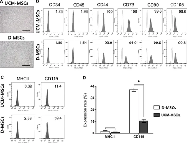
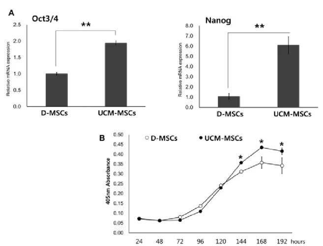
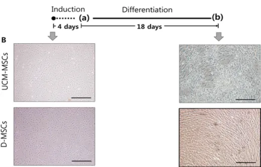
Meterials and Methods: Our study involved three stem cell sources-subacromial bursal tissue, bone marrow, and umbilical cord-derived stem cells. Under consent we