Original Article
Dosimetric Effects of Intrafractional Organ Motion in Field-in-Field Technique for Whole-Breast Irradiation
Chae-Seon Hong
1,2, Sang Gyu Ju
1, Doo Ho Choi
1, Youngyih Han
1, Seung Jae Huh
1, Won Park
1, Yong Chan Ahn
1, Jin Sung Kim
1,2, Do Hoon Lim
11Department of Radiation Oncology, Samsung Medical Center, Sungkyunkwan University School of Medicine, 2Department of Radiation Oncology, Yonsei Cancer Center, Yonsei University College of Medicine, Seoul, Korea
Received 23 September 2019 Revised 27 September 2019 Accepted 27 September 2019
Corresponding author Sang Gyu Ju ([email protected]) Tel: 82-2-3410-2612 Fax: 82-2-3410-2619 Corresponding author Doo Ho Choi
([email protected]) Tel: 82-2-3410-2612 Fax: 82-2-3410-2436
Purpose: We evaluated the motion-induced dosimetric effects on the field-in-field (FIF) technique for whole-breast irradiation (WBI) using actual patient organ motion data obtained from cine electronic portal imaging device (cine EPID) images during treatment.
Materials and Methods: Ten breast cancer patients who received WBI after breast-conserving surgery were selected. The static FIF (SFIF) plan involved the application of two parallel opposing tangential and boost FIFs. To obtain the amplitude of the internal organ motion during treatment, cine EPID images were acquired five times for each patient. The outside contour of the breast (OCB) and chest wall (CW) contour were tracked using in-house motion analysis software.
Intrafractional organ motion was analyzed. The dynamic FIF (DFIF) reflecting intrafractional organ motion incorporated into the SFIF plan was calculated and compared with the SFIF in terms of the dose homogeneity index (DHI90/10) for the target and V20 for the ipsilateral lung.
Results: The average motion amplitudes along the X and Y directions were 1.84±1.09 mm and 0.69±0.50 mm for OCB and 1.88±1.07 mm and 1.66±1.49 mm for CW, respectively. The maximum motion amplitudes along the X and Y directions were 5.53 and 2.08 mm for OCB and 5.22 and 6.79 mm for CW, respectively. Significant differences in DHI90/10 values were observed between SFIF and DFIF (0.94 vs 0.95, P<0.05) in statistical analysis. The average V20 for the lung in the DFIF was slightly higher than that of the SFIF in statistical analysis (19.21 vs 19.00, P<0.05).
Conclusion: Our findings indicate that the FIF technique can form a safe and effective treatment method for WBI. Regular monitoring using cine EPID images can be effective in reducing motion- induced dosimetric errors.
Keywords: Dose variation, Field-in-field technique, Organ motion, Whole-breast irradiation
Copyright © 2019 Korean Society of Medical Physics
CCThis is an Open-Access article distributed under the terms of the Creative Commons Attribution Non-Commercial License (http://creativecommons.org/licenses/by- nc/4.0) which permits unrestricted non-commercial use, distribution, and reproduction in any medium, provided the original work is properly cited.
Introduction
The whole-breast irradiation (WBI) technique has at- tracted considerable interest because breast cancer is one of the most prevalent cancers worldwide; the number of breast cancer patients has rapidly increased in recent times.
1)WBI has played an important role in minimizing the risk of ipsilateral recurrence after breast-conserving
surgery.
2-5)WBI has traditionally been performed with tan- gential irradiation (TI), which involves the application of parallel opposing half-beam wedge fields. Conventional TI is very simple and convenient, but dose inhomogeneity in the target volume resulting from tissue heterogeneity in the irradiated volume and the difference in beam path lengths is an unavoidable demerit. Overdosage can lead to un- wanted cosmetic outcomes and side effects.
6,7)In addition,
Progress in Medical Physics 30(3), September 2019 https://doi.org/10.14316/pmp.2019.30.3.65 eISSN 2508-4453
TI is not suitable to modify the beam intensity at specific regions, and modification is generally used for reducing the radiation dose to normal organs.
With this background, Oliver et al. employed intensity- modulated radiation therapy (IMRT) for WBI to obtain a homogeneous dose distribution in the target volume and reduce dosage to normal healthy tissue.
8-11)However, while IMRT is advantageous from the dosimetric point of view, the technique suffers from the demerits of long planning and treatment time, large number of MUs, and relative complexity of treatment compared with TI.
12)Mihai et al.
13)introduced the forward intensity-modulated radiation therapy (FIMRT) technique for WBI. FIMRT essentially involves the application of parallel opposing open fields to cover the entire target volume and sub-boost fields, which are shaped by a multi-leaf collimator to compensate for dose inhomogeneity within the target volume and re- duce dosage to normal organs. In comparison with IMRT, FIMRT offers simplicity of treatment technique, good dose homogeneity in the target volume, a small number of MUs, and reduced delivery time. Prabhakar et al. examined dif- ferent types of FIMRT and proposed the field-in-field (FIF) technique, which offers all the advantages of the IMRT, FIMRT, and TI techniques.
14,15)The target volume for WBI includes the tumor bed and the whole-breast tissue on the CW. The target volume is expected to be subject to geometrical uncertainty because of the influence of respiratory organ motion.
16)The IMRT, FIMRT, and FIF techniques are disadvantageous when considering organ motion in comparison with simple TI because their beams consist of many sub-fields of differ- ent radiation intensities to obtain the desirable dose dis- tribution within the treatment volume. Jain et al.
17)have reported that the organ motion effect is not significant from the dosimetric point of view when FIMRT is used for WBI. Song et al.
18)conducted similar research on the FIF technique and reported significant dose variation because of respiratory organ motion by applying speculative mo- tion values (1, 2, and 3 cm) instead of actual patient data.
However, to the best of our knowledge, no study has ad- dressed the dosimetric effects of target motion based on actual patient motion data with patient-specific treatment beam conditions. Thus, the dosimetric effects of respira-
tory organ motion need to be evaluated by applying actual patient data for the breast FIF technique to ensure better outcomes and safer treatment. We evaluated the motion- induced dosimetric effects on the breast FIF technique using actual patient organ motion data obtained from cine electronic portal imaging device (cine EPID) images during treatment.
Materials and Methods
1. Computed tomography simulation and treatment planning
Ten breast cancer patients were randomly selected (five right-breast and five left-breast cancer patients) for WBI in this study after breast-conserving surgery (details are listed in Table 1). A planning computed tomography (CT) was performed for each patient in the supine position with the arm up on the breast board (Medtech, USA). All CT images were transferred to the treatment planning system (TPS, Pinnacle3, Philips Medical Systems, Milpitas, CA, USA).
We delineated the clinical target volume (CTV) including
the tumor bed and breast tissue and organs at risk (OARs)
including the ipsilateral lung and heart. The FIF technique,
one of the FIMRT techniques, was applied in combination
with TI (Fig. 1a) and sub-boost fields (Fig. 1b, c) to improve
dose distribution, as described below. First, TI beams were
generated via the application of two open parallel oppos-
ing half-beams (6-MV photons, CL600, Varian, USA). The
initial dose distribution was calculated with equal beam
weights. Next, an isodose cloud was displayed on the digi-
tal reconstruction radiograph (DRR). Our plan criterion
for WBI was that the dose received by the CTV should lie
in the range of 95% to 107% of the prescribed dose. There-
fore, an isodose cloud outside this range was acquired,
and the dose was compensated manually using a multi-
leaf collimator (MLC) (Fig. 1). In other words, when the
CTV received a dose less than 95% of the prescribed dose,
a sub-field was added to compensate for the dose, and
when the CTV dose exceeded 107%, shielding was applied
via the MLC. The total number of sub-fields was limited to
less than two for the treatment plan of each patient. Fur-
thermore, we ensured that the total beam weight did not
exceed 10% of the entire beam weight. A total of 50 Gy with 25 fractions was prescribed for the CTV.
2. Measurement of intrafractional motion
Cine EPID images were used to measure the intrafrac- tional motion of the breast. Cine EPID imaging is used to obtain continuous images with the beam used in the actual treatment with an EPID (aS500, Varian, USA) during treat- ment, and the technique is suitable for real-time verifica- tion of organ motion.
19)Cine EPID images were obtained at a rate of 3.3 frames/s once a week per patient five times during the entire treatment schedule.
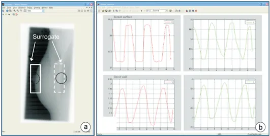
We developed an in-house motion analysis software
package to extract the motion of the breast from the ac- quired sequential cine EPID images (Matlab, MathWorks, USA) (Fig. 2). The outside contour of the breast (OCB) and the chest wall (CW) at the central axis of the beam were set as the region of interest (ROI), and the motion of the ROI in the sequential cine EPID was tracked by applying a pattern matching algorithm. Variations along the X and Y direc- tions were measured with respect to the vertical axis of the tangential beam direction, and based on these values, the maximum amplitude, mean amplitude, and the standard deviation were calculated.
a b c
Fig. 1. Design of field-in-field (FIF) treat ment plan. The FIF plan in volves the application of (a) two tan gen tial beams and (b, c) sub-boost fields to com pensate for the dose in homo ge- neity in the target and reduce the lung dose.
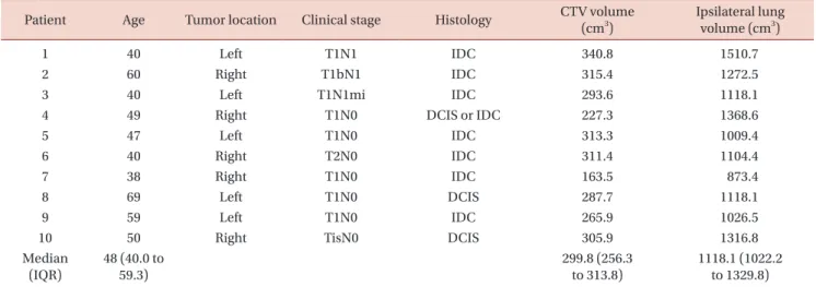
Table 1. Patients’ characteristics
Patient Age Tumor location Clinical stage Histology CTV volume
(cm3)
Ipsilateral lung volume (cm3)
1 40 Left T1N1 IDC 340.8 1510.7
2 60 Right T1bN1 IDC 315.4 1272.5
3 40 Left T1N1mi IDC 293.6 1118.1
4 49 Right T1N0 DCIS or IDC 227.3 1368.6
5 47 Left T1N0 IDC 313.3 1009.4
6 40 Right T2N0 IDC 311.4 1104.4
7 38 Right T1N0 IDC 163.5 873.4
8 69 Left T1N0 DCIS 287.7 1118.1
9 59 Left T1N0 IDC 265.9 1026.5
10 50 Right TisN0 DCIS 305.9 1316.8
Median (IQR)
48 (40.0 to 59.3)
299.8 (256.3 to 313.8)
1118.1 (1022.2 to 1329.8) CTV, clinical target volume; IDC, invasive ductal carcinoma; DCIS, ductal carcinoma in situ; IQR, interquartile range (Q1, Q3).
3. Analysis of motion-induced dosimetric effects
To analyze the dosimetric effects of the intrafractional variation due to respiration with regard to the FIF tech- nique, a dynamic FIF (DFIF) plan was generated; this plan reflects the motion measured by cine EPID onto the static FIF (SFIF) plan, which does not reflect organ motion. In other words, DFIF dose distribution was recalculated by adjusting the position of the sub-fields based on each patient’s specific movement values, as measured by cine EPID (the maximum amplitude of motion for the OBC and CW). We calculated the cumulative dose-volume histogram (DVH) for PTV and OARs from both SFIF and DFIF for all cases. Furthermore, for quantitative analysis of motion-induced dosimetric effects, the dose homogeneity index (DHI) for CTV and the percentage volume receiving over 20 Gy (V
20) for the ipsilateral lung were calculated and compared for both techniques.
18,20)DHI= D
90 D10DHI indicates dose uniformity within the CTV, and it is defined as the ratio of the treatment volume receiving 10%
of the prescription dose (D
10) to that receiving 90% (D
90). A DHI value of 1 is an ideal value that indicates uniform dose distribution within the CTV. To assess whether there is sig- nificant difference between the SFIF and DFIF, a Wilcoxon signed-rank test (version 9.4, SAS Institute, Cary, NC) was performed. A P-value of <0.05 was considered statistically significant.
Results
1. Measurement of intrafractional motion
We analyzed all 50 sets of cine EPID images obtained for the 10 patients and determined that the mean maximum motion amplitudes in all patients were 1.84±1.09 mm and 0.69±0.50 mm along the X and Y directions in the OCB, respectively, and 1.88±1.07 mm and 1.66±1.49 mm in the CW, respectively (Table 2). When the motion of the CW was slightly greater than that of the OCB, the error along
a b
Surrogate
Fig. 2. (a) In-house motion analysis soft ware and (b) the result of motion an alysis for whole-breast irradiation.
Solid-lines (breast skin contour) and dotted (chest wall contour) boxes re- pre sent surrogates for organ mo tion tracking based on a pattern match ing algorithm.
Table 2. Statistics of intrafractional movement from cine-EPID im- ages obtained using in-house motion analysis software for whole- breast irradiation
Patient
Movement of breast skin (mm)
Movement of chest wall (mm)
X Max. Y Max. X Max. Y Max.
1 5.53 0.84 5.22 1.69
2 3.13 2.08 2.61 6.79
3 1.57 1.55 2.09 2.58
4 2.68 0.52 2.23 1.07
5 1.57 1.04 1.57 3.66
6 1.65 0.450 2.13 1.00
7 2.61 2.08 2.08 2.08
8 0.54 0.54 1.17 0.66
9 3.24 0.92 3.35 1.73
10 1.69 0.45 1.73 0.76
Average 1.84 0.69 1.88 1.66
SD 1.09 0.50 1.07 1.49
Max 5.53 2.08 5.22 6.79
Max., maximum difference; X, perpendicular to tangential beam direction; Y, superior-inferior direction.
the X direction was greater than that along the Y direction.
The statistics of each patient’s motion showed a consider- able difference. In patient #2, the CW motion was 6.79 mm along the Y direction, whereas it was 0.66 mm in patient #8, which corresponded to a large difference of about 6 mm.
Moreover, patients with large OCB motion generally exhib- ited large CW motion as well. Among all the patients, the maximum OCB motion amplitudes were 5.53 and 2.08 mm and the maximum CW motion amplitudes were 5.22 and 6.79 mm along the X and Y directions, respectively.
2. Analysis of motion-induced dosimetric effects
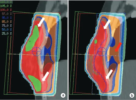
We used a total of 20 sub-fields in this study to perform the FIF plan, which corresponds to 2 sub-fields per patient on an average. In most patients, prominent improvement in the dose distribution, reduction in dose inhomogeneity within the target (white arrow in Fig. 3), and reduction in the lung dose (red arrow in Fig. 3b) were observed because of the addition of the sub-fields.
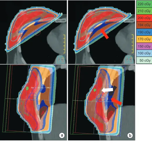
When the DFIF (Fig. 4b) was calculated by applying the maximum intrafractional movement measured from each patient to the SFIF (Fig. 4a), variations were observed not only in the dose distribution within the target but also in
the lung dose (red arrow). Such a dose change was evident in the DVH. In one extreme case, the ipsilateral lung dose and target dose slightly increased (Fig. 5).
The mean DHI
90/10value for the target with SFIF for all 10 patients was 0.94±0.01 (Table 3). The mean DHI
90/10with DFIF as generated for maximum movement along the +X direction was 0.94±0.01, and it was 0.95±0.01 along the -X direction. The mean DHI
90/10with DFIF as calculated for maximum movement along the +X and +Y directions si- multaneously was 0.94±0.01 (P>0.05), and it was 0.95±0.01 (P<0.05) for simultaneous maximum movement along the -X and -Y directions. However, the differences between all these values did not exceed 1%.
The V
20value of the ipsilateral lung with SFIF was 19.00±7.16. The V
20values of the ipsilateral lung with DFIF were 18.89±7.16 and 19.21±7.17 for the maximum move- ment along the +X and
−X directions, respectively. Further- more, the V
20value was 18.90±7.15 for simultaneous move- ment along the +X and +Y directions, and it was 19.20±7.18 for simultaneous movement along the -X and -Y directions (P<0.05). However, the differences between all these values did not exceed 1%.
a b
Fig. 3. Comparison of dose distribu- tion (sagittal view) between the appli ca tion of (a) a conventional tan- gen tial field and (b) a conventional field-in-field (FIF) boost technique.
Regions subject to high dosage with the conventional field exhibited a signi fi cant reduction with the FIF tech nique (white arrow) along with signi fi cant reduction in the lung dose also (red arrow).
Discussion
Many researchers have reported organ movement dur-
ing WBI. Saliou et al.
21)obtained portal films of TI during treatment, analyzed the central lung distance (CLD), and reported movements of 0.8~10 mm along the anterior–
posterior direction. Smith et al. obtained a total of 1,709 electronic portal images, analyzed the CLD, and observed a maximum CLD variation of 2.5 mm. This result indicates
220 cGy 210 cGy 200 cGy 194 cGy 190 cGy 170 cGy 150 cGy 100 cGy 50 cGy
a b
Fig. 4. Comparison of dose distribu- tion between (a) static FIF (SFIF) and (b) dynamic FIF (DFIF). A partial dose increase in the lung (red arrow) and dose decrease in the target (white arrow) were observed for the DFIF plan because of organ motion.
0 1.0 0.9 0.8 0.7 0.6 0.5 0.4 0.3 0.2 0.1
Norm.volume
Dose (cGy) 0.0
SFIF DFIF
250 200
150 100
50
Dose volume histogram
CTV
Ipsilateral lung
Fig. 5. An extreme example of dose-volume histogram (DVH) for target volume and lung. The ipsilateral lung dose and target dose increased slightly in dynamic FIF (DFIF, dotted line, applied maxi- mum of the organ motion along -X direction) compared to static FIF (SFIF).
Table 3. Dose variation due to organ motion Plan Movement Average DHI90/10
for target
Average V20
for lung (%)
Static FIF None 0.94±0.01 19.00±7.16
Dynamic FIF X+max 0.94±0.01 18.89±7.16 X-max 0.95±0.01 19.21±7.17 XY+max 0.94±0.01 18.90±7.15 XY-max 0.95±0.01 19.20±7.18 DHI90/10, dose homogeneity index; SFIF, static field-in-field;
DFIF+XYmax, dynamic field-in-field applied max. organ motion am- pli tude along +X and +Y directions; DFIF-XYmax, dynamic field-in- field applied max. organ motion amplitude along -X and -Y di rec- tions.
A significant dif fer ence in DHI90/10 for CTV and V20 for lung was observed bet ween the SFIF and DFIF (P<0.05) in statistical analysis, but the dif fer ence in values was clinically acceptable.
that little fluctuation was caused by breathing during treat- ment.
22)Baroni et al. attached markers on breast skin and monitored their motion in real time using an opto-elec- tronic system. Their analysis on motion caused by breath- ing during treatment showed that the median was consis- tent at 2-3 mm and exhibited the highest value along the anterior–posterior direction.
23)Richter et al. analyzed the motion of the chest using four-dimensional CT (4DCT) in 10 patients, and the maximum motion amplitude was less than 4 mm in all patients. Furthermore, they compared the motion amplitude obtained via cine EPID images to that obtained via 4DCT. The results were similar in both cas- es.
24)In our study, we obtained an average motion of less than 2 mm, which was consistent with the results of other studies. However, the maximum amplitude of motion was significantly different from patient to patient. In addition, considerable motion was observed in some patients.
One of the demerits of treatment techniques that use sub-fields, such as IMRT and FIF, is that they are more sen- sitive to dosimetric error by intrafractional motion than TI.
Although the FIF uses only a few sub-fields, their influence cannot be completely excluded. The interplay effect, which is the mismatch between the target motion and delivery timing of these multiple sub-fields, is strongly associated with not only organ motion but also patient-specific con- ditions including treatment techniques and anatomical geometry.
25,26)The changes in dose distribution because of respiratory motion have been investigated in many stud- ies. However, the results are inconsistent because these studies employed different types of information for do- simetric evaluation instead of actual patient data.
16,18,27,28)To overcome this issue, we evaluated the motion-induced dosimetric effects on the breast FIF technique by utilizing actual patient planning and organ motion data obtained from cine EPID images during treatment. Because of respi- ration, the DHI of the target volume exhibited a maximum difference of 1.06%, and the dosage of the ipsilateral lung exhibited a partial increase when the sub-field was applied along the direction of the chest wall close to the lung. How- ever, these differences were not clinically significant.
To assess the motion-induced dosimetric error, accurate measurement of motion needs to be performed regularly.
The cine EPID imaging used in this study does not require
special equipment because only the treatment beam is used, and the method does not trigger additional radia- tion exposure to patients. In particular, it is more effective for the breasts because the entire soft tissue of the breasts forms the treatment region and high-quality images can be obtained from the entire treatment field because of differences in density with respect to the lung behind the breasts.
We evaluated the effects of motion-induced dosimetric error by applying actual patient motion data that were ob- tained via cine EPID images for FIF WBI. Dose variation with regard to the FIF technique due to respiratory organ motion was observed in both the target and lung volumes as per statistical analysis; however, the difference in values was clinically acceptable. Our findings indicate that FIF can form a safe and effective treatment method for the radiation treatment of breasts as it can improve dose distri- bution within the target volume and reduce normal organ dose. We believe that the establishment of motion criteria and regular monitoring using cine EPID images can be ef- fective in reducing motion-induced dosimetric errors.
Acknowledgements
This work was supported by the research program, NRF- 2015R1C1A1A02036613.
Conflicts of Interest
The authors have nothing to disclose.
Availability of Data and Materials
All relevant data are within the paper and its Supporting Information files.
References
1. International Agency for Research on Cancer (IARC) and World Health Organization (WHO). GLOBOCAN 2012:
Estimated cancer incidence, mortality and prevalence worldwide 2012;Available at: http://globocan.iarc.fr/Pag- es/fact_sheets_cancer.aspx.
2. Blichert-Toft M, Rose C, Andersen JA, Overgaard M, Axels- son CK, Andersen KW, et al. Danish randomized trial comparing breast conservation therapy with mastectomy:
six years of life-table analysis. Danish Breast Cancer Co- operative Group. J Natl Cancer Inst Monogr 1992:19-25.
3. Fisher B, Anderson S, Bryant J, Margolese RG, Deutsch M, Fisher ER, et al. Twenty-year follow-up of a random- ized trial comparing total mastectomy, lumpectomy, and lumpectomy plus irradiation for the treatment of invasive breast cancer. N Engl J Med 2002;347:1233-1241.
4. Veronesi U, Cascinelli N, Mariani L, Greco M, Saccozzi R, Luini A, et al. Twenty-year follow-up of a randomized study comparing breast-conserving surgery with radical mastectomy for early breast cancer. N Engl J Med 2002;347:
1227-1232.
5. Fisher B, Anderson S, Redmond CK, Wolmark N, Wicker- ham DL, Cronin WM. Reanalysis and results after 12 years of follow-up in a randomized clinical trial comparing total mastectomy with lumpectomy with or without irradiation in the treatment of breast cancer. N Engl J Med 1995;333:
1456-1461.
6. Taylor ME, Perez CA, Halverson KJ, Kuske RR, Philpott GW, Garcia DM, et al. Factors influencing cosmetic results after conservation therapy for breast cancer. Int J Radiat Oncol Biol Phys 1995;31:753-764.
7. Park W, Huh SJ, Yang JH, Nam SJ, Kim JH, Choi JY, et al.
The implication of hot spots on bone scans within the ir- radiated field of breast cancer patients treated with mas- tectomy followed by radiotherapy. Ann Nucl Med 2008;22:
685-691.
8. Oliver M, Chen J, Wong E, Van Dyk J, Perera F. A treatment planning study comparing whole breast radiation therapy against conformal, IMRT and tomotherapy for accelerated partial breast irradiation. Radiother Oncol 2007;82:317- 323.
9. Prabhakar R, Haresh KP, Julka PK, Ganesh T, Rath GK, Joshi RC, et al. A study on contralateral breast surface dose for various tangential field techniques and the impact of set-up error on this dose. Australas Phys Eng Sci Med 2007;
30:42-45.
10. Harsolia A, Kestin L, Grills I, Wallace M, Jolly S, Jones C, et al. Intensity-modulated radiotherapy results in significant decrease in clinical toxicities compared with conventional
wedge-based breast radiotherapy. Int J Radiat Oncol Biol Phys 2007;68:1375-1380.
11. Kestin LL, Sharpe MB, Frazier RC, Vicini FA, Yan D, Matter RC, et al. Intensity modulation to improve dose uniformity with tangential breast radiotherapy: initial clinical experi- ence. Int J Radiat Oncol Biol Phys 2000;48:1559-1568.
12. Haciislamoglu E, Colak F, Canyilmaz E, Dirican B, Gurdal- li S, Yilmaz AH, et al. Dosimetric comparison of left-sided whole-breast irradiation with 3DCRT, forward-planned IMRT, inverse-planned IMRT, helical tomotherapy, and volumetric arc therapy. Radiother Oncol 2011;100:241-246.
13. Mihai A, Rakovitch E, Sixel K, Woo T, Cardoso M, Bell C, et al. Inverse vs. forward breast IMRT planning. Med Dosim 2005;30:149-154.
14. Onal C, Sonmez A, Arslan G, Oymak E, Kotek A, Efe E, et al. Dosimetric comparison of the field-in-field technique and tangential wedged beams for breast irradiation. Jpn J Radiol 2012;30:218-226.
15. Prabhakar R, Julka PK, Rath GK. Can field-in-field tech- nique replace wedge filter in radiotherapy treatment plan- ning: a comparative analysis in various treatment sites.
Australas Phys Eng Sci Med 2008;31:317-324.
16. Michalski A, Atyeo J, Cox J, Rinks M. Inter- and intra-frac- tion motion during radiation therapy to the whole breast in the supine position: a systematic review. J Med Imaging Radiat Oncol 2012;56:499-509.
17. Jain P, Marchant T, Green M, Watkins G, Davies J, McCar- thy C, et al. Inter-fraction motion and dosimetric conse- quences during breast intensity-modulated radiotherapy (IMRT). Radiother Oncol 2009;90:93-98.
18. Song T, Suh CO, Lee I, Jeong K, Keum K, Lee CG, et al. The effect of respiratory motion on forward intensity modu- lated radiotherapy for breast cancer. Technol Cancer Res Treat 2008;7:207-215.
19. Mitchell J, Formenti SC, DeWyngaert JK. Interfraction and intrafraction setup variability for prone breast radiation therapy. Int J Radiat Oncol Biol Phys 2010;76:1571-1577.
20. Kahan Z, Csenki M, Varga Z, Szil E, Cserhati A, Balogh A, et al. The risk of early and late lung sequelae after confor- mal radiotherapy in breast cancer patients. Int J Radiat Oncol Biol Phys 2007;68:673-681.
21. Saliou MG, Giraud P, Simon L, Fournier-Bidoz N, Fourquet A, Dendale R, et al. Radiotherapy for breast cancer: respi-
ratory and set-up uncertainties. Cancer Radiother 2005;9:
414-421.
22. Smith RP, Bloch P, Harris EE, McDonough J, Sarkar A, Kassaee A, et al. Analysis of interfraction and intrafrac- tion variation during tangential breast irradiation with an electronic portal imaging device. Int J Radiat Oncol Biol Phys 2005;62:373-378.
23. Baroni G, Ferrigno G, Orecchia R, Pedotti A. Real-time opto-electronic verification of patient position in breast cancer radiotherapy. Comput Aided Surg 2000;5:296-306.
24. Richter A, Sweeney R, Baier K, Flentje M, Guckenberger M. Effect of breathing motion in radiotherapy of breast cancer: 4D dose calculation and motion tracking via EPID.
Strahlenther Onkol 2009;185:425-430.
25. Berbeco RI, Pope CJ, Jiang SB. Measurement of the inter- play effect in lung IMRT treatment using EDR2 films. J Appl Clin Med Phys 2006;7:33-42.
26. Court LE, Seco J, Lu XQ, Ebe K, Mayo C, Ionascu D, et al.
Use of a realistic breathing lung phantom to evaluate dose delivery errors. Med Phys 2010;37:5850-5857.
27. Chui CS, Yorke E, Hong L. The effects of intra-fraction organ motion on the delivery of intensity-modulated field with a multileaf collimator. Med Phys 2003;30:1736-1746.
28. Cao J, Roeske JC, Chmura SJ, Salama JK, Shoushtari AN, Boyer AL, et al. Calculation and prediction of the effect of respiratory motion on whole breast radiation therapy dose distributions. Med Dosim 2009;34:126-132.