ICR 마우스 췌장 내분비세포에 대한 면역조직화학적 연구
구세광, 이형식1, 이재현2
동화약품 ㈜ 중앙연구소 약리독성연구실, 1경산대학교 자연과학부 생물학과
2경북대학교 수의과대학 조직학교실
(게재승인 : 2002년 1월 9일)
Immunohistochemical study of the pancreatic endocrine cells in the ICR mice
Sae-kwang Ku, Hyeung-sik Lee
1and Jae-hyun Lee
2Pharmacology & Toxicology Lab., Central Research Laboratories, Dong-Wha Pharm. Ind. Co
1
Department of Biology, Faculty of Natural Sciences, Kyungsan University
2
Department of Histology, College of Veterinary Medicine, Kyungpook National University
(Accepted : January 9, 2002)Abstract : The regional distribution and relative frequency of the pancreatic endocrine cells in the ICR mouse
were studied by immunohistochemical (PAP) method using four types of specific antisera against insulin, glucagon, somatostatin and human pancreatic polypeptide (PP). The pancreas of mice could be divided into three portions; pancreatic islets, exocrine and pancreatic ducts. Pancreatic islets, furthermore, were subdivided into three regions (central, mantle and peripheral region) according to their located types of immunoreactive cells. In the pancreatic islet portions, insulin-immunoreactive cells were located in the central and mantle regions but most of somatostatin-, glucagon- and PP-immunoreactive cells were detected in the mantle and peripheral regions with various frequencies. In addition, PP-immunoreactive cells were also found in the central regions of pancreatic islets of ICR mouse. In the exocrine portions, all four types of immunoreactive cells were demonstrated in the ICR mouse. In the pancreatic duct portions, insulin- and glucagon-immunoreactive cells were situated in the epithelial lining of ICR mouse with a few and rare frequencies, respectively. In addition, rare PP-immunoreactive cells were also demonstrated in the subepithelial regions of the pancreatic duct. However, no somatostatin-immunoreactive cells were demonstrated.Key words : ICR mouse, pancreatic endocrine cell, immunohistochemistry
국문초록 : ICR 마우스 췌장에 존재하는 내분비세포의 부위별 분포 및 상대적 빈도를 4 종류의 항혈 청 즉, insulin, glucagon, somatostatin 및 human pancreatic polypeptide (PP)을 사용하여 면역조직화학적 방법으로 관찰하였다. 마우스의 췌장은 췌장섬, 외분비부 및 췌관의 3 부분으로 구별되었으며, 췌장 섬은 이들 면역반응세포들의 출현 부위에 따라 다시 중심부분, mantle부분 및 가장자리부분으로 다 시 세분되었다. 췌장섬의 경우, insulin 면역반응세포들은 주로 췌장섬의 중심부분과 mantle 부분에서 주로 관찰되었으나, somatostatin, glucagon 및 PP 면역반응세포들은 다양한 출현빈도를 나타내며 주 로 mantle부분과 가장자리부분에 걸쳐 관찰되었다. 또한 극소수의 PP 면역반응세포들은 췌장섬의 중심 부분에서도 관찰되었다. 외분비부에서도 insulin, glucagon, somatostatin 및 PP 면역반응세포들 모두 관 찰되었으며, 주로 외분비 샘포 세포 사이공간에서 관찰되었다. 췌관에서는 insulin 및 glucagon 면역반 응세포들이 소수 또는 극소수의 빈도로 췌관 상피세포 사이공간에서 관찰되었으며, 극소수의 PP 면역 반응세포들 역시 췌관상피 아래부위에서 관찰되었으나, somatostatin 면역반응세포들은 이 부위에서 관 찰되지 않았다.
Key words : ICR mouse, pancreatic endocrine cell, immunohistochemistry
21
INTRODUCTION
The mouse, Mus musculus, is a rodent, order Rodentia, of the family Muridae. Commensal and wild mice have spread around the world, and the laboratory or house mice are kept occasionally as a pet. Since mice are small, prolific, maintained easily and economical in large populations, and possess great genetic diversity characterized anatomically and physiologically, they are the most widely used vertebrates in biochemical research and test. It is generally accepted that ICR, an abbreviated form of institute of cancer research, mouse is one of the most widely used strain and representative closed colony1.
It is generally known that pancreas of vertebrates is subdivided into two portions, one is exocrine portions where digestive enzymes are released and the other is endocrine portions where regulatory hormones such as insulin, glucagon, somatostatin and pancreatic polypeptide (PP) are released into blood circulation. The appearance, regional distribution and relative frequency of these regulatory hormones secreted by endocrine cells in the pancreas were well recognized by histochemistry2, immuno- fluorescence method3 and immunohistochemistry4. Except above regulatory hormones, peptide YY-, neuropeptide YY-5, motilin-6 and chromogranin family-7, 8 immunoreactive cells were also demonstrated in the vertebrate pancreas.
The pancreas has been treated as a valuable organ for endocrine studies and endocrine pancreas has been extensively studied, associated with diabetes9. In addition, the investigations of gastroenteropancraetic endocrine cells have been considered as an important part of a phylogenetic studies10.
Until now, the regional distribution and relative frequency of major four types of endocrine cells, insulin, glucagon, somatostatin and PP, were reported in the pancreas of the hamster11, wood mouse (Apodemus speciosus)12, C57BL/6 mouse13, preobese and obese yellow Avy/-mouse14, vole (Microtus arvalis)15, obese ob+/ob+ mouse16, sand rat (Psammomys obesus)17, herbivorous Japanese field vole (Microtus montebelli)18 and guinea pig19. In addition, angiotensin Ⅱ-immunoreactive cells were found in the pancreas of mouse20 and appearances of calcitonin gene- related peptide- and cholecystokinin-immunoreactive cells in the rat pancreas were also reported, respectively21, 22. With the increasing demands of diabetic animal models in many
fields, the regional distribution and relative frequency of pancreatic endocrine cells, especially insulin- and glucagon- producing cells in the laboratory animals have been a concern in recent years13, 14, 23. It had been accepted that insulin-immunoreactive cells were located in the central regions and the other cells such as glucagon-, somatostatin- and PP-immunoreactive cells were located in the peripheral or mantle regions. But many researchers suggested that species-dependent characteristic distribution of cells producing different hormones in the pancreas of each species of animals might be due to feeding habits, and now they are generally accepted24. In addition, it was also reported that different regional distribution and relative frequency of endocrine cells in the pancreatic islets were demonstrated in different portions of the pancreas even if they were the same pancreas of the same animal12. And strain-dependent characteristic distribution of these immunoreactive cells were also detected with the increasing produce of genetically mutated laboratory animals and breeding of specific laboratory animals having specific disease or unique nature, especially in rat and mouse12-14, 16, 23
. Although many studies have been elucidated the regional distribution and relative frequency of different endocrine cells in the pancreas of the various vertebrates including various species and strains of rodents, the reports are very seldom about immunohistochemical studies on the pancreatic endocrine cells in the ICR mouse in spite of their worldwide usefulness except for immunohistochemical study of the gastrointestinal endocrine cells.
The object of this study was to clarify the regional distribution and relative frequency of the endocrine cells in the pancreas of the ICR mouse by specific immuno- histochemistry using four types of specific antisera against insulin, glucagon, somatostatin and PP.
MATERIALS and METHODS
Five adult ICR mice (7 weeks old, 27-32g body weight upon receipt) were acquired from the Charles River Laboratories (Yokohama, Japan) and they were used in this study without sexual distinction, after acclimatization for one week. Animals were allocated 5 per polycarbonate cage in a temperature (20-25℃) and humidity (30-35%) controlled room during acclimatization periods. Light : dark cycle was 12hr : 12hr and feed (Samyang, Korea) and water was
supplied free to access.
After food restriction about 24 hours, the animals were phlebotomized to bleed under anesthetizing with ethyl ether.
Samples from the pancreas were fixed in Bouin's solution.
After paraffin embedding, 3-4㎛ serial sections were prepared. Representative sections of each tissue were stained with hematoxylin and eosin for light microscopic examination of the normal pancreatic architecture.
Each representative section was deparaffinized, rehydrated and immunostained with the peroxidase anti-peroxidase (PAP) method25. Blocking of nonspecific reaction was performed with normal goat serum prior to incubation with the specific antisera (Table 1). After rinsed in phosphate buffered saline (PBS; 0.01M, pH 7.4), the sections were incubated in secondary antiserum. They were then washed in PBS buffer and finally the PAP complex was prepared.
The peroxidase reaction was carried out in a solution 3,3′
-diaminobenzidine tetrahydrochloride containing 0.01% H2O2
in Tris-HCl buffer (0.05M, pH 7.6). After immunostained, the sections were lightly counterstained with Mayer's hematoxylin and the immunoreactive cells were observed under light microscope.
The specificity of each immunohistochemical reaction was determined as recommended by Sternberger25, inclu- ding the replacement of specific antiserum, which had been preincubated with its corresponding antigen and the relative
frequency of occurrence of each type of immunoreactive cells was placed into one of five categories according to their observed numbers as seen using light microscopy.
RESULTS
In this study, all four kinds of the immunoreactive endocrine cells were detected with the antisera against insulin, glucagon, somatostatin and PP in the pancreas of the ICR mice. The pancreatic islets of this study were distinguished into three distinct layers, central, mantle and peripheral regions with their composition of immunoreactive cells. According to the regions of the pancreas, different regional distribution and relative frequency of these immunoreactive cells were observed and these differences are shown in Table 2. Spherical to spindle or occasionally oval to round-shaped immunoreactive cells were located in the pancreas of the ICR mice.
In the pancreatic islet portion : Insulin-immunoreactive
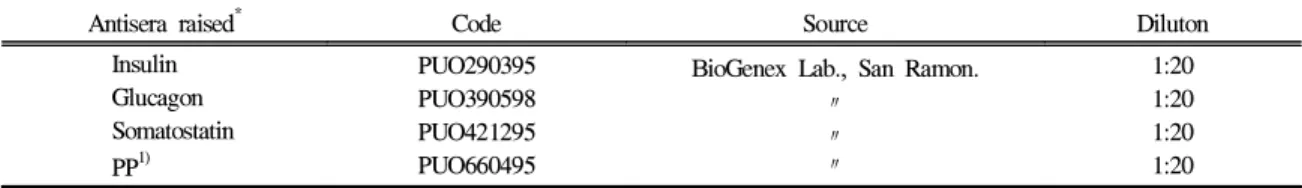
cells were located in the central regions with numerous frequency. In addition, cells with moderate frequency were also demonstrated in the mantle regions intermingled with other immunoreactive cells. However, no insulin- immunoreactive cells were found in the peripheral regions where predominant cell types were PP- immunoreactiveTable 1. Antisera used in this study
Antisera raised* Code Source Diluton
Insulin Glucagon Somatostatin PP1)
PUO290395 PUO390598 PUO421295 PUO660495
BioGenex Lab., San Ramon.
〃
〃
〃
1:20 1:20 1:20 1:20
* All antisera were raised in rabbits except for insulin which was raised in a guinea pig.
1) PP: human pancreatic polypeptide
Table 2. Regional distributions and relative frequencies of the endocrine cells in the pancreas of ICR mouse
Immunoreactive cells Pancreatic islets portionExocrine portion Pancreatic duct portion
Central Mantle Peripheral
Insulin Glucagon Somatostatin PP1)
+++
―
―
±
++
+++
++
±
―
+
±
++
±
±
++
+
+
±
―
±
* Relative frequencies; +++: numerous, ++: moderate, +: a few, ±: rare, : not detected
1) PP: human pancreatic polypeptide.
cells (Fig 1a, b). Glucagon-immunoreactive cells were located in the mantle and peripheral regions of pancreatic islets with numerous and a few frequencies, respectively. In mantle and peripheral regions, cytoplasmic processes of glucagon- immunoreactive cells were intermingled with other immunoreactive cells, especially somatostatin- and PP- immunoreactive cells. However, no glucagon-immunoreactive cells were demonstrated in the central regions where numerous insulin-immunoreactive cells were found (Fig 2a, b). Somatostatin-immunoreactive cells were located in the mantle and peripheral regions with moderate and rare frequencies, respectively. In mantle and peripheral regions, cytoplasmic processes of these immunoreactive cells were intermingled with other immunoreactive cells, especially glucagon- and PP-immunoreactive cells. However, no somatostatin-immunoreactive cells were demonstrated in the central regions where numerous insulin-immunoreactive cells were found (Fig 3a, b). PP-immunoreactive cells were located in the mantle and peripheral regions with rare and moderate frequencies, respectively (Fig 4a-c). Occasionally, rare frequencied round to oval shaped cells were also demonstrated in the central regions (Fig 4a, arrowhead).
In the exocrine portion : Insulin-, glucagon- (Fig 2c),
somatostatin- (Fig 3a, arrowhead) and PP-immunoreactive cells were demonstrated in this portion with rare, rare, moderate and a few frequencies, respectively. They were randomly scattered between pancreatic acinar cells.In the pancreatic duct portion : Insulin- and glucagon-
immunoreactive cells were situated in the lining epithelia with a few and rare frequencies, respectively (Fig 1c, 2d, arrowhead). In addition, PP-immunoreactive cells were demonstrated in subepithelial regions of the pancreatic duct with rare frequency (Fig 4c, arrowheads). However, no somatostatin-immunoreactive cells were found in the pancreatic duct of this study.DISCUSSION
Insulin is synthesized in the B cells of the pancreatic islets and regulates the serum glucose levels26. In the mammals, the regional distribution and relative frequency of insulin-immunoreactive cells in the pancreas were reported in the wood mouse12, hamster11, C57BL/6 mouse13,
voles15, three-toed sloth (Bradypus variegates)27, Australian brush-tailed possum28, opossum29 and laboratory animals24. From these previous reports11-13, 15, 24, 27-29
, it is well recognized that insulin-immunoreactive cells are situated in the central regions of pancreatic islets and other cells, such as glucagon-, somatostatin- and PP-immunoreactive cells, surround them. And they were also demonstrated frequently associated with acinar cells and pancreatic duct. However, somewhat different from other researchers, Reddy et al30 reported that these-immunoreactive cells were observed in most islets where they occurred as groups of cells peripherally and within the pancreatic islets of several marsupial species. In the present study, most of insulin- immunoreactive cells were restricted to the central regions of islets of the ICR mice similar to those of previous rodents11-15, 24 and some of these cells were also found in the lining epithelium of pancreatic duct and exocrine portions.
Glucagon is synthesized in the A cells of the pancreas and regulates glucose levels in blood26. In the present study, glucagon-immunoreactive cells were found in the mantle and peripheral regions of pancreatic islets. Although, glucagon- immunoreactive cells were located in the mantle and peripheral regions of mammalian pancreatic islets, exocrine portions and pancreatic duct11-15, 24, 27-29
, species- dependent variations were also reported. In the equine pancreas, A-cells demonstrated by anti-glucagon were found in the center of pancreatic islets where insulin- immunoreactive cells were numerously found in most vertebrates31. In addition, it was also reported that under specific disease conditions such as obese (diabetic condition) mouse, glucagon cells were intermingled with insulin- immunoreactive cells in the central regions of pancreatic islets, in contrast, normal non-obese littermates showed a peripheral localization16. Our results of distribution and frequency of glucagon-immunoreactive cells were well corresponded to those of previous reports11-15, 24, 27-29
. Somatostatin, which consisted of 14 amino acids, was isolated from hypothalamus of sheep for the first time. It could be divided into straight form and cyclic form32. This substance inhibited the secretion of the gastrin, cholecy- stokinin, secretin, glucagon, insulin, motilin and gastric acid33 and the absorption of amino acid, glucose and fatty acid in the gastrointestinal tract. So far as investigated, somatostatin-immunoreactive cells were located in the
peripheral regions of mammalian pancreatic islets and exocrine portions11-15, 24, 27-29
and well correspond to these previous studies, most of somatostatin cells were found in the mantle zones of the pancreatic islets, where they were intermingled with glucagon- and PP-immunoreactive cells and occupied outmost regions of pancreatic islets.
PP is a peptide hormone containing 36 amino acids, which is synthesized by F cells in the pancreatic islets. The specific function of this peptide is not clear, however, inhibition of food intake has been postulated as a possible function of this peptide26. Polak et al34 reported that they promoted the secretion of gastric acid and stimulated the glycolysis of liver in avian species. It has been revealed that PP-immunoreactive cells were conspicuously distributed in the peripheral regions of pancreatic islets and exocrine portions in mammalian species, if they occurred11-15, 24, 27-29
. In addition, colocalization of these cells with serotonin- immunoreactive cells in the pancreatic islets of the opossum29 and cattle35was also demonstrated. Anyway, da Mota et al27 reported that PP-immunoreactive cells were not found in the pancreas of the three-toed sloth. In the present study, well corresponding to previous studies11-15, 24,
27-29
, although cells with rare frequency were intermingled with other immunoreactive cells in the mantle zone where glucagon-immunoreactive cells were most predominant, most of PP-immunoreactive cells were detected in the outmost regions of pancreatic islets. However, quite different from other rodents11-15, 24, PP cells were also demonstrated in the central regions of pancreatic islets of ICR mouse, although they were showed relatively low frequency.
In conclusion, the regional distribution and relative frequency of pancreatic endocrine cells of the ICR mice were similar to those of other mammals, especially rodents.
However, some peculiar distributional patterns of pancreatic endocrine cells, such as PP-immunoreactive cells were also demonstrated.
LEGENDS for FIGURES
Fig 1. Insulin-immunoreactive cells in the pancreas of the
ICR mice; they were situated in the central and mantle regions of pancreatic islets (a, b) and occasionally located in the lining epithelium of the pancreatic duct (c) and exocrine portions.a, b: × 120, c: × 480, PAP method
Fig 2. Glucagon-immunoreactive cells in the pancreas of
the ICR mice; they were found in the mantle and peripheral regions of pancreatic islets (a, b) and they were also located in the exocrine (c) and pancreatic duct portions (d, arrowhead).a, b: × 120, c: × 480, d: × 240, PAP method
Fig 3. Somatostatin-immunoreactive cells in the pancreas of
the ICR mice; they were located in the similar regions to glucagon-immunoreactive cells in the pancreatic islets (a, b) and exocrine portions (a, arrowhead).a, b: × 120, PAP method
Fig 4. PP-immunoreactive cells in the pancreas of the ICR
mice; most of PP-immunoreactive cells were located in the outmost peripheral regions of pancreatic islets (a-c) but occasionally absolutely low frequencied cells were also found in the central regions of pancreatic islets (a, arrowhead). Some cells were demonstrated in the subepithelial regions of the pancreatic duct (c, arrowhead)a, b: × 120, c: × 240, PAP method
REFERENCES
1. Harkness JE, Wagner JE. The biology and medicine of rabbits and rodents. 4th ed, Wiliams & Wilkins, Baltimore, pp. 50-57, 1995.
2. Kobayashi K, Ali SS. Cell types of the endocrine pancreas in the shark, Scylliorhinus stellaris as revealed by correlative light and electron microscopy.
Cell Tissue Res. 215:475-490, 1981.
3. Orci L. Macro- and micro-domains in the endocrine pancreas. Diabetes. 31:538-564, 1982.
4. Sternberger LA, Hardy PH, Cuculis JJ, et al. The unlabeled antibody enzyme method of immunocytochemistry:
Preparation and properties of soluble antigen-antibody complex (Horseradish peroxidase-antihorseradish pero- xidase) and use in identification of spirochetes. J
Histochem Cytochem. 18:315-333, 1970.
5. Alli-Rachedi A, Varndell IM, Adrian TE, et al. Peptide YY (PYY) immunoreactivity is co-stored with glucagon- related immunoreactants in endocrine cells of the gut and pancreas. Histochemistry. 80:487-491, 1984.
6. Yamada J, Campos VJM, Kitamura N, et al. An immunohistochemical study of endocrine cells in the pancreas of Caiman latirostris (Alligatorinae), with special reference to pancreatic motilin cells. Biomed
Res. 7:199-208, 1986.
7. Rindi G, Buffa R, Sessa F, et al. Chromogranin A, B and C immunoreactivities of mammalian emdocrine cells:
Distribution from costored hormones/prohormones and relationship with argyrophil component of secretory granules. Histochemistry. 85:19-28, 1986.
8. Ito H, Hashimoto Y, Kitagawa H, et al. Distribution of chromogranin containing cells in the porcine gastroen- teropancreatic endocrine system. Jpn J Vet Sci. 50:395- 404, 1987.
9. Jansson L, Sandler S. The influence of cyclosporin A on the vascular permeability of the pancreatic islets and on diabetes induced by multiple low dose of streptozotocin in the mouse. Virchows Archiv A Pathol
Anat Histopathol. 412:225-230, 1988.
10. DEste L, Buffa R, Pelagi M, et al. Immuno- histochemical localization of chromogranin A and B in the endocrine cells of the alimentary tract of the green frog, Rana esculanta. Cell Tissue Res. 277:341-349, 1994.
11. Camihort G, Del Zotto H, Gomez Dumm CL, et al.
Quantitative ultrastructural changes induced by sucrose administration in the pancreatic B cells of normal hamsters. Biocell. 24:31-37, 2000.
12. Yukawa M, Takeuchi T, Watanabe T, et al.
Proportions of various endocrine cells in the pancreatic islets of wood mice (Apodemus speciosus). Anat Histol
Embryol. 28:13-16, 1999.
13. Gomez-Dumm CL, Console GM, Lunna GC, et al.
Quantitative immunohistochemical changes in the endocrine pancreas of nonobese diabetic (NOD) mice.
Pancreas. 11:396-401, 1995.
14. Warbritton A, Gill AM, Yen TT, et al. Pancreatic islet cells in preobese yellow Avy/- mice: relation to adult hyperinsulinemia and obesity. Proc Soc Exp Biol Med.
206:145- 151, 1994.
15. Sasaki M, Arai T, Usui T, et al. Immunohistochemical, ultrastructural, and hormonal studies on the endocrine pancreas of voles (Microtus arvalis) with monosodium aspartate-induced diabetes. Vet Pathol. 28:497-505, 1991.
16. Starich GH, Zafirova M, Jabelenska R, et al. A morphological and immunohistochemical investigation of endocrine pancreas from obese ob+/ob+ mice. Acta
Histochem. 90:93-101, 1991.
17. Donev S, Petkov P, Marquie G, et al. Immuno- histochemical investigations of the endocrine pancreas in normoglycemic sand rat (Psammomys obesus). Acta
Diabetol Lat. 26:309- 313, 1989.
18. Ohara N, Kitamura N, Yamada J, et al. Immunohisto- chemical study of gastroenteropancreatic endocrine cells of the herbivorous Japanese field vole, Microtus
montebelli. Res Vet Sci. 41:21-27, 1986.
19. Reddy SN, Bibby NJ, Elliott RB. Cellular distribution of insulin, glucagon, pancreatic polypeptide hormone and somatostatin in the fetal and adult pancreas of the guinea pig: a comparative immunohistochemical study.
Eur J Cell Biol. 38:301-305, 1985.
20. Leung PS, Chan HC, Wong PY. Immunohistochemical localization of angiotensin Ⅱ in the mouse pancreas.
Histochem J. 30:21-25, 1998.
21. Ding WG, Guo LD, Kitasato H, et al. Phylogenic study of calcitonin gene-related peptide-immunoreactive structures in the pancreas. Histochem Cell Biol.
109:103-109, 1998.
22. Shimizu K, Kato Y, Shiratori K, et al. Evidence for existence of CCK-producing cells in rat pancreatic islets. Endocrinology. 139:389-396, 1998.
23. Fu Q, Honda M, Ohgawara H, et al. Morphological analysis of pancreatic endocrine cells in newborn animals delivered by experimental diabetic rats. Diabetes
Res Clin Pract. 31:57-62, 1996.
24. Wieczorek G, Pospischil A, Perentes EA. Comparative immunohistochemical study of pancreatic islets in laboratory animals (rats, dogs, minipigs, nonhuman primates). Exp Toxicol Pathol. 50:151-172, 1998.
25. Sternberger LA. The unlabeled antibody peroxidase- antiperoxidase (PAP) method. In: Immunocytochemistry (Sternberger LA, ed), John Wiley & Sons, New York, pp. 1040-169, 1979.
26. Hsu WH, Crump MH. The endocrine pancreas. In:
Veterinary endocrinology and reproduction (McDonald LE, Pineda MH, eds), Lea & Febiger, Philadelphia, pp.
186-201, 1989.
27. da Mota DL, Yamada J, Gerge LL, et al. An immuno- histochemical study on the pancreatic endocrine cells of
the three-toed sloth, Bradypus variegatus. Arch Histol
Cytol. 55:203-209. 1992.
28. Leigh CM, Edwin NA. Light-microscopic immunocy- tochemical study of the endocrine pancreas in the Australian brush-tailed possum (Trichosurus vulpecula).
Eur J Histochem. 36:237-241, 1992.
29. Krause WJ, Cutts JH 3rd, Cutts JH, et al. Immuno- histochemical study of the developing endocrine pancreas of the opossum (Didelphis virginiana). Acta
Anat (Basel). 135:84-96, 1989.
30. Reddy S, Bibby NJ, Fisher SL, et al. Immuno- localization of insulin, glucagon, pancreatic polypeptide and somatostatin in the pancreatic islets of the possum,
Trichosurus vulpecula. Gen Comp Endocrinol. 64:157-
162, 1986.31. Helmstaedter V, Feurle GE, Forssmann WG. Insulin-, glucagon- and somatostatin-immunoreactive cells in the
equine pancreas. Cell Tissue Res. 172:447-454, 1976.
32. Brazeau P, Vale W, Burgurs R, et al. Hypothalamic polypeptide that inhibits the secretion of immunoreactive pituitary growth hormone. Science. 179:77-79, 1973.
33. Kitamura N, Yamada J, Calingasan NY, et al.
Immunocytochemical distribution of endocrine cells in the gastrointestinal tract of the horse. Equine Vet J.
16:103-107, 1984.
34. Polak JM, Adrian TE, Bryant MG, et al. Pancreatic polypeptide in the insulomas, gastrinomas and gluca- gonomas. Lancet. 1:328-330, 1976.
35. Nakajima S, Kitamura N, Yamada J, et al. Immuno- histochemical study on the endocrine pancreas of cattle with special reference to coexistence of serotonin and glucagon or bovine pancreatic polypeptide. Acta Anat