http://dx.doi.org/10.6111/JKCGCT.2015.25.3.098
Effect of surface-treatments on flexibility and guided bone regeneration of titanium barrier membrane
Jin-Tae Kim, Byoung Soo Kim, Hee Seok Jeong, Young Ku Heo, Sang-Wan Shin*, Jeong-Yol Lee*, Young Ho Shim* and Deuk Yong Lee**
,†Dental Materials Research Center, Neobiotech Co., Ltd., Seoul 152-582, Korea
*Institute for Clinical Dental Research, Korea University, Seoul 136-701, Korea
**Department of Biomedical Engineering, Daelim University, Anyang 431-715, Korea (Received April 7, 2015)
(Revised May 6, 2015) (Accepted May 8, 2015)
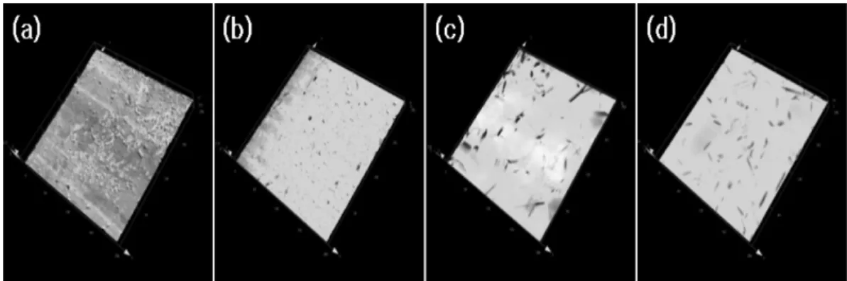
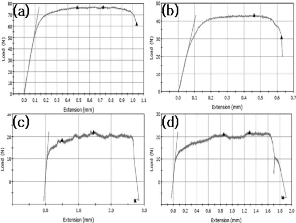
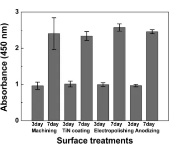
Abstract Titanium barrier membranes are prepared to investigate the effect of surface-treatments, such as machining, electropolishing, anodizing, and electropolishing + TiN coating, on the biocompatibility and physical properties of the membranes. The surface roughness (Ra) of the membrane decreases from machining (0.37 ± 0.09 µm), TiN coating (0.22 ± 0.09 µm), electropolishing (0.20 ± 0.03 µm), to anodizing (0.15 ± 0.03 µm). The highest ductility (24.50 %) is observed for the electropolished Ti membrane. No evidence of causing cell lysis or toxicity is found for the membranes regardless of the surface-treatments. Cell adhesion results of L-929 and MG-63 show that the machined Ti membrane exhibits the highest cell adhesion while the electropolished membrane is the best membrane for the L-929 cell proliferation after 7 days. However, no appreciable difference in MG-63 cell proliferation among variously surface-treated membranes is detected, suggesting that the electropolished Ti membrane is likely to be the best membrane due to the synergic combination of tailored flexibility and excellent fibroblast proliferation.
Key words Pure titanium, Surface-treatment, Roughness, Biocompatibility, Cytotoxicity, Cell proliferation
1. Introduction
Commercially pure titanium ( Cp Ti) has been consid- ered as the biocompatible metallic materials for orthope- dic and dentistry implants because of its excellent mechanical properties, corrosion resistance, and biocom- patibility [1-3]. The excellent biocompatibility of Ti is associated with its stable and inert oxide layer, leading to low level of electronic conductivity, high corrosion resistance, thermodynamic state at physiological pH val- ues, and low ion-formation tendency in aqueous envi- ronment [1]. Ti has been widely used for the implant devices replacing failed hard tissue.
When tooth extraction is required it typically results in additional bone loss in buccal or lingual cortical plate.
The guided bone regeneration (GBR) using the barrier membrane is useful in bone augmentation [4-6]. The GBR and the implant fixation at an infected site are fre- quently complicated by soft-tissue dehiscence, mem- brane exposure, and implant failure due to deficient
mature bone mass and quality to support the implant.
The principle of the GBR is the creation and mainte- nance of sufficient space underneath the barrier.
The mechanical properties of the membranes should avoid collapse of the membrane and provide a constant volume underneath it. The porous areas are small enough to prevent soft tissue penetration through the membrane with no compromise in diffusion of interstitial fluid [4- 6]. The non-absorbable or the resorbable barrier mem- branes are likely to collapse due to improper mechani- cal properties, resulting in insufficient space underneath the membrane for new bone to form [4]. The barriers should be left in place for up to 6 months for bone regeneration. Among the membranes, a Ti membrane with microperforation is used as the barrier because of the rigidity of Ti membrane. It makes the barrier suit- able to maintain the space and cells and fluids necessary for nourishment can be passed through the perforated holes. The porous membranes are attributed to their osteoconductive properties to facilitate the migration of osteoblasts from surrounding bone into the implant site [7, 8]. However, the Ti barrier membranes need a sec- ondary surgery to remove the barrier. In addition, it is difficult to insert the Ti membrane than flexible mem-
†
Corresponding author
†
Tel: +82-31-467-4835
†
Fax: +82-31-467-4432
†
E-mail: [email protected]
2. Materials and Methods 2.1. Materials
Ti specimens with a dimension of 10( φ) × 0.15(t) mm
2are prepared from grade 2 titanium [1]. The specimens for in-vitro test are then surface-treated (electropolishing, anodizing, electropolishing + TiN coating) to investigate the effect of surface roughness on the biocompatibility of the Ti membrane. The samples without any surface treatments are used as control group. Prior to the test, the specimens are γ-ray sterilized [9,10].
The electrolyte for electropolishing is prepared by mixing H
2SO
4, H
3PO
4, and distilled water. The speci- mens are then electropolished for 20~40 s at 75~85
oC with a voltage of 7 V and currents of 40~50 A. Then, they are thoroughly rinsed with distilled water and neu- tralized with NaOH, followed by rinsing with distilled water and drying. Prior to anodizing, the specimens are cleaned by dipping in trichloroethylene for 15 min, fol- lowed by surface activation in a solution mixture of HF and HNO
3for 30 s. The Ti specimen is then anodized for 20~30 s in potentiostatic mode (4~5 V and 3~5 A) at room temperature in the electrolytes (H
3PO
4, H
2SO
4, H
2O). After anodizing, the specimens are neutralized with NaOH and then dried. For TiN coating, the as-elec- tropolished specimens are first surface cleaned by soni- cating for 10 min using ethanol and acetone, followed by Ar plasma treatment for 10 min under vacuum (3 × 10
−5torr). Then, the TiN coated specimens are obtained for 5 min at 300
oC in N
2gas atmosphere (10~20 mtorr).
2.2. Surface roughness and tension test
The laser surface roughness tester (Lext OLS4100, Olympus, Japan) and the electron microscopy (CX-100S, Coxem, Korea) are employed to evaluate the roughness of the surface-treated Ti specimens. The specimens with a dimension of 2 × 25 × 0.15(t) mm
3are surface-treated and then mechanical properties are examined by using a tension tester (E3000, Instron, UK).
2