ⓒ 2016 The Korean Medicine Society For The Herbal Formula Study
This paper is available at http://www.formulastudy.com which permits unrestricted non-commercial use, distribution, and reproduction in any medium, provided the original work is properly cited.
Official Journal of The Korean Medicine Society For The Herbal Formula Study Available at http://www.formulastudy.comHFS
Original Article / 원저
비알코올성 지방간 세포 모델에서 소분청음의 지방증 완화 효능 연구
이혜인, 김지수, 김천중, 김하나, 양태준, 정상준, 최창원*
동신대학교 한의과대학
Research on Anti-lipogenic Effect of Sobuncheong-eum on Experimental Cellular Model of Non-alcoholic Fatty Liver Disease
Hye-in Lee, Ji-su Kim, Cheon-jung Kim, Ha-na Kim, Tae-jun Yang, Sang-jun Jeong, Chang-Won Choi*
College of Oriental Medicine, Dongsin University
ABSTRACT
Objectives : The purpose of this study is to evaluate the anti-lipogenic effect of Sobuncheong-eum on non-alcoholic fatty liver disease in free fatty acid induced cellular model.
Methods : HepG2 cells were treated with palmitate for 24h to overload intracellular triglyceride (TG) content in the presence or absence of Sobuncheong-eum extract. After palmitate treatment, Intracellular TG content was measured with TG assay kit. Several lipogenesis related markers, including AMP-activated protein kinase (AMPK), sterol regulatory element-binding transcription factor-1c (SREBP-1c), acetyl-CoA carboxylase (ACC), and fatty acid synthase (FAS), were assessed using Western-blot analyses and RT-PCR.
Results : Palmitate markedly increased intracellular TG in HepG2 cells, and which were alleviated by coadministered Sobuncheong-eum extract. Sobuncheong-eum extract activated AMPK, which plays a key role in reducing hepatic lipid accumulation, and reduced lipogenic fators, SREBP-1c, ACC, and FAS.
Conclusions : Taken together, it is conceivable that Sobuncheong-eum has an potential to alleviate steatosis, and which may be mediated by activating AMPK at least in part.
Keywords : Sobuncheong-eum, non-alcoholic fatty liver disease, AMPK, HepG2 cell
비알코올성 지방간 (non-alcoholic fatty liver disease)이란 간 내 지방 함량이 증가한 상태로서, 음주, C형 간염, 약물 등의 특정 유발 원인이 배제 된 경우를 말한다1). 비만 및 당뇨와 연관되어 발생 하며2), 특히 내장 지방 (visceral fat)이 증가할수록 발생 위험이 높아지는 것으로 알려져 있다3).
약 20%의 비알코올성 지방간 환자에서는 염증 소 견이 동반되는데 이러한 상태를 비알코올성 지방간 염 (non-alcoholic steatohepatitis)이라고 한다4). 비알코올성 지방간염 환자의 약 25%는 간섬유화 (liver fibrosis) 또는 간경변 (liver cirrhosis)으로 진행하고, 그 중 약 30%는 결국 말기 간질환 (end stage liver disease)으로 진행한다4).
지방증의 완화와 간섬유화를 막기 위해서는 적절 한 체중 감량이 요구되며, 치료제로는 지방흡수를 억제하는 orlistat, 염증을 완화시키는 pentoxifylline, 인슐린 분비를 유도하는 liraglutide 등 다양한 약 물이 평가되고 있으나, 아직 표준 치료로 인정되고 있는 치료제는 없다5),6).
이에 저자들은 비알코올성 지방간에 적용 가능한 한 약 들 을 탐 색 하 던 중 , 소 분 청 음 (小 分 淸 飮 )이 HepG2 cell에 지방산으로 유도한 비알코올성 지방 간 세포 모델에서 지방증 (steatosis)을 완화시키는 효능을 확인하고, 그 기전을 조사하여 보고하는 바 이다.
1) 소분청음 (小分淸飮) 추출물 제조
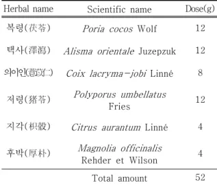
소분청음 52 g에 증류수 500 cc를 넣고 100 ℃ 에서 1시간 동안 가열 추출한 후, 추출액을 여과하 여 잔사를 제거하고, 감압 농축 및 동결 건조하여 3.9 g의 건조추출물을 얻었다 (수율 7.5 %). 소분 청음의 약재 구성은 Table 1과 같다7).
Table 1. Prescription of Sobuncheong-eum (SBCE) Herbal name Scientific name Dose(g)
복령(茯苓) Poria cocos Wolf 12 택사(澤瀉) Alisma orientale Juzepzuk 12 의이인(薏苡仁) Coix lacryma-jobi Linné 8
저령(猪苓) Polyporus umbellatus
Fries 12
지각(枳殼) Citrus aurantum Linné 4
후박(厚朴) Magnolia officinalis
Rehder et Wilson 4 Total amount 52
2) 재료
3 ‐ ( 4 , 5 ‐ d i m e t h y l t h i a z o l ‐ 2 ‐ y l ) ‐ 2 , 5 ‐ diphenyltetrazolium bromide (MTT), Palmitate 은 Sigma‐Aldrich Co. (St. Louis, MO, USA)에서 구입하였다. EnzyChromTM Triglyceride Assay Kit는 BioAssay Systems (Hayward, CA, USA)에 서 구입하였다. Phospho-AMPK antibody는 Cell Signaling Technology Inc. (Danvers, MA, USA) 에서 구입하였다. SREBP-1c antibody, β-Actin antibody, 및 horseradish peroxidase (HRP)‐
conjugated goat anti‐rabbit IgG는 Santa Cruz Biotechnology Inc. (Santa Cruz, CA, USA)에서 구입하였다.
3) 세포 배양
HepG2 cell은 human hepatocellular carcinoma 기원 세포로서 American Type Culture Collection (Manassas, VA, USA)에서 구입하였다. 100 U/ml penicillin, 100 μg/ml streptomycin, 및 10%
heat‐inactivated fetal bovine serum을 첨가한 Dulbecco's modified Eagle's medium (DMEM)을 이용하여, 37℃, 5% CO2 환경에서 배양하였다.
*Corresponding Author : Chang-Won Choi, Dongshin university oriental hospital, 331, Isu-ro, Suncheon-si, Jeollanam-d0, 57949, Republic of Korea.
Tel : +82-61-729-7133, Fax : +82-61-725-1717, E-mail : [email protected]
∙Received : March 24, 2016 / Revised : May 27, 2016 / Accepted : May 27, 2016
4) MTT assay
소분청음 추출물이 세포 활성도를 떨어뜨리는지 확인하기 위하여 MTT assay를 시행하였다. HepG2 Cell을 96-well plate에 1 × 10⁴ cells/well의 밀 도로 투입하고 24 시간 후 소분청음 추출물을 여러 가지 농도 (0, 50, 100, 300, 500, 700, 1,000, 1,500 μg/ml)로 투약하고 다시 24 시간 배양하였 다. 각 well에 MTT 500 μg/ml를 4 시간 동안 처 리하였다. 세포들이 생성한 formazan crystal을 DMSO를 사용하여 용해한 다음 570 nm에서 흡광 도 (absorbance)를 측정하였다.
5) 세포 내 중성 지방 측정
HepG2 cell을 6-well plate에 1 × 10⁶ cells/well의 밀도로 투입하고 palmitate 0.5 mM 과 1% bovine serum albumin을 함유한 DMEM으로 24시간 배양 하여 지방증 (steatosis)을 유도하였다. 실험군에서 는 DMEM에 소분청음 추출물도 함께 투여하였다.
지방증이 유도된 HepG2 cell을 trypsin 처리 후 회 수하여 1.5 mL Eppendorf tube에 담고 centrifuge로 3,000 rpm × 5 분간 침전시켰다. 세포덩어리 (cell pellet)들을 PBS로 세척 후, microsmashing tube에 옮겨 담고 ultrasonication하여 분쇄한 다음, EnzyChromTM Triglyceride Assay Kit을 이용하여 제공된 프로토콜에 따라 세포내 triglyceride 함량 을 측정하였다.
6) Western blot analysis
Phospho-AMPK protein과 SREBP-1c protein level을 western blot analysis로 측정하였다. 먼 저 HepG2 cell을 Protease inhibitor Cocktail (Roche, Indianapolis, IN, USA)를 포 함 한 radioimmunoprecipitation assay (RIPA) buffer (50 mM Tris-HCl (pH 8.0), 150 mM NaCl, 2 mM EDTA, 1% sodium orthovanadate, 1%
Triton X-100, 0.5% deoxycholate, 0.1% SDS)로 처리하여 total cell lysate를 추출하였다. 다음으로 NE-PER™ nuclear extraction kit (Thermo Scientific, IL, USA)로 nuclear protein 부분을 추출하였다. Bradford assay로 단백량을 정량하고, sodium dodecyl sulfate polyacrylamide gel electrophoresis (SDS-PAGE)로 단백을 분리하여,
Polyvinylidene fluoride (PVDF) membrane에 전 사하였다. 각각의 specific primary antibody를 처 리하고 4℃에서 24 시간 반응 후, horseradish peroxidase (HRP)가 결합되어 있는 2차 항체와 상 온 에 서 1시 간 반 응 시 켰 다 . SuperSignal®
chemiluminescence detection kit (Thermo Scientific, IL, USA)로 각각의 band를 검출하였 다. 각 band들은 image J 1.50e software를 이용 하여 정량화하였다.
7) semi-quantitative RT-PCR
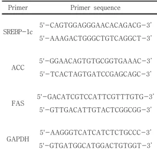
SREBP-1c, ACC 및 FAS의 mRNA 발현 정도를 측정하기 위하여 semi-quantitative RT-PCR을 시 행하였다. 먼저 RNeasy Mini Kit (Qiagen, Hilden, Germany)로 total RNA를 추출하였다. M-MLV Reverse Transcriptase (Promega, Madison, WI, USA)를 이용하여 추출된 total RNA로부터 cDNA 를 합성하였다. 각각의 primer (Bioneer)와 TaqPCRx DNA Polymerase (Invitrogen, Carlsbad, CA, USA)를 사용하여 cDNA를 증폭하였다. 증폭된 DNA를 1.2% agarose gel에서 전기영동한 다음 ethidium bromide 염색 후 자외선에서 band를 확 인하였다. 각 band들은 image J 1.50e software를 이용하여 정량화하였다. 각각의 primer는 Table 2 와 같다.
Table 2. Primer Sequence of Each Gene Detected in Semi-quantitative RT-PCR
Primer Primer sequence
SREBP-1c
5'-CAGTGGAGGGAACACAGACG-3’
5'-AAAGACTGGGCTGTCAGGCT-3’
ACC 5'-GGAACAGTGTGCGGTGAAAC-3’
5'-TCACTAGTGATCCGAGCAGC-3’
FAS
5'-GACATCGTCCATTCGTTTGTG-3’
5'-GTTGACATTGTACTCGGCGG-3’
GAPDH 5'-AAGGGTCATCATCTCTGCCC-3’
5'-GTGATGGCATGGACTGTGGT-3’
8) 통계 분석
통계 분석은 IBM SPSS statistics 23K (SPSS Inc, Chicago, IL, USA)을 사용하여 시행하였다.
측정값들은 평균과 표준오차로 표현하였다. One-way analysis of variance test와 Student’s t-test로 그룹 간 평균을 비교하여, P<0.05일 때 통계적으 로 유의한 차이가 있는 것으로 판정하였다.
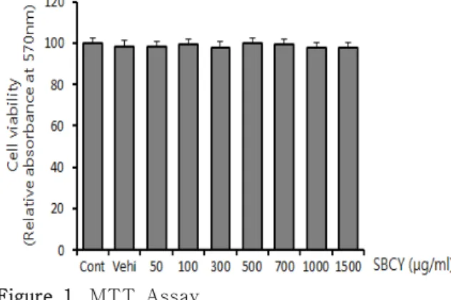
1) 소분청음 추출물의 세포독성 (cytotoxicity) 평가 HepG2 cell에 대하여 소분청음 추출물이 세포독 성을 나타내지 않는 농도 범위를 측정하기 위하여, HepG2 cell에 다양한 농도의 소분청음 추출물을 처 리하고 MTT assay를 통해 HepG2 cell의 세포 활 성도를 측정하였다. 소분청음 추출물은 1,500 μ g/ml 이하의 농도에서는 세포 활성도를 떨어뜨리지 않았다 (Figure 1).
Figure 1. MTT Assay
To evaluate cytotoxicity of SBCE, HepG2 cells was administered with various concentration of SBCE for 24h prior to MTT assay. Data are represented as mean±SEM (n=3).
2) 소분청음 추출물이 세포 내 지질량에 미치는 영향 평가
HepG2 cell에 palmitate를 처리하여 세포 내 지 질 축적을 유도하였으며, palmitate와 함께 소분청 음 추출물을 투여하였을 때 세포 내 지질 축적이 감소하는지를 확인하였다. 함께 투여한 소분청음 추 출물의 농도가 증가할수록 세포 내 지질량이 점점 감소하였으며, 소분청음 추출물 농도가 300 μg/ml 이상일 때 통계적으로 유의한 차이가 확인되었다 (Figure 2).
Figure 2. The Effect of SBCE on TG accumulation in Steatotic HepG2 Cells
HepG2 cells were treated with palmitate (0.5 mM) with various concentration of SBCE (0, 50, 100, 300, 500, 700, 1,000 g/ml) for 24h.
Intracellular TG content was measured according to the experimental procedures described in the ‘Material and Methods’
part, and expressed as a relative ratio to control. Data are represented as mean±SEM (n=3). The * mark indicates significant difference compared to only palmitate treated cells without SBCE (P<0.05).
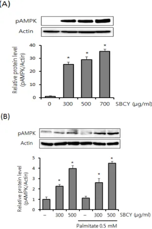
3) 소분청음 추출물이 AMPK 활성화에 미치는 영향 평가
Phospho-AMPK는 AMPK의 활성형으로, 여러 가지 lipogenic genes의 발현을 억제하여 세포 내 지질 축적을 완화시키는 역할을 하는 것으로 알려 져 있다8). 먼저 normal HepG2 cell에 소분청음 추 출물을 투여하고, phospho-AMPK level을 측정하
여, 소분청음 추출물의 농도가 증가할수록
phospho-AMPK level이 증가하는 것을 확인하였다 (Figure 3A). 다음으로 palmitate로 HepG2 cell에 지방증을 유도할 때, 소분청음 추출물을 함께 투여 하고, phospho-AMPK level을 측정하였다. 지방증 이 유도된 HepG2 cell에서도 소분청음 추출물에 의 하여 phospho-AMPK level이 증가하는 것을 확인 하였다 (Figure 3B).
Figure 3. Effect of SBCE on AMPK Activation in HepG2 Cells
The pAMPK protein level was measured by western blot analysis, escalating SBCE concentration in normal HepG2 cells (A), and in steatotic HepG2 cells (B). The intensity of each band was assessed with image J 1.50e software, and converted into relative ratio to control. Data are represented as mean±SEM (n=3). The * mark indicates significant difference compared to only palmitate treated cells without SBCE (P<0.05).
4) 소분청음 추출물이 SREBP-1c 발현에 미치는 영향 평가
SREBP-1c는 lipogenesis를 조절하는 주요 전사 인자이다9). HepG2 cell에 palmitate를 처리할 때, 실험군에는 소분청음 추출물을 함께 투여하였다.
24 시간 배양한 다음, SREBP-1c mRNA 및 SREBP-1c protein level을 측정하였다. Palmitate 에 의해 SREBP-1c mRNA 및 SREBP-1c protein level이 증가하는 것을 확인하였으며, 소분청음 추
출물을 함께 투여한 경우 SREBP-1c mRNA 및 SREBP-1c protein level 상승 정도가 둔화된 것을 확인하였다 (Figure 4).
Figure 4. Effect of SBCE on SREBP-1c in HepG2 Cells
The SREBP-1c mRNA level was measured by RT-PCR (A), and the SREBP-1c protein level by western blot analysis (B). The intensity of each band was assessed with image J 1.50e software, and converted into relative ratio to control. Data are represented as mean±SEM (n=3). The * mark indicates significant difference compared to only palmitate treated cells without SBCE (P
<0.05).
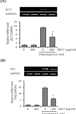
5) 소분청음 추출물이 ACC, FAS에 미치는 영향 ACC와 FAS는 SREBP-1c에 의해 발현이 증가하 여 중성 지방의 합성에 관여하는 효소들이다10). HepG2 cell에 palmitate을 처리하면 SREBP-1c이 발현되면서 ACC과 FAS mRNA 또한 증가한다. 위
에서 기술한 것과 같은 방법으로 palmitate를 처리 할 때, 실험군에는 소분청음 추출물을 함께 투여하 였다. 소분청음 추출물을 함께 처리할 경우 palmitate에 의한 ACC, FAS mRNA의 발현이 뚜렷하게 감소하 였다 (Figure 5).
Figure 5. Effect of SBCE on ACC and FAS in HepG2 Cells
The ACC (A) and FAS (B) mRNA levels were measured by RT-PCR. The intensity of each band was assessed with image J 1.50e software, and converted into relative ratio to control. Data are represented as mean±SEM (n=3). The * mark indicates significant difference compared to only palmitate treated cells without SBCE (P<0.05).
비알코올성 지방간에 대한 표준 치료로 인정된 치료제는 아직 없는 실정이며, 최근 발표된 연구에
서는 한약 처방들이 기존 치료제들에 비하여 지질 축적 및 염증 소견을 완화시키는 효능이 오히려 우 수한 것으로 보고되었다11). 비알코올성 지방간은 한 의학에서 “積聚”의 범주에 속하며, 한의이론으로 는 肝失疎泄 및 脾失健運으로 인해 濕濁內結하여 발생한 것으로 해석된다12). 소분청음(小分淸飮)은
『景岳全書』에 최초 수록된 처방으로 그 효능을
“治小水不利, 濕滯腫脹, 不能受補等證, 此方主之”
라 하여, 濕滯傷中으로 인한 제반 증상에 사용하는 것으로 기록되어 있다7). 따라서, 소분청음은 濕濁內 結하여 발생한 비알코올성 지방간에도 치료 효능이 있을 것으로 추론하였다.
ACC, FAS는 상기한 바와 같이 간세포 내에서 지 질 합성에 관여하는 효소들이며10), SREBP-1c는 이 러한 지질 합성 관련 인자들을 조절하는 주요 전사 인자 (transcription factor)의 하나이다9). AMPK 는 신체의 지질 대사 및 에너지 대사를 조절하는 핵심 조절 인자 (key regulator)로 2형 당뇨, 비만, 및 고지혈증 등 각종 대사 질환들에 주요 therapeutic target으로 판단되고 있다13). AMPK가 활성화되어 phospho-AMPK가 되면, 동화 작용 (anabolic pathway)을 억제하고, 이화작용 (catabolic pathways)을 촉진하여 ATP 생산을 증가시키는 역 할을 담당한다14). 간세포 내에서 AMPK는 지질합성 을 억제할 뿐만 아니라, 지방산의 산화를 촉진시킨 다8). 간세포 내에서 활성화된 AMPK는 SREBP-1c 에 결합하여, SREBP-1c의 target 인자들인 ACC, FAS 같은 지질 합성 효소의 발현을 억제한다15),16). 본 연구에서는 비알코올성 지방간에 대한 소분청 음의 치료 효능을 확인해보고자, HepG2 cell에 지 방산을 처리하여 유도한 비알코올성 지방간 세포 모델에서, 소분청음이 세포 내 중성지방 (triglyceride) 함량에 미치는 변화를 관찰하였다. 아울러, 소분청 음이 lipogenesis를 억제하는 AMPK의 활성화에 미 치는 영향과 lipogenic genes인 SREBP-1c, ACC, 및 FAS 발현에 대한 영향을 조사하였다. 그 결과 소분청음은 HepG2 cell에서 palmitate에 의한 세 포 내 지질 축적을 뚜렷히 완화하였으며, 세포내 지 질 축적에 관여하는 SREBP-1c, ACC, 및 FAS의 발현을 감소시켰다. 또한, 이러한 anti-lipogenic effect를 유발하는 AMPK를 활성화시키는 것이 확 인되었다. 향후, 후속 동물 실험을 계획하고 있으
며, 동물 실험 중 안전성에 대한 평가도 함께 계획 하고 있다.
이상의 결과를 요약하면, 소분청음 추출물은 1,500 μg/ml 이하의 농도에서는 HepG2 cell의 세 포활성도를 억제하지 않으며, 300 ~ 1,000 μg/ml 의 농도에서 HepG2 cell의 세포 내 중성 지방 함량 을 통계적으로 의미있게 감소시켰다. 또한, 300 μ g/ml 농도에서 phospho-AMPK를 증가시켜, palmitate로 유발된 SREBP-1c, ACC, 및 FAS의 발현을 감소시켰다.
이 논문은 동신대학교 학술연구비 (2015)에 의하 여 연구되었음.
1. Chalasani N, Younossi Z, Lavine JE, Diehl AM, Brunt EM, Cusi K, et al. The diagnosis and management of non-alcoholic fatty liver disease: practice Guideline by the American Association for the Study of Liver Diseases, American College of Gastroenterology, and the American Gastroenterological Association.
Hepatology. 2012;55(6):2005-23.
2. Angulo P. Nonalcoholic fatty liver disease.
Revista de gastroenterologia de Mexico.
2005;70 Suppl 3:52-6.
3. Matteoni CA, Younossi ZM, Gramlich T, Boparai N, Liu YC, McCullough AJ.
Nonalcoholic fatty liver disease: a spectrum of clinical and pathological severity.
Gastroenterology. 1999;116(6):1413-9.
4. Argo CK, Caldwell SH. Epidemiology and natural history of non-alcoholic steatohepatitis.
Clinics in liver disease. 2009;13(4):511-31.
5. Popov VB, Lim JK. Treatment of Nonalcoholic Fatty Liver Disease: The Role of Medical, Surgical, and Endoscopic Weight Loss.
Journal of clinical and translational hepatology.
2015;3(3):230-8.
6. Yeon JE. Nonalcoholic Fatty Disease : pathogenesis and treatment. J of KAHP.
2006;4:132-9.
7. Lee NG. Hyeontojuseog Gyeongagjeonseo.
Seoul: Bubin Publishers Co, 2007:1021.
8. Viollet B, Foretz M, Guigas B, Horman S, Dentin R, Bertrand L, et al. Activation of AMP-activated protein kinase in the liver:
a new strategy for the management of metabolic hepatic disorders. The Journal of physiology. 2006;574(Pt 1):41-53.
9. Repa JJ, Liang G, Ou J, Bashmakov Y, Lobaccaro JM, Shimomura I, et al.
Regulation of mouse sterol regulatory element-binding protein-1c gene (SREBP-1c) by oxysterol receptors, LXRalpha and LXRbeta. Genes & development. 2000;14(22):
2819-30.
10. Pettinelli P, O bregon A M , V idela LA . Molecular mechanisms of steatosis in nonalcoholic fatty liver disease. Nutricion hospitalaria. 2011;26(3):441-50.
11. Shi KQ, Fan YC, Liu WY, Li LF, Chen YP, Zheng MH. Traditional Chinese medicines benefit to nonalcoholic fatty liver disease:
a systematic review and meta-analysis.
Molecular biology reports. 2012;39(10):9715-22.
12. Traditional Chinese Association. Guidelines for Diagnosis and Treatment of Common Internal Diseases in Chinese Medicine Diseases of Modern Medicine. Beijing:
China Traditional China Medicine Publisher, 2008:153.
13. Steinberg GR, Kemp BE. AMPK in Health and Disease. Physiological reviews. 2009;89(3):
1025-78.
14. Viollet B, Guigas B, Leclerc J, Hebrard S, Lantier L, Mounier R, et al. AMP-activated protein kinase in the regulation of hepatic energy metabolism: from physiology to therapeutic perspectives. Acta physiologica.
2009;196(1):81-98.
15. Pasarin M, Abraldes JG, Rodriguez-Vilarrupla A, La Mura V, Garcia-Pagan JC, Bosch J.
Insulin resistance and liver microcirculation
in a rat model of early NAFLD. Journal of hepatology. 2011;55(5):1095-102.
16. Soetikno V , Sari FR , Sukum aran V , Lakshmanan AP, Harima M, Suzuki K, et al.
Curcumin decreases renal triglyceride accumulation through AMPK-SREBP signaling pathway in streptozotocin-induced type 1 diabetic rats. The Journal of nutritional biochemistry. 2013;24(5):796-802.