Hye-jin Yoon, Bong Soo Cha
Hye-jin Yoon, Bong Soo Cha, Department of Internal Medi-cine, Yonsei University College of MediMedi-cine, Seoul 120-752, South Korea
Author contributions: Yoon HJ and Cha BS equally contributed
to this paper.
Correspondence to: Bong Soo Cha, MD, PhD, Department
of Internal Medicine, Yonsei University College of Medicine, 50 Yonsei-ro, Seodaemoon-gu, Seoul 120-752,
South Korea. [email protected]
Telephone: +82-2-22281962 Fax: +82-2-3936884 Received: July 23, 2014 Revised: August 27, 2014
Accepted: October 14, 2014
Published online: November 27, 2014
Abstract
Non-alcoholic fatty liver disease affects approximately
one-third of the population worldwide, and its incidence
continues to increase with the increasing prevalence of
other metabolic disorders such as type 2 diabetes. As
non-alcoholic fatty liver disease can progress to liver
cirrhosis, its treatment is attracting greater attention.
The pathogenesis of non-alcoholic fatty liver disease is
closely associated with insulin resistance and
dyslipid-emia, especially hypertriglyceridemia. Increased serum
levels of free fatty acid and glucose can cause
oxida-tive stress in the liver and peripheral tissue, leading
to ectopic fat accumulation, especially in the liver. In
this review, we summarize the mechanism underlying
the progression of hepatic steatosis to steatohepatitis
and cirrhosis. We also discuss established drugs that
are already being used to treat non-alcoholic fatty liver
disease, in addition to newly discovered agents, with
respect to their mechanisms of drug action, focusing
mainly on hepatic insulin resistance. As well, we review
clinical data that demonstrate the efficacy of these
drugs, together with improvements in biochemical or
histological parameters.
© 2014 Baishideng Publishing Group Inc. All rights reserved.
Key words: Non-alcoholic fatty liver disease; Insulin
re-sistance; Drugs; Pathogenesis
Core tip: In this review, we summarize the
pathogen-esis underlying the progression of hepatic steatosis to
steatohepatitis and cirrhosis. We also discuss
estab-lished drugs that are already being used to treat
non-alcoholic fatty liver disease, in addition to newly
discov-ered agents, with respect to their mechanisms of drug
action, focusing mainly on hepatic insulin resistance.
As well, we review clinical data that demonstrate the
efficacy of these drugs, together with improvements in
biochemical or histological parameters. Furthermore,
we introduced future treatment option for non-alcoholic
fatty liver disease.
Yoon HJ, Cha BS. Pathogenesis and therapeutic approaches for non-alcoholic fatty liver disease. World J Hepatol2014; 6(11): 800-811 Available from: URL: http://www.wjgnet. com/1948-5182/full/v6/i11/800.htm DOI: http://dx.doi. org/10.4254/wjh.v6.i11.800
INTRODUCTION
Non-alcoholic fatty liver disease (NAFLD), the
accumula-tion of lipid within hepatocytes, is a common disease
[1].
The worldwide prevalence of NAFLD is estimated to
be 20%-30%
[2], although increasing to 57%-74% among
obese patients
[3]. NAFLD refers to a wide spectrum of
fatty degenerative disorders of the liver in the absence of
alcohol intake, ranging from simple steatosis to
steatohepa-titis and cirrhosis
[4]. Nonalcoholic steatohepatitis (NASH)
is histologically characterized by inflammatory cell
recruit-ment. NASH is a significant risk factor for hepatic cirrhosis,
compared with simple steatosis
[5], and 4%-27% of cases
of NASH progress to hepatocellular carcinoma after the
development of cirrhosis
[6]. In one study, NAFLD was
pres-TOPIC HIGHLIGHT
DOI: 10.4254/wjh.v6.i11.800 © 2014 Baishideng Publishing Group Inc. All rights reserved.Pathogenesis and therapeutic approaches for non-alcoholic
fatty liver disease
ent in 75% of obese [body mass index (BMI)
≥
30 kg/m
2]
patients, 16% of non-obese patients, and 34%-74% of
patients with type 2 diabetes
[7]. Another study reported
di-agnoses of fatty liver in 39% of obese (BMI
≥
30 kg/m
2)
patients, 41% of patients with known type 2 diabetes, and
32% of patients with dyslipidemia
[8]. Patients with NAFLD
are not only insulin resistant, but also tend to present with
alterations in plasma triglyceride (TG) levels
[9]. NAFLD
is strongly associated with metabolic syndrome, especially
insulin resistance, central obesity, and dyslipidemia.
There-fore, NAFLD is regarded as a difficult to treat component
of metabolic syndrome
[10]. In this review, we investigate the
mechanisms of hepatic fat accumulation, focusing on the
role of insulin resistance therein, and review current
thera-peutic options and new candidate drugs for the treatment
of NAFLD.
PATHOGENESIS
Insulin resistance - free fatty acid flux and hyperinsulinemia
Hepatic steatosis is caused by an imbalance in
triglyc-eride movement through the liver cell. Triglyctriglyc-eride is
composed of free fatty acid (FFA) and glycerol. Total
FFA is derived from three sources, the diet (15%),
de novo
synthesis (26%), and circulating FFA (56%)
[11]. A
high-fat diet is known to lead to the development of hepatic
steatosis. However, estimates suggest that approximately
60% of liver fat is derived from circulating
nonesteri-fied fatty acids (NEFAs) in individuals who eat a normal
fat-containing diet
[11]. Obesity is associated with insulin
resistance and an elevated leptin level. In particular,
in-creased visceral fat correlates with peripheral and hepatic
insulin resistance
[12,13]. Insulin resistance in skeletal muscle
and adipose tissue results in increased levels of NEFAs
through increased lipid oxidation in adipose tissue (Figure
1). Accordingly, NEFA flux plays an important role in
hepatic fat accumulation
[14]. An increase in hepatocellular
diacylglycerol is associated with decreased tyrosine
phos-phorylation
of insulin receptor substrate 2 (IRS-2)
[15,16].
In turn, the decreased activity of IRS-2 and PI3K leads
to increased hepatic glucose production
[17].
Hyperinsu-linemia also arises in response to insulin resistance in
adi-pose tissue, leading not only to downregulation of IRS-2
in the liver, but also to a continued increase in the level of
sterol regulatory element binding protein-1c (SREBP-1c)
via the insulin signaling pathway involving AKT2, liver X
receptor (LXR) and mammalian target of rapamycin
[18,19].
Elevated levels of SREBP-1c up-regulate lipogenic gene
expression, increase fatty acid synthesis, and accelerate
hepatic fat accumulation
[20]. Additionally, overexpression
of SREBP-1c represses IRS-2 expression
[21].
Glucose-stimulated lipogenesis is mediated by
carbohydrate-responsive element-binding protein (ChREBP) in the
liver. Like SREBP-1c, ChREBP increases lipogenesis by
inducing lipogenic gene expression during consumption
of a diet high in carbohydrates
[22,23].
Endoplasmic reticulum stress
The endoplasmic reticulum (ER) is an intracellular
organ-elle that plays an important role in the synthesis, folding,
and trafficking of proteins. Cellular nutrient status and
energy condition highly influence the function of the
ER, and dysfunction in the ER causes accumulation of
unfolded proteins therein, triggering an unfolded
pro-tein response (UPR)
[24]. Under stress, such as hypoxia,
inflammation and energy excess, UPR is characterized by
adaptive cellular processes of increased degradation of
proteins and translational arrest of protein synthesis to
restore normal function of the ER. As well, UPR
medi-ates metabolic and immune responses that aggravate
insulin resistance
[25-27]. Both PKR-like kinase and the
α-subunit of translation initiation factor 2 (eIF2α),
well-known ER stress markers, are increased in hepatocytes
of ob/ob mice, compared with control mice
[26]. Obesity
causes ER stress that leads to
suppression of insulin
sig-naling through serine phosphorylation of insulin receptor
substrate-1 (IRS-1) and activation of the c-Jun N-terminal
kinase (JNK) pathway
[26]. Among subjects with metabolic
syndrome, those with NASH showed higher levels
phos-phorylated JNK protein, compared to subjects with
sim-ple hepatic steatosis. Furthermore, subjects with NASH
did not generate spliced manipulation of X-box–binding
protein-1 (sXBP-1), which is a key regulator in ER stress
in relation to insulin action
[24,26]. Additionally, weight
reduction in obese subjects has been shown to induce
improvement in ER stress
via suppression of
phosphory-lated JNK and eIF2α in adipose tissue and the liver
[28].
Role of oxidative stress - mitochondrial dysfunction
The two-hit hypothesis is a key concept of NAFLD
pathogenesis. In fatty livers, simple hepatic steatosis
(first hit) sensitizes the liver to inflammatory cytokines
or oxidative stress (second hit), leading to development
of steatohepatitis
[29]. Oxidative stress is resulted from a
serious imbalance between the limited antioxidant
de-fenses and excessive formation of reactive species such
as reactive oxygen species (ROS) or reactive nitrogen
spe-cies (RNS)
[30]. ROS is an integrated term that describes a
variety of species of free radicals derived from
molecu-lar oxygen, such as superoxide, hydrogen peroxide, and
hydroxyl
[31]. In cells, mitochondria are a major source
of ROS generation. The important factor modulating
mitochondrial ROS generation is the redox state of the
respiratory chain
[32,33]. FFAs are metabolized
via the
mito-chondrial β-oxidation pathway and the tricarboxylic acid
(TCA) cycle, which generates citrate that in turn inhibits
glycolysis. As a result, glucose oxidation and glucose
up-take
via glucose transporter type 4 (GLUT4) in skeletal
muscle are reduced
[34,35]. To compensate for the
exces-sive fat storage in the liver, increased hepatic FFA uptake
stimulates hepatic oxidation of fatty acids in obese
indi-viduals. Mitochondrial FFA oxidation is maintained until
mitochondrial respiration becomes severely impaired
[36,37].
However, accelerated β-oxidation not only causes
exces-sive electron flux in the electron transport chain, but also
leads to increased production of ROS, and can lead to
mitochondrial dysfunction
[38]. Excessive ROS
the mitochondrial membrane and DNA and can impair
mitochondrial metabolic functions
[33]. The increase in
hepatic lipogenesis in NASH results in increased
produc-tion of malonyl-CoA. Inhibiproduc-tion of carnitine
palmitoyl-transferase-I (CPT-1) by malonyl-CoA leads to decreased
entry of long chain fatty acid into the mitochondria, and
causes reduced β-oxidation and enhanced triglyceride
ac-cumulation in the liver
[38-40]. The nuclear receptor
peroxi-some proliferator-activated receptor α (PPAR-α) plays
an important role in the transcriptional control of many
enzymes involved in mitochondrial fatty acid β-oxidation.
Peroxisome proliferator-activated receptor-gamma
co-activator (PGC) -1α cooperates with PPAR-α and
regu-lates genes that encode mitochondrial fatty acid oxidation
enzymes, such as CPT-1 and medium chain acyl-CoA
dehydrogenase
[40]. Previously, a PPAR-α-deficient mouse
model showed a lack of hepatic peroxisome proliferation
and dyslipidemia with obesity and hepatic steatosis
[41].
Inflammation and adipokines
Overall obesity is correlated with NAFLD, and
accu-mulation of intra-abdominal fat in particular is believed
to play an important role in the development of insulin
resistance
[12,13]. Meanwhile, hepatic fat accumulation is
associated with insulin resistance independent of
intra-abdominal fat accumulation and overall obesity. Even
in normal weight subjects, hepatic steatosis has been
shown to be related to various parameters of insulin
resistance, such as basal glucose level or serum FFA
level
[42]. In addition to being a major organ of triglyceride
deposition, adipose tissue acts an endocrine organ that
secretes several hormones
[43]. Adipocytes secrete
adipo-nectin and leptin, in addition to the other adipokines,
such as retinol-binding protein, tumor necrosis factor-α
(TNF-α), interleukin 6 (IL-6), and plasminogen activator
inhibitor-1
[43]. Adiponectin stimulates phosphorylation
of AMP-activated protein kinase (AMPK) and
acetyl-CoA carboxylase (ACC) in the liver and muscles, thereby,
increasing glucose utilization and fatty-acid oxidation
[44].
In a previous study, serum adiponectin levels decreased
with an increases in obesity, in particular increases in
intra-abdominal fat mass
[45,46]. In another study,
adiponec-tin knockout mice fed a high-fat diet exhibited increased
incidences of obesity, hyperinsulinemia, and
steatohepa-titis. These experimental data indicate that adiponectin
may play a key protective role against the progression of
NASH
[47]. Reportedly, adipose tissue in obese individuals
stimulates a shift in macrophage activation from the
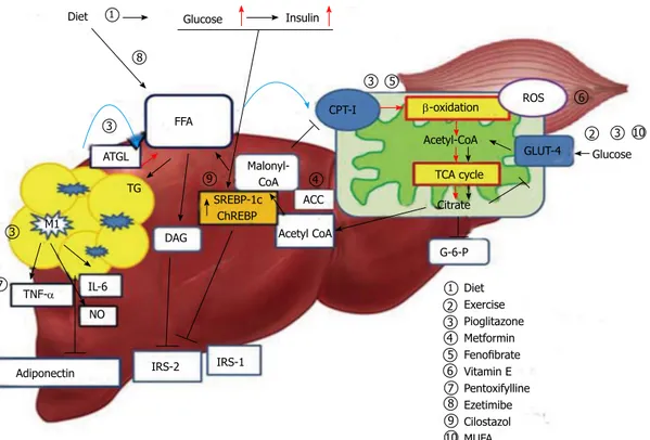
al-Figure 1 Mechanism of hepatic insulin resistance and the key pathway of drug action. Delivery of FFAs to the liver and skeletal muscle is increased in insulin
resistance conditions, and these are metabolized via mitochondrial β-oxidation. Consequently, hyperglycemia and increased hepatic FFA uptake reduce glucose up-take and oxidation in skeletal muscle. Diet and exercise are the main treatment strategies for this pathogenesis; insulin sensitizers and MUFA may contribute to reduc-ing peripheral insulin resistance. Pioglitazone and fenofibrate act on β-oxidation of mitochondria and reduce hepatic steatosis. Accelerated β-oxidation also causes increased production of ROS. Vitamin E can reduce oxidative stress. Adipose tissue inflammation of the liver leads to inflammatory activation of hepatic Kupffer cells via classic response (M1) and produce inflammatory cytokines. This is also associated with decreased adiponectin levels and promotes hepatic steatohepatitis. Pent-oxifylline inhibits TNF-α and alleviates steatohepatitis. Hyperglycemia caused by insulin resistance up-regulates lipogenic gene expression, such as SREBP-1c and ChREBP, and induces lipogenesis in hepatocytes. Cilostasol may inhibit SREBP-1c. FFA: Free fatty acid; TG: Triglyceride; CPT-Ⅰ: Carnitine
palmitoyltransferase-Ⅰ; ACC: Acetyl-CoA carboxylase; ATGL: Adipose triglyceride lipase; ChREBP: Carbohydrate responsive element binding protein; SREBP-1c: Sterol regulatory ele-ment binding protein-1c; TCA: Tricarboxylic acid; ROS: Reactive oxygen species; IRS: Insulin receptor substrate; DAG: Diacylglycerol; G-6-P: Glucose 6-phosphate; TNF-α: Tumor necrosis factor- α; MUFA: Monosaturated fatty acids; M1: Kupffer cells activated via classic pathway.
Diet Glucose Insulin
FFA ATGL TG M1 DAG TNF-α IL-6 NO
Adiponectin IRS-2 IRS-1 SREBP-1c ChREBP Malonyl-CoA Acetyl CoA ACC CPT-I β-oxidation Acetyl-CoA TCA cycle Citrate GLUT-4 ROS Glucose Diet Exercise Pioglitazone Metformin Fenofibrate Vitamin E Pentoxifylline Ezetimibe Cilostazol MUFA 5 6 7 8 9 10 1 1 2 3 3 3 3 3 2 4 4 5 6 7 8 9 10 G-6-P
TREATMENT
Life style modification - diet and exercise
Weight loss due to diet and exercise has been
demon-strated to alleviate hepatic steatosis
[61]. Body weight
re-duction and exercise are important independent factors
for improvement of hepatic steatosis
[62]. In obese women,
hepatic fat content measured by magnetic resonance
im-aging was shown to decrease in response to weight loss
interventions
[63]. Several studies have shown a significant
reduction in alanine transaminase (ALT) levels and
im-provement in biochemical markers following intervention
with a calorie-restricted diet combined with exercise
[63,64].
A few studies have also shown histologic improvement
with increased exercise and weight reduction
[65,66](Table
1). Exercise improves insulin sensitivity in skeletal muscle
via GLUT4 expression and increases glucose utilization.
Thus, exercise decreases levels of serum glucose and
in-sulin
[67]. An improvement in hyperinsulinemia can result
in decreased liver fat mass, because hyperinsulinemia
stimulates hepatic steatosis
via the SREBP-1c pathway
[19].
In particular, NAFLD patients with metabolic syndrome
show a great improvement in hepatic steatosis after
weight loss
[68].
Insulin sensitizer-thiazolidinedione, metformin
Thiazolidinedione: Thiazolidinediones (TZDs) are
in-sulin-sensitizing agents that have been shown to improve
not only hepatic steatosis, but also whole body insulin
resistance
[69]. Improvements in insulin resistance and
his-tologic and biochemical parameters were reported with
TZD treatment
[70-74]. Rosiglitazone is one TZD and is
as-ternative response (M2) to the classic response (M1), and
these classically activated macrophages (CAMs) secrete a
variety of inflammatory cytokines, such as TNF-α, IL-6,
and NO
[48]. Additionally, studies showed that
inflamma-tory activation of hepatic Kupffer cells in ob/ob mice
promotes hepatotoxicity, resulting in hepatic insulin
resis-tance and steatohepatitis
[49,50]. Thus, increases in TNF-α
and IL-6 in obese subjects may play an important role in
insulin resistance and hepatic steatosis
[51,52].
Gut-microbial alternation and TLRs stimulation
As mentioned above, obesity is often associated with
NASH and systemic inflammation characterized increases
in inflammatory cytokine levels. Obesity also can cause
in-creased intestinal mucosa permeability and endotoxin
lev-els in portal circulation that can contribute to
hepatocel-lular damage
[53,54]. Kupffer cells in the liver play a key role
in clearing endotoxin and are activated through Toll like
receptor 2,3,4 and 9 signaling in the presence of
endotox-in. In particular, activation of Toll like receptor4 (TLR4) is
reportedly associated with stimulation of
lipopolysaccha-ride (LPS)
[55-57]. Previously, animal model studies showed
that TLRs 2, 4 and 9 may contribute to the pathogenesis
of NAFLD
[55,58]. Activated Kupffer cells induce
expres-sion of pro-inflammatory cytokines, such TNF-α, IL-6,
IL-18 and IL-12 as well as anti-inflammatory cytokines
[59].
TLRs including TLRs 2,4 and 9 are activated
via a MyD88
dependent pathway. This pathway consists of the
activa-tion of serine kinase IL-1R-associated kinase and
TBF-receptor-associated factor 6 and is involved in the
activa-tion of the transcripactiva-tion factor NF-κB, which is related to
inflammatory cytokine production
[60].
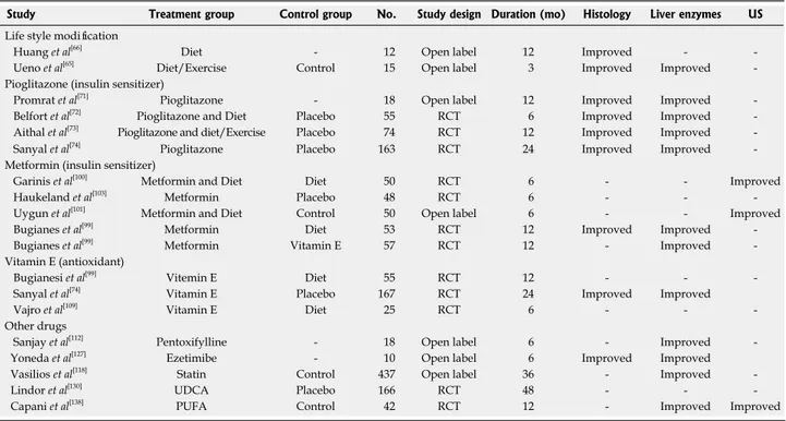
Study Treatment group Control group No. Study design Duration (mo) Histology Liver enzymes US
Life style modification
Huang et al[66] Diet - 12 Open label 12 Improved -
Ueno et al[65] Diet/Exercise Control 15 Open label 3 Improved Improved
Pioglitazone (insulin sensitizer)
Promrat et al[71] Pioglitazone - 18 Open label 12 Improved Improved
Belfort et al[72] Pioglitazone and Diet Placebo 55 RCT 6 Improved Improved
Aithal et al[73] Pioglitazone and diet/Exercise Placebo 74 RCT 12 Improved Improved
Sanyal et al[74] Pioglitazone Placebo 163 RCT 24 Improved Improved
Metformin (insulin sensitizer)
Garinis et al[100] Metformin and Diet Diet 50 RCT 6 - - Improved
Haukeland et al[103] Metformin Placebo 48 RCT 6 - -
Uygun et al[101] Metformin and Diet Control 50 Open label 6 - - Improved
Bugianes et al[99] Metformin Diet 53 RCT 12 Improved Improved
Bugianes et al[99] Metformin Vitamin E 57 RCT 12 - Improved
Vitamin E (antioxidant)
Bugianesi et al[99] Vitemin E Diet 55 RCT 12 - -
Sanyal et al[74] Vitamin E Placebo 167 RCT 24 Improved Improved
Vajro et al[109] Vitamin E Diet 25 RCT 6 - -
Other drugs
Sanjay et al[112] Pentoxifylline - 18 Open label 6 - Improved
Yoneda et al[127] Ezetimibe - 10 Open label 6 Improved Improved
Vasilios et al[118] Statin Control 437 Open label 36 - Improved
Lindor et al[130] UDCA Placebo 166 RCT 48 - -
Capani et al[138] PUFA Control 42 RCT 12 - Improved Improved
Table 1 Treatment outcomes of variable regimens
sociated with an increased risk of myocardial infarction
and cardiovascular death
[75]. Meanwhile, pioglitazone is
re-garded as safe in regards to cardiovascular outcomes and
is not associated with increased cardiovascular risk
[76,77]. In
patients with type 2 diabetes, pioglitazone has been
rec-ommended for the treatment of steatohepatitis proven
by liver biopsy; however, its role in non-diabetic patients
has not been established. The American Association for
the Study of Liver Disease (AASLD) introduced
piogli-tazone as a first-line treatment of NAFLD in patients
with type 2 diabetes
[78]. TZDs increase glucose utilization
of peripheral tissue and improve whole body insulin
sen-sitivity as measured by the hyperinsulinemic euglycemic
clamp technique, in patients with type 2 diabetes.
More-over, serum adiponectin levels increase and serum insulin
levels decreases after treatment with pioglitazone
[79,80]. An
increase in serum adiponectin contributes to alleviation
of hepatic steatosis and improves hepatic and peripheral
insulin resistance
[79]. As mentioned above, adiponectin
increases lipid oxidation of FFA by ACC
phosphoryla-tion in the liver
[44], and promotes the activation of
anti-inflammatory M2 macrophages rather than M1
mac-rophages
[81]. Obesity is closely related to an increase in
NAFLD risk
[82]. Increased levels of inflammatory adipose
tissue macrophages (ATMs) and their secreted cytokines
in a mouse model were shown to be related to systemic
insulin resistance, which is associated with NAFLD
de-velopment
[15,83]. According to previous studies, ATMs are
increased in obese subjects
[84], and pioglitazone treatment
results in not only a decrease in ATM content, but also in
the inflammatory markers, TNF-α, IL-6, and inducible
nitric oxide synthase (iNOS)
[85,86]. TZDs also promote the
alternative activation of monocytes into macrophages
with anti-inflammatory properties, as opposed to the
pro-inflammatory phenotype
[87]. Although the pathogenesis
of NAFLD development is closely related to obesity,
the distribution of fat is more important than overall
obesity. Excessive visceral fat accumulation plays an
im-portant role in the development of insulin resistance and
NAFLD by acting as a source of FFA
[12]. Pioglitazone is
strongly associated with fat redistribution, increases in
subcutaneous fat area decreases in visceral fat area
(vis-ceral to subcutaneous fat ratio)
[88]. Another study showed
that the ratio of visceral fat thickness to subcutaneous
fat thickness decreases after pioglitazone treatment and
is correlated with a change in high sensitivity C-reactive
protein levels
[89]. TZD treatment results revealed a
de-crease in serum FFA levels, which in turn reduced FFA
supply to the liver and led to a decrease in hepatic
triglyc-eride content
[90]. Recent studies have focused on the role
of sirtuin-6 (SIRT-6) in the glucose and lipid metabolism
associated with TZDs. TZD treatment reduced hepatic
fat accumulation and increased expression of SIRT-6 and
PGC1-α in rat livers
[91]. Also, liver-specific SIRT-6
knock-out mice exhibited fatty liver formation
[92], leading to
NASH
[93].
Metformin: Metformin improves insulin resistance and
hyperinsulinemia by increasing peripheral glucose uptake
and decreasing hepatic gluconeogenesis
[94]. Metformin
activates AMP kinase
via a LKB-1 dependent mechanism
in skeletal muscle. Also it can activate AMPK by
stimu-lating AMP accumulation in hepatocytes. The increase
in AMP interferes with glucagon action and decreases
cAMP levels, leading to decreased production of hepatic
glucose
[95,96]. Activation of AMPK results in decreased
hepatic triglyceride synthesis and increased fatty acid
oxidation
[97], as well as attenuated hepatic steatosis due to
decreased SREBP-1c activity
[98]. A randomized controlled
trial showed that subjects treated with metformin exhibit
significant improvement in ALT levels, compared with those
who were on a restricted diet or were treated with vitamin
E, as well as improvements in histology after a 12 mo of
treatment
[99,100]. Many studies have shown that metformin
treatment normalizes transaminase levels and decreases
hepatic steatosis as determined by follow-up ultrasound;
nevertheless, histologic data remain limited
[100-103]. As
NASH is closely associated with development of HCC
and liver fibrosis, metformin may be limited in the
reduc-tion of these severe outcomes, including mortality
[104].
Antioxidant - vitamin E (α-tocopherol), pentoxifylline
As mentioned above, oxidative stress contributes to the
progression of NASH from simple hepatic steatosis.
A recent study reported that subjects who were treated
with vitamin E (α-tocopherol) showed improvement in
hepatic steatosis and serum aminotransferase levels
com-pared to a placebo group
[74]. Vitamin E (α-tocopherol)
has been used to treat non-diabetic NASH patients
di-agnosed by liver biopsy
[78]. Meta-analyses of vitamin E
have revealed an increase in all-cause mortality with high
dose (
≥
400 IU/d) vitamin E supplement use, especially
in subjects with chronic disease or at high risk for
cardio-vascular disease events, such as type 2 diabetes. However,
these results are uncertain in healthy subjects
[105,106]. Two
pilot studies reported improved ALT levels with vitamin
E treatment
[107,108]. However, two randomized controlled
trials failed to show the efficacy of vitamin E treatment
in NAFLD
[109,110]. Pentoxifylline, a TNF-α inhibitor, has
also been considered for the treatment of hepatic
ste-atosis, since it plays an important role in the progression
of simple hepatic steatosis to steatohepatitis. In previous
studies, administration of pentoxifylline generated
im-provements in biochemical markers, such as
aminotrans-ferase and Homa-IR, in patients with NASH
[111,112].
Nev-ertheless, further study is needed to prove the efficacy of
pentoxifylline with respect to histologic improvement of
NAFLD.
Lipid-lowering agents - fibrates, ezetimibe and statins
Hypertriglyceridemia is a major component of metabolic
syndrome and is strongly associated with NAFLD.
In-creased FFA delivery to the liver causes accumulation of
hepatic fat
[9]. Many different lipid-lowering agents have
been investigated for the treatment of NAFLD. Patients
treated with gemfibrozil, one type of fibrate, showed
decreased ALT levels, compared to the control group
[113].
NAFLD
[114]. PPAR-α modulates not only FFA transport
and β-oxidation to decrease triglyceride in hepatocytes,
but also glucose and amino acid metabolism in liver and
skeletal muscle. PPAR-α activation is involved in
lipopro-tein metabolism by increasing lipolysis, thus reducing the
production of triglyceride-rich particles
[115]. Fenofibrate
increased levels of PPAR-α and decreased hepatic
steato-sis in an APOE2KI mouse model that represented
diet-induced NASH
[116]. A prospective study using atorvastatin
reported significant reductions in serum transaminase
level
[117,118]. Atorvastatin induces hepatic low-density
lipo-protein receptor-related lipo-protein 1 (LRP-1) that plays an
important role in clearance of circulating triglyceride in
the liver
[119]. In disposal of chylomicron in hepatocytes,
interaction of LRP-1 receptors and ApoE play important
roles
[120]. Thus, ApoE-deficient mice showed
develop-ment of hepatic steatosis even when they were fed a
nor-mal chow-diet. Accordingly, ApoE may play a key role in
intracellular metabolism and control of VLDL
produc-tion by hepatocytes
[121]. Statins are very important drugs
to treat dyslipidemia in subjects with both insulin
resis-tance and NAFLD. However, there is continued concern
about the use of statins in subjects with established liver
disease. According to several randomized controlled
stud-ies and retrospective studstud-ies, statin rarely induces serious
liver injury
[122-125]. Ezetimibe, a potent inhibitor of
choles-terol absorption, has been reported to improve hepatic
steatosis in obese Zucker fatty rats
[126]. In a randomized
controlled study, six months of treatment with ezetimibe
led to improvements in serum ALT levels and histologic
observations
[127,128].
Ursodeoxycholic acid
Ursodeoxycholic acid (UDCA) is widely used in subjects
with abnormal liver function. Several studies have
in-vestigated the efficacy of UDCA as a treatment drug of
NAFLD, reporting that UDCA treatment attenuated
he-patic steatosis, including histologic improvement
[114,129,130].
However, in a placebo controlled, randomized control
trial, UDCA exhibited limited efficacy in histologic
im-provement in subjects with NASH and imim-provements
in liver enzyme did not differ in the UDCA group,
com-pared to the placebo group
[130]. Accordingly, AASLD does
not recommend UDCA for the treatment of NAFLD
[78].
Other treatment options - future candidates
Cilostazol: SREBP-1c is a key regulator of lipogenic
gene expression in hepatocytes. Recent data have shown
that cilostazol, a selective type
Ⅲ
phosphodiesterase
in-hibitor, inhibits SREBP-1c expression
via the suppression
of LXR and Sp1 activity
[131]. Cilostazol also decreases
serum triglyceride levels by increasing lipoprotein lipase
(LPL) activity in STZ-induced diabetic rats
[132]. Also,
ex-perimental data show that cilostazol stimulates LRP1
pro-moter activity in hepatocytes, leading to increased hepatic
LRP1 expression
[133]. In a study that used two
experimen-tal NAFLD models, both high-fat/high-calorie (HF/HC)
diet mice and the choline-deficient/L-amino acid-defined
(CDAA) diet mice, cilostazol generated improvement in
hepatic steatosis in both mice models
[134]. Cilostazol
ex-hibits the potential for improvement of hepatic steatosis,
and further data on its role in NAFLD are needed.
Polyunsaturated fatty acids and monounsaturated
fatty acids: Polyunsaturated fatty acids (PUFAs) are
found primarily in safflower, corn, soybean, cottonseed,
sesame, and sunflower oils. Omega-3 fatty acids are
rep-resentative of PUFA. A marked increase in long-chain
PUFA n-6/n-3 ratio is observed in NAFLD patients and
is associated with increased production of
pro-inflamma-tory eicosanoids and dysregulation of liver and adipose
tissue function
[135]. PPAR-α activity is impaired in
condi-tions in which levels of circulating n-3 PUFA are
de-creased and the n-6/n-3 fatty acid ratio is inde-creased
[136,137].
Treatment with n-3 PUFA was shown to improve
bio-chemical parameters and alleviated hepatic steatosis by
ultrasound follow-up
[138,139]. Monounsaturated fatty
ac-ids (MUFAs) are comprised in olive oil. In a rat model,
supplementation with MUFA resulted in improved insulin
sensitivity, compared to rats fed a saturated fatty acid (SFA)
diet. Additionally, GLUT4 translocation in skeletal muscle
was decreased in rats fed a SFA diet, but not in those fed
a MUFA diet. Increased GLUT4 translocation is related
to an improvement in insulin sensitivity
[140]. In obese rats,
MUFA diet attenuated hepatic steatosis and altered
he-patic fatty acid levels
[141]. The beneficial effects of dietary
MUFA in NAFLD patients should be investigated.
GLP-1 analogue: Exenatide is the synthetic form of
exendin4 and it stimulates endogenous insulin secretion,
leading to decreases in blood glucose. In one animal study,
treatment of exendin4 resulted in a decrease of hepatic
fat content, as well as reduction of fatty acid synthesis, in
the liver of ob/ob mice
[142]. In patients with type 2
dia-betes, an exenatide treatment group showed greater
im-provements in liver enzymes, attenuating hepatic steatosis,
than the metformin treatment group. However, this study
had limitations of a lack of histologic confirmation of the
liver
[143]. To prove the efficacy of glucagon like peptide-1
(GLP-1) analogue in treatment of NAFLD, randomized
controlled trials over a longer period are required.
MK615: MK615 is extracted from Japanese apricots, and
can suppress the production of inflammatory cytokines
such as TNF-α and IL-6 by inactivating NF-κB
[144,145].
MK615 is regarded as a hepatoprotective agent, as it has
been shown that a MK615 treatment group exhibited
greater decreases in liver enzyme levels, compared with
control groups. In rat models, MK615 treatment mice
showed more improved liver histology than control
mice
[146]. Thus, further studies are required to clarify the
effects of MK615 in subjects with NAFLD.
CONCLUSION
NAFLD is a common disease that can progress to liver
cirrhosis. Moreover, NAFLD is strongly associated with
type 2 diabetes and insulin resistance. NAFLD is the
result of complex interactions among diet, metabolic
components, adipose tissue inflammation, and
mitochon-drial dysfunction. The pathogenesis of hepatic steatosis
has not yet been fully determined. In this review, we
outlined previously known mechanisms of NAFLD,
as well as introduced new mechanisms that have been
recently discovered. Above all, we reviewed the
mecha-nisms of drugs matched to the pathogenesis of NAFLD.
Furthermore, we introduced future treatment option for
NAFLD. TZDs play a key role in restoring insulin
sensi-tivity and decreasing adipose tissue inflammation,
gener-ating histologic improvements in hepatic steatohepatitis.
Pioglitazone can be used to treat NASH in patients with
type 2 diabetes with biopsy-proven NAFLD; meanwhile,
non-diabetic patients can be treated with vitamin E.
Met-formin is a well-known insulin sensitizer; however,
fur-ther study is needed to prove histologic improvements in
patients with NAFLD. Additionally, the
cholesterol-low-ering agent ezetimibe has also shown histologic
improve-ments. Cilostazol acts on SREBP-1c and can improve
dyslipidemia; however, further research is needed to
clarify the relationship between NAFLD and cilostazol.
Finally, there is an outstanding need for effective
preven-tive and therapeutic regimens to overcome NAFLD.
REFERENCES
1 Teli MR, James OF, Burt AD, Bennett MK, Day CP. The
natural history of nonalcoholic fatty liver: a follow-up study.
Hepatology 1995; 22: 1714-1719 [PMID: 7489979]
2 Milić S, Stimac D. Nonalcoholic fatty liver
disease/steato-hepatitis: epidemiology, pathogenesis, clinical presentation and treatment. Dig Dis 2012; 30: 158-162 [PMID: 22722431 DOI: 10.1159/000336669]
3 Angulo P. Nonalcoholic fatty liver disease. N Engl J Med
2002; 346: 1221-1231 [PMID: 11961152 DOI: 10.1056/NEJM-ra011775]
4 Byrne CD, Olufadi R, Bruce KD, Cagampang FR, Ahmed
MH. Metabolic disturbances in non-alcoholic fatty liver disease. Clin Sci (Lond) 2009; 116: 539-564 [PMID: 19243311 DOI: 10.1042/cs20080253]
5 Argo CK, Northup PG, Al-Osaimi AM, Caldwell SH.
Sys-tematic review of risk factors for fibrosis progression in non-alcoholic steatohepatitis. J Hepatol 2009; 51: 371-379 [PMID: 19501928 DOI: 10.1016/j.jhep.2009.03.019]
6 Starley BQ, Calcagno CJ, Harrison SA. Nonalcoholic fatty
liver disease and hepatocellular carcinoma: a weighty con-nection. Hepatology 2010; 51: 1820-1832 [PMID: 20432259 DOI: 10.1002/hep.23594]
7 Browning JD, Szczepaniak LS, Dobbins R, Nuremberg P,
Horton JD, Cohen JC, Grundy SM, Hobbs HH. Prevalence of hepatic steatosis in an urban population in the United States: impact of ethnicity. Hepatology 2004; 40: 1387-1395 [PMID: 15565570 DOI: 10.1002/hep.20466]
8 Fan JG, Zhu J, Li XJ, Chen L, Lu YS, Li L, Dai F, Li F, Chen
SY. Fatty liver and the metabolic syndrome among Shanghai adults. J Gastroenterol Hepatol 2005; 20: 1825-1832 [PMID: 16336439 DOI: 10.1111/j.1440-1746.2005.04058.x]
9 Cassader M, Gambino R, Musso G, Depetris N, Mecca F,
Ca-vallo-Perin P, Pacini G, Rizzetto M, Pagano G. Postprandial triglyceride-rich lipoprotein metabolism and insulin sensi-tivity in nonalcoholic steatohepatitis patients. Lipids 2001; 36: 1117-1124 [PMID: 11768156]
10 Zelber-Sagi S, Nitzan-Kaluski D, Halpern Z, Oren R. Preva-lence of primary non-alcoholic fatty liver disease in a
popu-lation-based study and its association with biochemical and anthropometric measures. Liver Int 2006; 26: 856-863 [PMID: 16911469 DOI: 10.1111/j.1478-3231.2006.01311.x]
11 Donnelly KL, Smith CI, Schwarzenberg SJ, Jessurun J, Boldt MD, Parks EJ. Sources of fatty acids stored in liver and secreted via lipoproteins in patients with nonalcoholic fatty liver disease. J Clin Invest 2005; 115: 1343-1351 [PMID: 15864352 DOI: 10.1172/jci23621]
12 Cnop M, Landchild MJ, Vidal J, Havel PJ, Knowles NG, Carr DR, Wang F, Hull RL, Boyko EJ, Retzlaff BM, Walden CE, Knopp RH, Kahn SE. The concurrent accumulation of intra-abdominal and subcutaneous fat explains the association between insulin resistance and plasma leptin concentrations : distinct metabolic effects of two fat compartments. Diabetes 2002; 51: 1005-1015 [PMID: 11916919]
13 Miyazaki Y, Glass L, Triplitt C, Wajcberg E, Mandarino LJ, DeFronzo RA. Abdominal fat distribution and peripheral and hepatic insulin resistance in type 2 diabetes mellitus.
Am J Physiol Endocrinol Metab 2002; 283: E1135-E1143 [PMID:
12424102 DOI: 10.1152/ajpendo.00327.2001]
14 Bugianesi E, Gastaldelli A, Vanni E, Gambino R, Cassader M, Baldi S, Ponti V, Pagano G, Ferrannini E, Rizzetto M. Insulin resistance in non-diabetic patients with non-alcoholic fatty liver disease: sites and mechanisms. Diabetologia 2005; 48: 634-642 [PMID: 15747110 DOI: 10.1007/s00125-005-1682-x] 15 Samuel VT, Liu ZX, Qu X, Elder BD, Bilz S, Befroy D,
Ro-manelli AJ, Shulman GI. Mechanism of hepatic insulin resis-tance in non-alcoholic fatty liver disease. J Biol Chem 2004; 279: 32345-32353 [PMID: 15166226 DOI: 10.1074/jbc.M313478200] 16 Erion DM, Shulman GI. Diacylglycerol-mediated insulin
resistance. Nat Med 2010; 16: 400-402 [PMID: 20376053 DOI: 10.1038/nm0410-400]
17 Preiss D, Sattar N. Non-alcoholic fatty liver disease: an over-view of prevalence, diagnosis, pathogenesis and treatment considerations. Clin Sci (Lond) 2008; 115: 141-150 [PMID: 18662168 DOI: 10.1042/cs20070402]
18 Shimomura I, Matsuda M, Hammer RE, Bashmakov Y, Brown MS, Goldstein JL. Decreased IRS-2 and increased SREBP-1c lead to mixed insulin resistance and sensitivity in livers of lipodystrophic and ob/ob mice. Mol Cell 2000; 6: 77-86 [PMID: 10949029]
19 Li S, Brown MS, Goldstein JL. Bifurcation of insulin signal-ing pathway in rat liver: mTORC1 required for stimulation of lipogenesis, but not inhibition of gluconeogenesis. Proc
Natl Acad Sci USA 2010; 107: 3441-3446 [PMID: 20133650
DOI: 10.1073/pnas.0914798107]
20 Horton JD, Goldstein JL, Brown MS. SREBPs: activators of the complete program of cholesterol and fatty acid syn-thesis in the liver. J Clin Invest 2002; 109: 1125-1131 [PMID: 11994399 DOI: 10.1172/jci15593]
21 Shimano H. SREBP-1c and TFE3, energy transcription factors that regulate hepatic insulin signaling. J Mol Med (Berl) 2007;
85: 437-444 [PMID: 17279346 DOI: 10.1007/s00109-007-0158-5]
22 Uyeda K, Repa JJ. Carbohydrate response element binding protein, ChREBP, a transcription factor coupling hepatic glucose utilization and lipid synthesis. Cell Metab 2006; 4: 107-110 [PMID: 16890538 DOI: 10.1016/j.cmet.2006.06.008] 23 Browning JD, Horton JD. Molecular mediators of hepatic
steatosis and liver injury. J Clin Invest 2004; 114: 147-152 [PMID: 15254578 DOI: 10.1172/jci22422]
24 Puri P, Mirshahi F, Cheung O, Natarajan R, Maher JW, Kel-lum JM, Sanyal AJ. Activation and dysregulation of the un-folded protein response in nonalcoholic fatty liver disease.
Gastroenterology 2008; 134: 568-576 [PMID: 18082745 DOI:
10.1053/j.gastro.2007.10.039]
25 Hummasti S, Hotamisligil GS. Endoplasmic reticulum stress and inflammation in obesity and diabetes. Circ Res 2010; 107: 579-591 [PMID: 20814028 DOI: 10.1161/circre-saha.110.225698]
26 Ozcan U, Cao Q, Yilmaz E, Lee AH, Iwakoshi NN, Ozdelen E, Tuncman G, Görgün C, Glimcher LH, Hotamisligil GS.
En-doplasmic reticulum stress links obesity, insulin action, and type 2 diabetes. Science 2004; 306: 457-461 [PMID: 15486293 DOI: 10.1126/science.1103160]
27 Schwabe RF, Uchinami H, Qian T, Bennett BL, Lemasters JJ, Brenner DA. Differential requirement for c-Jun NH2-terminal kinase in TNFalpha- and Fas-mediated apoptosis in hepatocytes. FASEB J 2004; 18: 720-722 [PMID: 14766793 DOI: 10.1096/fj.03-0771fje]
28 Gregor MF, Yang L, Fabbrini E, Mohammed BS, Eagon JC, Hotamisligil GS, Klein S. Endoplasmic reticulum stress is re-duced in tissues of obese subjects after weight loss. Diabetes 2009; 58: 693-700 [PMID: 19066313 DOI: 10.2337/db08-1220] 29 Day CP, James OF. Steatohepatitis: a tale of two “hits”?
Gas-troenterology 1998; 114: 842-845 [PMID: 9547102]
30 Robertson G, Leclercq I, Farrell GC. Nonalcoholic steatosis and steatohepatitis. II. Cytochrome P-450 enzymes and oxi-dative stress. Am J Physiol Gastrointest Liver Physiol 2001; 281: G1135-G1139 [PMID: 11668021]
31 Halliwell B. Biochemistry of oxidative stress. Biochem Soc
Trans 2007; 35: 1147-1150 [PMID: 17956298 DOI: 10.1042/
bst0351147]
32 Skulachev VP. Role of uncoupled and non-coupled oxida-tions in maintenance of safely low levels of oxygen and its one-electron reductants. Q Rev Biophys 1996; 29: 169-202 [PMID: 8870073]
33 Murphy MP. How mitochondria produce reactive oxygen species. Biochem J 2009; 417: 1-13 [PMID: 19061483 DOI: 10.1042/bj20081386]
34 Roden M, Price TB, Perseghin G, Petersen KF, Rothman DL, Cline GW, Shulman GI. Mechanism of free fatty acid-induced insulin resistance in humans. J Clin Invest 1996; 97: 2859-2865 [PMID: 8675698 DOI: 10.1172/jci118742]
35 García-Ruiz C, Baulies A, Mari M, García-Rovés PM, Fer-nandez-Checa JC. Mitochondrial dysfunction in non-alco-holic fatty liver disease and insulin resistance: cause or con-sequence? Free Radic Res 2013; 47: 854-868 [PMID: 23915028 DOI: 10.3109/10715762.2013.830717]
36 Sanyal AJ, Campbell-Sargent C, Mirshahi F, Rizzo WB, Contos MJ, Sterling RK, Luketic VA, Shiffman ML, Clore JN. Nonalcoholic steatohepatitis: association of insulin resistance and mitochondrial abnormalities. Gastroenterology 2001; 120: 1183-1192 [PMID: 11266382 DOI: 10.1053/gast.2001.23256] 37 Miele L, Grieco A, Armuzzi A, Candelli M, Forgione A,
Gas-barrini A, GasGas-barrini G. Hepatic mitochondrial beta-oxidation in patients with nonalcoholic steatohepatitis assessed by 13C-octanoate breath test. Am J Gastroenterol 2003; 98: 2335-2336 [PMID: 14572600 DOI: 10.1111/j.1572-0241.2003.07725.x] 38 Rolo AP, Teodoro JS, Palmeira CM. Role of oxidative stress
in the pathogenesis of nonalcoholic steatohepatitis. Free Radic
Biol Med 2012; 52: 59-69 [PMID: 22064361 DOI: 10.1016/j.free
radbiomed.2011.10.003]
39 McGarry JD, Foster DW. Importance of experimental con-ditions in evaluating the malonyl-CoA sensitivity of liver carnitine acyltransferase. Studies with fed and starved rats.
Biochem J 1981; 200: 217-223 [PMID: 7340831]
40 Vega RB, Huss JM, Kelly DP. The coactivator PGC-1 cooper-ates with peroxisome proliferator-activated receptor alpha in transcriptional control of nuclear genes encoding mitochon-drial fatty acid oxidation enzymes. Mol Cell Biol 2000; 20: 1868-1876 [PMID: 10669761]
41 Costet P, Legendre C, Moré J, Edgar A, Galtier P, Pineau T. Peroxisome proliferator-activated receptor alpha-isoform deficiency leads to progressive dyslipidemia with sexu-ally dimorphic obesity and steatosis. J Biol Chem 1998; 273: 29577-29585 [PMID: 9792666]
42 Seppälä-Lindroos A, Vehkavaara S, Häkkinen AM, Goto T, Westerbacka J, Sovijärvi A, Halavaara J, Yki-Järvinen H. Fat accumulation in the liver is associated with defects in insulin suppression of glucose production and serum free fatty ac-ids independent of obesity in normal men. J Clin Endocrinol
Metab 2002; 87: 3023-3028 [PMID: 12107194 DOI: 10.1210/
jcem.87.7.8638]
43 Trayhurn P, Beattie JH. Physiological role of adipose tissue: white adipose tissue as an endocrine and secretory organ.
Proc Nutr Soc 2001; 60: 329-339 [PMID: 11681807]
44 Yamauchi T, Kamon J, Minokoshi Y, Ito Y, Waki H, Uchida S, Yamashita S, Noda M, Kita S, Ueki K, Eto K, Akanuma Y, Froguel P, Foufelle F, Ferre P, Carling D, Kimura S, Nagai R, Kahn BB, Kadowaki T. Adiponectin stimulates glucose utili-zation and fatty-acid oxidation by activating AMP-activated protein kinase. Nat Med 2002; 8: 1288-1295 [PMID: 12368907 DOI: 10.1038/nm788]
45 Arita Y, Kihara S, Ouchi N, Takahashi M, Maeda K, Miya-gawa J, Hotta K, Shimomura I, Nakamura T, Miyaoka K, Kuriyama H, Nishida M, Yamashita S, Okubo K, Matsubara K, Muraguchi M, Ohmoto Y, Funahashi T, Matsuzawa Y. Paradoxical decrease of an adipose-specific protein, adipo-nectin, in obesity. Biochem Biophys Res Commun 1999; 257: 79-83 [PMID: 10092513]
46 Cnop M, Havel PJ, Utzschneider KM, Carr DB, Sinha MK, Boyko EJ, Retzlaff BM, Knopp RH, Brunzell JD, Kahn SE. Relationship of adiponectin to body fat distribution, insulin sensitivity and plasma lipoproteins: evidence for inde-pendent roles of age and sex. Diabetologia 2003; 46: 459-469 [PMID: 12687327 DOI: 10.1007/s00125-003-1074-z]
47 Asano T, Watanabe K, Kubota N, Gunji T, Omata M, Kad-owaki T, Ohnishi S. Adiponectin knockout mice on high fat diet develop fibrosing steatohepatitis. J Gastroenterol
Hepatol 2009; 24: 1669-1676 [PMID: 19788607 DOI: 10.1111/
j.1440-1746.2009.06039.x]
48 Odegaard JI, Chawla A. Alternative macrophage activation and metabolism. Annu Rev Pathol 2011; 6: 275-297 [PMID: 21034223 DOI: 10.1146/annurev-pathol-011110-130138] 49 Odegaard JI, Ricardo-Gonzalez RR, Red Eagle A, Vats D,
Morel CR, Goforth MH, Subramanian V, Mukundan L, Fer-rante AW, Chawla A. Alternative M2 activation of Kupffer cells by PPARdelta ameliorates obesity-induced insulin resistance. Cell Metab 2008; 7: 496-507 [PMID: 18522831 DOI: 10.1016/j.cmet.2008.04.003]
50 Li Z, Diehl AM. Innate immunity in the liver. Curr Opin
Gas-troenterol 2003; 19: 565-571 [PMID: 15703606]
51 Kern PA, Ranganathan S, Li C, Wood L, Ranganathan G. Adipose tissue tumor necrosis factor and interleukin-6 ex-pression in human obesity and insulin resistance. Am J
Physi-ol EndocrinPhysi-ol Metab 2001; 280: E745-E751 [PMID: 11287357]
52 Carter-Kent C, Zein NN, Feldstein AE. Cytokines in the pathogenesis of fatty liver and disease progression to ste-atohepatitis: implications for treatment. Am J
Gastroen-terol 2008; 103: 1036-1042 [PMID: 18177455 DOI: 10.1111/
j.1572-0241.2007.01709.x]
53 Brun P, Castagliuolo I, Di Leo V, Buda A, Pinzani M, Palù G, Martines D. Increased intestinal permeability in obese mice: new evidence in the pathogenesis of nonalcoholic ste-atohepatitis. Am J Physiol Gastrointest Liver Physiol 2007; 292: G518-G525 [PMID: 17023554 DOI: 10.1152/ajpgi.00024.2006] 54 Li Z, Yang S, Lin H, Huang J, Watkins PA, Moser AB,
Desimone C, Song XY, Diehl AM. Probiotics and antibodies to TNF inhibit inflammatory activity and improve nonalco-holic fatty liver disease. Hepatology 2003; 37: 343-350 [PMID: 12540784 DOI: 10.1053/jhep.2003.50048]
55 Rivera CA, Adegboyega P, van Rooijen N, Tagalicud A, Allman M, Wallace M. Toll-like receptor-4 signaling and Kupffer cells play pivotal roles in the pathogenesis of non-alcoholic steatohepatitis. J Hepatol 2007; 47: 571-579 [PMID: 17644211 DOI: 10.1016/j.jhep.2007.04.019]
56 Thobe BM, Frink M, Hildebrand F, Schwacha MG, Hub-bard WJ, Choudhry MA, Chaudry IH. The role of MAPK in Kupffer cell toll-like receptor (TLR) 2-, TLR4-, and TLR9-me-diated signaling following trauma-hemorrhage. J Cell Physiol 2007; 210: 667-675 [PMID: 17117477 DOI: 10.1002/jcp.20860]
57 Jiang W, Sun R, Wei H, Tian Z. Toll-like receptor 3 ligand attenuates LPS-induced liver injury by down-regulation of toll-like receptor 4 expression on macrophages. Proc Natl
Acad Sci USA 2005; 102: 17077-17082 [PMID: 16287979 DOI:
10.1073/pnas.0504570102]
58 Thuy S, Ladurner R, Volynets V, Wagner S, Strahl S, König-srainer A, Maier KP, Bischoff SC, Bergheim I. Nonalcoholic fatty liver disease in humans is associated with increased plasma endotoxin and plasminogen activator inhibitor 1 concentrations and with fructose intake. J Nutr 2008; 138: 1452-1455 [PMID: 18641190]
59 Su GL. Lipopolysaccharides in liver injury: molecular mech-anisms of Kupffer cell activation. Am J Physiol Gastrointest
Liver Physiol 2002; 283: G256-G265 [PMID: 12121871 DOI:
10.1152/ajpgi.00550.2001]
60 Aderem A, Ulevitch RJ. Toll-like receptors in the induction of the innate immune response. Nature 2000; 406: 782-787 [PMID: 10963608 DOI: 10.1038/35021228]
61 Suzuki A, Lindor K, St Saver J, Lymp J, Mendes F, Muto A, Okada T, Angulo P. Effect of changes on body weight and lifestyle in nonalcoholic fatty liver disease. J Hepatol 2005; 43: 1060-1066 [PMID: 16140415 DOI: 10.1016/j.jhep.2005.06.008] 62 Xiao J, Guo R, Fung ML, Liong EC, Tipoe GL. Therapeutic
approaches to non-alcoholic fatty liver disease: past achieve-ments and future challenges. Hepatobiliary Pancreat Dis Int 2013; 12: 125-135 [PMID: 23558065]
63 Tiikkainen M, Bergholm R, Vehkavaara S, Rissanen A, Häk-kinen AM, Tamminen M, Teramo K, Yki-Järvinen H. Effects of identical weight loss on body composition and features of insulin resistance in obese women with high and low liver fat content. Diabetes 2003; 52: 701-707 [PMID: 12606511] 64 Palmer M, Schaffner F. Effect of weight reduction on hepatic
abnormalities in overweight patients. Gastroenterology 1990;
99: 1408-1413 [PMID: 2210247]
65 Ueno T, Sugawara H, Sujaku K, Hashimoto O, Tsuji R, Ta-maki S, Torimura T, Inuzuka S, Sata M, Tanikawa K. Thera-peutic effects of restricted diet and exercise in obese patients with fatty liver. J Hepatol 1997; 27: 103-107 [PMID: 9252081] 66 Huang MA, Greenson JK, Chao C, Anderson L, Peterman
D, Jacobson J, Emick D, Lok AS, Conjeevaram HS. One-year intense nutritional counseling results in histological improvement in patients with non-alcoholic steatohepatitis: a pilot study. Am J Gastroenterol 2005; 100: 1072-1081 [PMID: 15842581 DOI: 10.1111/j.1572-0241.2005.41334.x]
67 Richter EA, Hargreaves M. Exercise, GLUT4, and skeletal muscle glucose uptake. Physiol Rev 2013; 93: 993-1017 [PMID: 23899560 DOI: 10.1152/physrev.00038.2012]
68 Dixon JB, Bhathal PS, Hughes NR, O’Brien PE. Nonalcoholic fatty liver disease: Improvement in liver histological analy-sis with weight loss. Hepatology 2004; 39: 1647-1654 [PMID: 15185306 DOI: 10.1002/hep.20251]
69 de Souza CJ, Eckhardt M, Gagen K, Dong M, Chen W, Lau-rent D, Burkey BF. Effects of pioglitazone on adipose tissue remodeling within the setting of obesity and insulin resis-tance. Diabetes 2001; 50: 1863-1871 [PMID: 11473050]
70 Neuschwander-Tetri BA, Brunt EM, Wehmeier KR, Oliver D, Bacon BR. Improved nonalcoholic steatohepatitis after 48 weeks of treatment with the PPAR-gamma ligand rosigli-tazone. Hepatology 2003; 38: 1008-1017 [PMID: 14512888 DOI: 10.1053/jhep.2003.50420]
71 Promrat K, Lutchman G, Uwaifo GI, Freedman RJ, Soza A, Heller T, Doo E, Ghany M, Premkumar A, Park Y, Liang TJ, Yanovski JA, Kleiner DE, Hoofnagle JH. A pilot study of pioglitazone treatment for nonalcoholic steatohepatitis.
Hepatology 2004; 39: 188-196 [PMID: 14752837 DOI: 10.1002/
hep.20012]
72 Belfort R, Harrison SA, Brown K, Darland C, Finch J, Har-dies J, Balas B, Gastaldelli A, Tio F, Pulcini J, Berria R, Ma JZ, Dwivedi S, Havranek R, Fincke C, DeFronzo R, Bannayan GA, Schenker S, Cusi K. A placebo-controlled trial of
piogli-tazone in subjects with nonalcoholic steatohepatitis. N Engl
J Med 2006; 355: 2297-2307 [PMID: 17135584 DOI: 10.1056/
NEJMoa060326]
73 Aithal GP, Thomas JA, Kaye PV, Lawson A, Ryder SD, Spendlove I, Austin AS, Freeman JG, Morgan L, Webber J. Randomized, placebo-controlled trial of pioglitazone in non-diabetic subjects with nonalcoholic steatohepatitis.
Gastroen-terology 2008; 135: 1176-1184 [PMID: 18718471 DOI: 10.1053/
j.gastro.2008.06.047]
74 Sanyal AJ, Chalasani N, Kowdley KV, McCullough A, Diehl AM, Bass NM, Neuschwander-Tetri BA, Lavine JE, Tonascia J, Unalp A, Van Natta M, Clark J, Brunt EM, Kleiner DE, Hoofnagle JH, Robuck PR. Pioglitazone, vitamin E, or pla-cebo for nonalcoholic steatohepatitis. N Engl J Med 2010; 362: 1675-1685 [PMID: 20427778 DOI: 10.1056/NEJMoa0907929] 75 Nissen SE, Wolski K. Effect of rosiglitazone on the risk of
myocardial infarction and death from cardiovascular causes.
N Engl J Med 2007; 356: 2457-2471 [PMID: 17517853 DOI:
10.1056/NEJMoa072761]
76 Mannucci E, Monami M, Lamanna C, Gensini GF, Mar-chionni N. Pioglitazone and cardiovascular risk. A compre-hensive meta-analysis of randomized clinical trials. Diabetes
Obes Metab 2008; 10: 1221-1238 [PMID: 18505403 DOI:
10.1111/j.1463-1326.2008.00892.x]
77 Shah P, Mudaliar S. Pioglitazone: side effect and safety pro-file. Expert Opin Drug Saf 2010; 9: 347-354 [PMID: 20175701 DOI: 10.1517/14740331003623218]
78 Chalasani N, Younossi Z, Lavine JE, Diehl AM, Brunt EM, Cusi K, Charlton M, Sanyal AJ. The diagnosis and manage-ment of non-alcoholic fatty liver disease: practice Guideline by the American Association for the Study of Liver Diseases, American College of Gastroenterology, and the Ameri-can Gastroenterological Association. Hepatology 2012; 55: 2005-2023 [PMID: 22488764 DOI: 10.1002/hep.25762] 79 Bajaj M, Suraamornkul S, Piper P, Hardies LJ, Glass L,
Cersosimo E, Pratipanawatr T, Miyazaki Y, DeFronzo RA. Decreased plasma adiponectin concentrations are closely re-lated to hepatic fat content and hepatic insulin resistance in pioglitazone-treated type 2 diabetic patients. J Clin
Endocri-nol Metab 2004; 89: 200-206 [PMID: 14715850 DOI: 10.1210/
jc.2003-031315]
80 Hirose H, Kawai T, Yamamoto Y, Taniyama M, Tomita M, Matsubara K, Okazaki Y, Ishii T, Oguma Y, Takei I, Saruta T. Effects of pioglitazone on metabolic parameters, body fat distribution, and serum adiponectin levels in Japanese male patients with type 2 diabetes. Metabolism 2002; 51: 314-317 [PMID: 11887166]
81 Lovren F, Pan Y, Quan A, Szmitko PE, Singh KK, Shukla PC, Gupta M, Chan L, Al-Omran M, Teoh H, Verma S. Adi-ponectin primes human monocytes into alternative anti-in-flammatory M2 macrophages. Am J Physiol Heart Circ Physiol 2010; 299: H656-H663 [PMID: 20622108 DOI: 10.1152/ajp-heart.00115.2010]
82 Luyckx FH, Desaive C, Thiry A, Dewé W, Scheen AJ, Gielen JE, Lefèbvre PJ. Liver abnormalities in severely obese sub-jects: effect of drastic weight loss after gastroplasty. Int J Obes
Relat Metab Disord 1998; 22: 222-226 [PMID: 9539189]
83 Moraes-Vieira PM, Yore MM, Dwyer PM, Syed I, Aryal P, Kahn BB. RBP4 activates antigen-presenting cells, leading to adipose tissue inflammation and systemic insulin resistance.
Cell Metab 2014; 19: 512-526 [PMID: 24606904 DOI: 10.1016/
j.cmet.2014.01.018]
84 Weisberg SP, McCann D, Desai M, Rosenbaum M, Leibel RL, Ferrante AW. Obesity is associated with macrophage accu-mulation in adipose tissue. J Clin Invest 2003; 112: 1796-1808 [PMID: 14679176 DOI: 10.1172/jci19246]
85 Koppaka S, Kehlenbrink S, Carey M, Li W, Sanchez E, Lee DE, Lee H, Chen J, Carrasco E, Kishore P, Zhang K, Hawkins M. Reduced adipose tissue macrophage content is associ-ated with improved insulin sensitivity in
thiazolidinedione-treated diabetic humans. Diabetes 2013; 62: 1843-1854 [PMID: 23349486 DOI: 10.2337/db12-0868]
86 Marx N, Kehrle B, Kohlhammer K, Grüb M, Koenig W, Hombach V, Libby P, Plutzky J. PPAR activators as antiin-flammatory mediators in human T lymphocytes: implica-tions for atherosclerosis and transplantation-associated arte-riosclerosis. Circ Res 2002; 90: 703-710 [PMID: 11934839] 87 Bouhlel MA, Derudas B, Rigamonti E, Dièvart R, Brozek J,
Haulon S, Zawadzki C, Jude B, Torpier G, Marx N, Staels B, Chinetti-Gbaguidi G. PPARgamma activation primes hu-man monocytes into alternative M2 macrophages with anti-inflammatory properties. Cell Metab 2007; 6: 137-143 [PMID: 17681149 DOI: 10.1016/j.cmet.2007.06.010]
88 Miyazaki Y, Mahankali A, Matsuda M, Mahankali S, Har-dies J, Cusi K, Mandarino LJ, DeFronzo RA. Effect of piogli-tazone on abdominal fat distribution and insulin sensitivity in type 2 diabetic patients. J Clin Endocrinol Metab 2002; 87: 2784-2791 [PMID: 12050251 DOI: 10.1210/jcem.87.6.8567] 89 Moon JH, Kim HJ, Kim SK, Kang ES, Lee BW, Ahn CW,
Lee HC, Cha BS. Fat redistribution preferentially reflects the anti-inflammatory benefits of pioglitazone treatment.
Metabolism 2011; 60: 165-172 [PMID: 20092860 DOI: 10.1016/
j.metabol.2009.12.007]
90 Mayerson AB, Hundal RS, Dufour S, Lebon V, Befroy D, Cline GW, Enocksson S, Inzucchi SE, Shulman GI, Petersen KF. The effects of rosiglitazone on insulin sensitivity, lipoly-sis, and hepatic and skeletal muscle triglyceride content in patients with type 2 diabetes. Diabetes 2002; 51: 797-802 [PMID: 11872682]
91 Park CY, Park SW. Role of peroxisome proliferator-activated receptor gamma agonist in improving hepatic steatosis: Pos-sible molecular mechanism. J Diabetes Investig 2012; 3: 93-95 [PMID: 24843551 DOI: 10.1111/j.2040-1124.2012.00204.x] 92 Kim HS, Xiao C, Wang RH, Lahusen T, Xu X, Vassilopoulos
A, Vazquez-Ortiz G, Jeong WI, Park O, Ki SH, Gao B, Deng CX. Hepatic-specific disruption of SIRT6 in mice results in fatty liver formation due to enhanced glycolysis and triglyc-eride synthesis. Cell Metab 2010; 12: 224-236 [PMID: 20816089 DOI: 10.1016/j.cmet.2010.06.009]
93 Xiao C, Wang RH, Lahusen TJ, Park O, Bertola A, Maruyama T, Reynolds D, Chen Q, Xu X, Young HA, Chen WJ, Gao B, Deng CX. Progression of chronic liver inflammation and fi-brosis driven by activation of c-JUN signaling in Sirt6 mutant mice. J Biol Chem 2012; 287: 41903-41913 [PMID: 23076146 DOI: 10.1074/jbc.M112.415182]
94 Marchesini G, Brizi M, Bianchi G, Tomassetti S, Zoli M, Melchionda N. Metformin in non-alcoholic steatohepatitis.
Lancet 2001; 358: 893-894 [PMID: 11567710]
95 Shaw RJ, Lamia KA, Vasquez D, Koo SH, Bardeesy N, Depinho RA, Montminy M, Cantley LC. The kinase LKB1 mediates glucose homeostasis in liver and therapeutic effects of metformin. Science 2005; 310: 1642-1646 [PMID: 16308421 DOI: 10.1126/science.1120781]
96 Pernicova I, Korbonits M. Metformin--mode of action and clinical implications for diabetes and cancer. Nat Rev
Endo-crinol 2014; 10: 143-156 [PMID: 24393785 DOI:
10.1038/nren-do.2013.256]
97 Zhou G, Myers R, Li Y, Chen Y, Shen X, Fenyk-Melody J, Wu M, Ventre J, Doebber T, Fujii N, Musi N, Hirshman MF, Goodyear LJ, Moller DE. Role of AMP-activated protein kinase in mechanism of metformin action. J Clin Invest 2001;
108: 1167-1174 [PMID: 11602624 DOI: 10.1172/jci13505]
98 Li Y, Xu S, Mihaylova MM, Zheng B, Hou X, Jiang B, Park O, Luo Z, Lefai E, Shyy JY, Gao B, Wierzbicki M, Verbeuren TJ, Shaw RJ, Cohen RA, Zang M. AMPK phosphorylates and inhibits SREBP activity to attenuate hepatic steatosis and atherosclerosis in diet-induced insulin-resistant mice.
Cell Metab 2011; 13: 376-388 [PMID: 21459323 DOI: 10.1016/
j.cmet.2011.03.009]
99 Bugianesi E, Gentilcore E, Manini R, Natale S, Vanni E,
Vil-lanova N, David E, Rizzetto M, Marchesini G. A randomized controlled trial of metformin versus vitamin E or prescrip-tive diet in nonalcoholic fatty liver disease. Am J
Gastroen-terol 2005; 100: 1082-1090 [PMID: 15842582 DOI: 10.1111/
j.1572-0241.2005.41583.x]
100 Garinis GA, Fruci B, Mazza A, De Siena M, Abenavoli S, Gulletta E, Ventura V, Greco M, Abenavoli L, Belfiore A. Metformin versus dietary treatment in nonalcoholic hepatic steatosis: a randomized study. Int J Obes (Lond) 2010; 34: 1255-1264 [PMID: 20179669 DOI: 10.1038/ijo.2010.40] 101 Uygun A, Kadayifci A, Isik AT, Ozgurtas T, Deveci S, Tuzun
A, Yesilova Z, Gulsen M, Dagalp K. Metformin in the treat-ment of patients with non-alcoholic steatohepatitis. Alitreat-ment
Pharmacol Ther 2004; 19: 537-544 [PMID: 14987322]
102 Nar A, Gedik O. The effect of metformin on leptin in obese patients with type 2 diabetes mellitus and nonalcoholic fatty liver disease. Acta Diabetol 2009; 46: 113-118 [PMID: 18839053 DOI: 10.1007/s00592-008-0067-2]
103 Haukeland JW, Konopski Z, Eggesbø HB, von Volkmann HL, Raschpichler G, Bjøro K, Haaland T, Løberg EM, Birke-land K. Metformin in patients with non-alcoholic fatty liver disease: a randomized, controlled trial. Scand J Gastroenterol 2009; 44: 853-860 [PMID: 19811343 DOI: 10.1080/00365520902 845268]
104 Vernon G, Baranova A, Younossi ZM. Systematic review: the epidemiology and natural history of non-alcoholic fatty liver disease and non-alcoholic steatohepatitis in adults.
Ali-ment Pharmacol Ther 2011; 34: 274-285 [PMID: 21623852 DOI:
10.1111/j.1365-2036.2011.04724.x]
105 Miller ER, Pastor-Barriuso R, Dalal D, Riemersma RA, Ap-pel LJ, Guallar E. Meta-analysis: high-dosage vitamin E sup-plementation may increase all-cause mortality. Ann Intern
Med 2005; 142: 37-46 [PMID: 15537682]
106 Bjelakovic G, Nikolova D, Gluud LL, Simonetti RG, Gluud C. Mortality in randomized trials of antioxidant supple-ments for primary and secondary prevention: systematic review and meta-analysis. JAMA 2007; 297: 842-857 [PMID: 17327526 DOI: 10.1001/jama.297.8.842]
107 Hasegawa T, Yoneda M, Nakamura K, Makino I, Terano A. Plasma transforming growth factor-beta1 level and efficacy of alpha-tocopherol in patients with non-alcoholic steato-hepatitis: a pilot study. Aliment Pharmacol Ther 2001; 15: 1667-1672 [PMID: 11564008]
108 Lavine JE. Vitamin E treatment of nonalcoholic steatohepa-titis in children: a pilot study. J Pediatr 2000; 136: 734-738 [PMID: 10839868]
109 Vajro P, Mandato C, Franzese A, Ciccimarra E, Lucariello S, Savoia M, Capuano G, Migliaro F. Vitamin E treatment in pediatric obesity-related liver disease: a randomized study. J
Pediatr Gastroenterol Nutr 2004; 38: 48-55 [PMID: 14676594]
110 Kugelmas M, Hill DB, Vivian B, Marsano L, McClain CJ. Cytokines and NASH: a pilot study of the effects of lifestyle modification and vitamin E. Hepatology 2003; 38: 413-419 [PMID: 12883485 DOI: 10.1053/jhep.2003.50316]
111 Adams LA, Zein CO, Angulo P, Lindor KD. A pilot trial of pentoxifylline in nonalcoholic steatohepatitis. Am J
Gastro-enterol 2004; 99: 2365-2368 [PMID: 15571584 DOI: 10.1111/
j.1572-0241.2004.40064.x]
112 Satapathy SK, Garg S, Chauhan R, Sakhuja P, Malhotra V, Sharma BC, Sarin SK. Beneficial effects of tumor necrosis fac-tor-alpha inhibition by pentoxifylline on clinical, biochemi-cal, and metabolic parameters of patients with nonalcoholic steatohepatitis. Am J Gastroenterol 2004; 99: 1946-1952 [PMID: 15447754 DOI: 10.1111/j.1572-0241.2004.40220.x]
113 Basaranoglu M, Acbay O, Sonsuz A. A controlled trial of gemfibrozil in the treatment of patients with nonalcoholic steatohepatitis. J Hepatol 1999; 31: 384 [PMID: 10453959] 114 Laurin J, Lindor KD, Crippin JS, Gossard A, Gores GJ,
Lud-wig J, Rakela J, McGill DB. Ursodeoxycholic acid or clofi-brate in the treatment of non-alcohol-induced steatohepatitis: