Tuberc Respir Dis 2011;71:266-270
CopyrightⒸ2011. The Korean Academy of Tuberculosis and Respiratory Diseases. All rights reserved.
조기 폐암환자에서 광역동치료의 효과
1
울산대학교 의과대학 서울아산병원 호흡기내과학교실,
2한림대학교 의과대학 한림대성심병원 호흡기내과학교실
이영석1, 오연목1, 심태선1, 김우성1, 안정선2, 최창민1, 장승훈2The Clinical Outcomes of Photodynamic Therapy in Early Lung Cancer Patients
Young Seok Lee, M.D.
1, Yeon-Mok Oh, M.D., Ph.D.
1, Tae Sun Shim, M.D., Ph.D.
1, Woo Sung Kim, M.D., Ph.D.
1, Jung Sun An, M.D.
2, Chang-Min Choi, M.D., Ph.D.
1, Seung Hun Jang, M.D., Ph.D.
21
Department of Pulmonary and Critical Care Medicine, Asan Medical Center, University of Ulsan College of Medicine, Seoul,
2
Division of Pulmonary, Allergy and Critical Care Medicine, Hallym University Sacred Heart Hospital, Hallym University College of Medicine, Anyang, Korea
Background: Photodynamic therapy (PDT) is effective in managing small superficial early lung cancer patients who were deemed nonsurgical candidates. However, we do not have any previous report on the usefulness of PDT in early lung cancer in South Korea. Thus we report here our experience of PDT in early lung cancer patients.
Methods: 10 patients who underwent PDT for managing early lung cancer between June 2006 and July 2010 were analyzed. PDT was carried out 48 hours after photosensitizer injection. Re-bronchoscopy was carried out 48 hours after PDT in order to remove a necrotic tissue from the PDT site. For evaluation of PDT response, bronchoscopy and chest computed tomography (CT) were performed after 3 months.
Results: The median age of patients was 69 (49∼77) and all patients were male. The smoking history of patients was 48 (20∼75) pack-year and the median follow up of patients was 25 (11∼52) months. Complete remission was observed in 10 patients and the recurrence of lung cancer was observed in 3 patients. Out of 10 patients, 3 patients died (one case of lung cancer progression and two cases of pneumonia).
Conclusion: The PDT is a safe and effective treatment in early lung cancer patients who are not suitable for surgical resection. The PDT in clinical practice is an attractive option in the treatment of early lung cancer.
Key Words: Lung Neoplasm; Photochemotherapy; Therapeutics
Address for correspondence: Seung Hun Jang, M.D., Ph.D.
Division of Pulmonary, Allergy and Critical Care Medicine, Hallym University Sacred Heart Hospital, Hallym University College of Medicine, 896, Pyeongan-dong, Dongan-gu, Anyang 431-796, Korea
Phone: 82-31-380-3718, Fax: 82-31-380-3973 E-mail: [email protected]
Co-correspondence: Chang-Min Choi, M.D., Ph.D.
Department of Pulmonary and Critical Care Medicine, Asan Medical Center, University of Ulsan College of Medicine, 86, Asanbyeongwon-gil, Songpa-gu, Seoul 138-736, Korea Phone: 82-2-3010-5902, Fax: 82-2-3010-6968
E-mail: [email protected] Received: Jul. 19, 2011 Accepted: Sep. 1, 2011
서 론
폐암은 전세계적으로 암사망의 주요한 원인 중의 하나 이고, 다른 고형암과는 달리 치료성적의 개선이 매우 더디 다1. 폐암의 가장 좋은 치료법은 수술이지만, 수술을 고려 할 만큼 조기에 발견되는 경우가 많지 않고, 조기 폐암이 라 할지라도 심폐기능의 저하로 인해 수술을 시행하지 못 하거나 환자가 수술을 원치 않거나 동시 다발성 기관지 병변(synchronous multiple bronchial lesion)인 경우 차 선책이 필요한데, 이 때 광역동치료(photodynamic ther- apy, PDT)를 고려해 볼 수 있다2,3. PDT는 정상조직에 비 하여 암조직에 선택적으로 축적되는 광과민제를 투여한 후, 광과민제의 흡수도와 일치하는 파장의 레이저를 조사
하면 자유라디칼(free radical)이 생성되어 암세포 자체의 광손상과 암세포에 영양을 공급하는 혈관의 파괴, 면역반 응으로 인해 항암효과를 나타내는 치료법이다4-6. 1970년 대 PDT의 유용성이 알려진 이후 PDT는 여러 연구를 통해 수술을 할 수 없는 조기 폐암환자에서 유용하고 안전한 치료법으로 인정받았다2,4,7. 국내에서 PDT에 관련된 연구 는 폐암으로 인한 기관지폐쇄환자의 완화요법에 대한 것 으로 조기 폐암에 대한 근치적 치료성적 보고는 거의 없다8. 이에 저자들은 수술을 할 수 없는 조기 폐암환자에서의 PDT치료 성적을 문헌고찰과 함께 보고하고자 한다.
대상 및 방법
본 연구는 2006년 6월부터 2010년 7월까지 서울아산병 원과 한림대학교 성심병원에서 조직학적으로 진단된 비 소세포암환자 중 PDT시술을 받은 10명을 대상으로 임상 지표와 치료성적을 후향적으로 조사하였다. 대상자들은 모두 심폐기능이 나쁜 경우, 환자가 수술을 거부한 경우, 동시 다발성 기관지 병변인 경우 등으로 수술을 못하는 조기 폐암환자들이었다. PDT는 각 병원의 시행방법에 따 라, 4명의 환자는 광과민제로 PhotogemⓇ (Photogem, Moscow, Russia)을 체중에 관계없이 200 mg 정주하였고, 나머지 6명은 PhotofrinⓇ을 2 mg/kg로 정주하였으며, 모 두 Ceralas PDT diode laser system (630 nm 파장)을 이용 하여 시행하였다. 광과민제 정주 48시간 후에 광섬유 (OptiguideTM DCYL 220; diffuser length 2 cm, cylindrical type 800 mW)를 통해 diffuser 1 cm당 100∼200 J의 레이 저를 병변에 조사하였고, PDT 시행 48시간 후 기관지내시 경으로 병변 관찰 후 추가로 PDT를 시행할지를 결정하였 다. 광과민제의 부작용인 일광화상을 예방하기 위하여 퇴 원 전까지 자연광에 대한 차폐를 시행하였고, 퇴원 후 4주 까지는 외출을 삼가고, 외출 시에는 모자착용 등 빛에 노 출되지 않아야 함을 환자에게 교육하였다. 시술 1개월 후 기관지내시경으로 추적관찰 하였고, 이후로는 흉부 전산 화 단층촬영과 기관지내시경으로 3개월마다 추적관찰 하 였다. 치료효과는 완전관해(complete remission, CR), 3년 무진행 생존율(disease free survival rate)과 3년 전체 생 존율(overall survival rate)로 기술하였다. 완전관해는 최 소 4주 이상 PDT시행 병소의 조직 검사나 세포진 검사에 서 암세포가 발견되지 않고, 국소 및 원격 전이가 발견되 지 않은 경우로 정의하였다. 무진행 생존기간은 PDT시행 일로부터 최초로 재발이 발견된 시점까지, 전체생존기간
은 PDT시행일로부터 모든 원인에 의한 사망 시점까지로 계산하였다9,10. 연속변수는 중앙값으로 표시하였다.
결 과
1. 환자의 특성
환자의 나이는 69세(범위, 49∼77세)였고, 모두 남자였 으며, 흡연력은 48갑년(범위, 20∼75갑년)이었다. 환자의 FEV1은 2.2 L (범위, 1.4∼3.9 L)였고, 상피내암종(carci- noma in situ)이 70% (7/10), 1 cm 이하 두께의 기관지 돌출 병변이 30% (3/10)였다. 이들의 추적관찰기간은 25 개월(범위, 11∼52개월)이었고, 조직형은 모두 편평상피 암(squamous cell carcinoma)이었다. PDT시행 이유를 보 면, 주병소와 다른 부위에 부병소가 있어 주병소는 수술적 치료를 하고 부병소는 PDT를 시행한 동시 다발성 기관지 병변 환자가 40% (4/10)이고, 이들 중 50% (2/4)는 PDT를 시행한 부위 외 다른 부위 암에 대해서 수술을 시행하였으 며, 나머지 50% (2/4)는 수술과 보조 항암치료를 병행하 였다. 또한 내과적 제한점 때문에 PDT를 시행한 환자가 60% (6/10)인데, 이들 중 좌측 주기관지 병변이 있고 폐기 능이 충분하지 못하여 근치적 절제가 불가능했던 경우가 33.3% (2/6), 환자가 수술을 거부한 경우가 33.3% (2/6), 예전에 폐암으로 수술하고 재발했는데 이전 수술로 인해 재수술이 불가능했던 경우가 16.7% (1/6), 협심증과 폐결 핵으로 수술이 불가능했던 경우가 16.7% (1/6)였다. 광과 민제로는 40% (4/10)가 PhotogemⓇ을 사용하였고, 60%
(6/10)가 PhotofrinⓇ을 사용하였다(Table 1).
2. 치료효과
동시 다발성 기관지 병변 환자 4명에서 PDT를 시행한 부위의 치료성과는, 모두 CR이었으며 치료부위의 재발은 없었지만, 25% (1/4)에서 PDT 치료 6개월 만에 종격동 임파선 전이 형태로 재발하여 항암치료 도중 폐렴과 폐암 의 진행으로 사망하였다(Table 2). 내과적 제한점 때문에 수술을 시행하지 못했던 환자 6명에서도 PDT를 시행한 부위의 치료성과는, 모두 CR이었으나, 33.3% (2/6)에서 PDT를 시행한 지 각각 3개월과 7개월에 PDT시행 부위에 서 재발하였고, 재발한 병변에 대해서는 모두 방사선치료 를 시행하였다. 그 외 다른 부위의 재발은 발견되지 않았 다. 이 환자들 중 33.3% (2/6)가 폐렴으로 사망하였다 (Table 2). PDT를 시행한 10명 중에 PDT부작용이 발생한 환자는 없었고, 모든 환자가 CR이었으며 20% (2/10)는
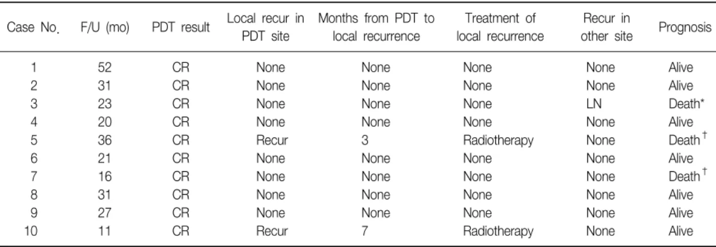
Table 2. The outcome in PDT patients
Case No. F/U (mo) PDT result Local recur in PDT site
Months from PDT to local recurrence
Treatment of local recurrence
Recur in
other site Prognosis
1 52 CR None None None None Alive
2 31 CR None None None None Alive
3 23 CR None None None LN Death*
4 20 CR None None None None Alive
5 36 CR Recur 3 Radiotherapy None Death
†6 21 CR None None None None Alive
7 16 CR None None None None Death
†8 31 CR None None None None Alive
9 27 CR None None None None Alive
10 11 CR Recur 7 Radiotherapy None Alive
*Death due to lung cancer progression,
†Death due to pneumonia.
F/U: follow-up period; PDT: photodynamic therapy; CR: complete remission; LN: cancer recurrence in mediastinal lymph node; mo:
month.
Table 1. Clinical characteristics of patients
Case
No.
Age
(yr) Sex Smoking
(PY) Histology TNM Site FEV
1(L) Cause Other site
Other treatment
Other histology
Photo sensitizer
1 72 M 75 Sqcc TisN0M0 LUL 2.17 Double* RLL OP+CTX Adeno
carcinoma
Photogem
2 70 M 40 Sqcc TisN0M0 RUL 2.28 Double LLL OP+CTX Sqcc Photogem
3 69 M 50 Sqcc TisN0M0 LLL 2.12 Double RLL OP Sqcc Photogem
4 58 M 30 Sqcc T1aN0M0 LUL 3.03 Double RLL OP Sqcc Photofrin
5 77 M 50 Sqcc TisN0M0 RLL 1.97 Refuse
†N N N Photogem
6 49 M 60 Sqcc TisN0M0 Lt. main 3.98 Lt. main
‡N N N Photofrin
7 56 M 45 Sqcc T1aN0M0 LLL 1.43 Postop.
§N N N Photofrin
8 69 M 46 Sqcc TisN0M0 RML 2.4 Refuse N N N Photofrin
9 69 M 50 Sqcc TisN0M0 LLL 2.29 CRI N N N Photofrin
10 65 M 20 Sqcc T1aN0M0 Lt. main 1.95 Lt. main N N N Photofrin
*Synchronous multiple bronchial lesion,
†Operation refuse,
‡Cancer on Lt. main bronchus,
§Cancer after previous lung cancer oper- ation (pneumonectomy).
PY: pack-year; TNM: the TNM Classification of Malignant tumors; FEV
1: forced expiratory volume in one second; sqcc: squamous cell carcinoma; CRI: cardiorespiratory insufficiency; LUL: left upper lobe; RUL: right upper lobe; RLL: right lower lobe; LLL: left lower lobe; N: no other site cancer; OP+CTX: operation and chemotherapy.
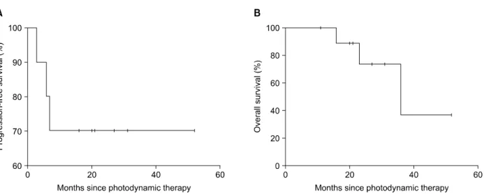
PDT를 시행한 부위에서 재발이 있었고, 10% (1/10)는 종 격동 임파선 전이의 형태로 재발하였으며, 10% (1/10)는 암의 재발 후 진행으로 항암치료를 시행하였으나 사망하 였고, 20% (2/10)는 단순폐렴으로 사망하였다. 우리 환자 에서 3년 무진행 생존율은 70%였고, 3년 전체 생존율은 75%였다(Figure 1).
고 찰
광역동 치료는 1 cm 이하의 다른 부위에 전이가 없는 중심부 조기 폐암 환자에서 전신 상태나 다른 이유로 인해 수술이 불가능할 때 유용한 치료이다2. 본 연구의 환자들 도 수술이 불가능한 동시 다발성 병변을 가지고 있거나 수술을 거부하는 등 조기 폐암임에도 불구하고 수술을 시 행하지 못하는 환자들을 대상으로 하였다. 기존의 연구보 고와 마찬가지로 모두 편평상피암이었고, 남자였으며, 흡
Figure 1. Progression-free survival and overall survival in the PDT patients. Kaplan-Meier curves for progression-free survival (A) and overall survival (B) in PDT patients are shown. In (A) and (B), tick marks indicate patients for whom data were censored at the data cutoff point. The median of progression-free survival is N.R. (not reached) and the median of overall survival is 36 months. PDT: photodynamic therapy.
연자였다. 조기 폐암에서 PDT의 효과는 여러 논문에서 증명되었다2,4,11-15. 대부분의 환자들은 수술의 적응증이 안 되는 조기 폐암환자들을 대상으로 하였고, 완전관해는 64
∼86%, 재발 13∼39%, 5년 생존율은 43.4∼68.4%로 다 양하였지만, 평균적으로 완전관해는 75%, 재발은 30% 정 도였다13. 폐암의 침윤 정도에 따라 치료효과가 결정되기 때문에 침윤 정도는 중요한데16, 최근 초음파 기관지내시 경 검사(endobronchial ultrasonography, EBUS)의 이용 이 증가하면서 PDT의 적용이 늘어나고 있다. 중심부 초기 폐암의 진단에 있어서 EBUS가 유용하다는 보고가 있었 고, 이 연구에서는 일반적인 기관지내시경과 고해상도 흉 부 전산화 단층촬영보다 EBUS에서 폐암의 침윤 정도를 정확히 파악할 수 있으므로 PDT치료에서 중요한 검사법 이라고 보고하였다17. 조기 폐암환자에서 생존율을 가장 증가시킬 수 있는 치료법은 수술이지만, 비용 대비 효과 (cost-effectiveness) 면에서 수술과 PDT를 비교하였을 때 PDT가 더 우월하다는 연구도 있었다18.
폐암에서 동시 다발성 기관지 병변인 경우는 0.2∼20%
정도 된다고 알려져 있고19-21, 이 경우에도 수술을 하는 것이 생존율을 높일 수 있는 방법인데, 수술에 관련된 사 망률이 나이와 동반 질환에 따라 1.1∼11%까지 다양하여 수술적 치료를 선택할 때는 나이와 동반 질환에 대한 고려 가 필요하다22. 폐암환자군이 고령인 점을 감안하면23 수 술적 치료가 힘든 경우가 많고, 수술을 시행했다고 하더라 도 재발하는 경우가 많아24,25, 적절한 적응증만 된다면
PDT는 효과적인 치료법이 될 수 있다. 동시 다발성 기관 지 병변 환자에서 PDT의 효용성에 대한 보고는 있지만26,27, 아직까지 명확하지는 않다. 향후 이 부분에 대해서 많은 연구가 필요할 것이라 생각된다.
조기 폐암에서 PDT의 효용성에 관한 국내 보고는 거의 없다. 본 연구가 대상자가 적고, 여러 병원이 참여한 연구 가 아니라 결론을 내기 힘든 것이 사실이나 앞에서 본 바 와 같이 적절한 적응증의 환자만 선택된다면, PDT는 효과 적인 치료법이며, 조기 폐암환자에게 치료법의 일환으로 제시할 수 있을 것이라 생각된다.
결론적으로 PDT는 수술적응증이 되지 않는 중심부 조 기 폐암환자를 대상으로 하고 있고, 재발할 수 있으나 환 자들이 편안하게 시술받을 수 있으며, 한 번 시술 후에도 여러 차례 다시 시술할 수 있다는 등의 장점을 가진 치료 법이다. 아직 우리나라에서는 경험이 적어 그 적용에 제 한이 있으나 적절한 적응증인 환자를 선택할 수 있고, 적 절한 방법으로 시술한다면 조기 폐암환자에서 효과적인 치료법이 될 수 있을 것이라 생각된다.
참 고 문 헌
1. Welch HG, Schwartz LM, Woloshin S. Are increasing 5-year survival rates evidence of success against can- cer? JAMA 2000;283:2975-8.
2. Kennedy TC, McWilliams A, Edell E, Sutedja T, Downie G, Yung R, et al. Bronchial intraepithelial neoplasia/
early central airways lung cancer: ACCP evidence- based clinical practice guidelines (2nd edition). Chest 2007;132(3 Suppl):221S-233S.
3. Moghissi K. Role of bronchoscopic photodynamic ther- apy in lung cancer management. Curr Opin Pulm Med 2004;10:256-60.
4. Maziak DE, Markman BR, MacKay JA, Evans WK;
Cancer Care Ontario Practice Guidelines Initiative Lung Cancer Disease Site Group. Photodynamic therapy in nonsmall cell lung cancer: a systematic review. Ann Thorac Surg 2004;77:1484-91.
5. Nowis D, Makowski M, Stokƚosa T, Legat M, Issat T, Goƚab J. Direct tumor damage mechanisms of photo- dynamic therapy. Acta Biochim Pol 2005;52:339-52.
6. Kim JO, Jung MK, Jung SS. Photodynamic therapy (PDT) in lung cancer. Tuberc Respir Dis 2007;62:
175-83.
7. Moghissi K, Dixon K. Is bronchoscopic photodynamic therapy a therapeutic option in lung cancer? Eur Respir J 2003;22:535-41.
8. Yoon SH, Han KT, Kim GN, Lee SI. Effect of photo- dynamic therapy in lung cancer. Tuberc Respir Dis 2004;57:358-63.
9. Endo C, Miyamoto A, Sakurada A, Aikawa H, Sagawa M, Sato M, et al. Results of long-term follow-up of pho- todynamic therapy for roentgenographically occult bronchogenic squamous cell carcinoma. Chest 2009;
136:369-75.
10. Okunaka T, Kato H, Tsutsui H, Ishizumi T, Ichinose S, Kuroiwa Y. Photodynamic therapy for peripheral lung cancer. Lung Cancer 2004;43:77-82.
11. Corti L, Toniolo L, Boso C, Colaut F, Fiore D, Muzzio PC, et al. Long-term survival of patients treated with photodynamic therapy for carcinoma in situ and early non-small-cell lung carcinoma. Lasers Surg Med 2007;
39:394-402.
12. Furukawa K, Kato H, Konaka C, Okunaka T, Usuda J, Ebihara Y. Locally recurrent central-type early stage lung cancer <1.0 cm in diameter after complete re- mission by photodynamic therapy. Chest 2005;128:
3269-75.
13. Mathur PN, Edell E, Sutedja T, Vergnon JM; American College of Chest Physicians. Treatment of early stage non-small cell lung cancer. Chest 2003;123(1 Suppl):
176S-180S.
14. Moghissi K, Dixon K, Thorpe JA, Stringer M, Oxtoby C. Photodynamic therapy (PDT) in early central lung cancer: a treatment option for patients ineligible for surgical resection. Thorax 2007;62:391-5.
15. Usuda J, Kato H, Okunaka T, Furukawa K, Tsutsui H, Yamada K, et al. Photodynamic therapy (PDT) for lung cancers. J Thorac Oncol 2006;1:489-93.
16. Nakamura H, Kawasaki N, Hagiwara M, Ogata A, Kato H. Endoscopic evaluation of centrally located early squamous cell carcinoma of the lung. Cancer 2001;91:
1142-7.
17. Miyazu Y, Miyazawa T, Kurimoto N, Iwamoto Y, Kanoh K, Kohno N. Endobronchial ultrasonography in the assessment of centrally located early-stage lung cancer before photodynamic therapy. Am J Respir Crit Care Med 2002;165:832-7.
18. Kato H, Okunaka T, Tsuchida T, Shibuya H, Fujino S, Ogawa K. Analysis of the cost-effectiveness of photo- dynamic therapy in early stage lung cancer. Diagn Ther Endosc 1999;6:9-16.
19. Carey FA, Donnelly SC, Walker WS, Cameron EW, Lamb D. Synchronous primary lung cancers: preva- lence in surgical material and clinical implications.
Thorax 1993;48:344-6.
20. Ferguson MK, DeMeester TR, DesLauriers J, Little AG, Piraux M, Golomb H. Diagnosis and management of synchronous lung cancers. J Thorac Cardiovasc Surg 1985;89:378-85.
21. Wu SC, Lin ZQ, Xu CW, Koo KS, Huang OL, Xie DQ.
Multiple primary lung cancers. Chest 1987;92:892-6.
22. Jung EJ, Lee JH, Jeon K, Koh WJ, Suh GY, Chung MP, et al. Treatment outcomes for patients with synchro- nous multiple primary non-small cell lung cancer. Lung Cancer 2011;73:237-42.
23. National Cancer Information Center (NCIC). Cancer in- cidence rate 2008. Goyang: ICIC; c2010 [cited 2011 Sep 30]. Available from: http://www.cancer.go.kr/ncic/cics_f/
01/011/index.html.
24. Johnson BE. Second lung cancers in patients after treat- ment for an initial lung cancer. J Natl Cancer Inst 1998;90:1335-45.
25. Johnson BE, Cortazar P, Chute JP. Second lung cancers in patients successfully treated for lung cancer. Semin Oncol 1997;24:492-9.
26. Moghissi K, Dixon K. Photodynamic therapy for syn- chronous occult bronchial cancer 17 years after pneumonectomy. Interact Cardiovasc Thorac Surg 2005;4:327-8.
27. Usuda J, Ichinose S, Ishizumi T, Hayashi H, Ohtani K, Maehara S, et al. Management of multiple primary lung cancer in patients with centrally located early cancer lesions. J Thorac Oncol 2010;5:62-8.