INTRODUCTION
Low-energy stimulation of tissues by lasers or light- emitting diode(LED) have shown to increase cellular activity during wound healing by increasing collagen production and angiogenesis. The previous reports sug- gest that biostimulation of near-infrared(NIR) lasers or LED influenced cell proliferation by increasing mito- chondria respiration through stimulation of cytochrome oxidase. In other experiments, photo-irradiation by NIR- light-emitting diode increased the production of cytochrome oxidase in cultured primary neurons and
reversed the reduction of cytochrome oxidase activity produced by metabolic inhibitors
1). In this report, there are significant upregulation of gene expression in path- ways involved in mitochondrial energy production and antioxidant cellular protection. In connection with those results, we hypothesized LED can be used as a non-inva- sive therapeutic approach for cell death by modulating cellular signalling mechanisms
2-4).
Apoptosis is a gene-regulated mechanism of cell death, playing a pivotal role in physiological and pathological processes
5). Hypoxic / ischemic condition has been rec- ognized for a long time as an important mediator or modulator of apoptosis because this condition is accom- panied by the production of reactive oxygen species (ROS) which can attack nucleic acids, proteins and mem- brane phospholipids
6-8). Hypoxia / ischemia - induced apoptosis is a major concern in various clinical entities such as ischemic diseases, organ transplantations and
* Corresponding author
Sun-Youl Ryu
Dept. of OMFS, College of Dentistry, Chonnam National Univ.
5 Hak-Dong, Dong-Ku, Gwangju, 501-757, Korea
Tel: 82-62-220-5430 Fax: 82-62-228-8712
E-mail: [email protected]
Effects of LED irradiation on the expression of apoptosis-related molecules in human SH-SY5Y neuroblastoma cells
Kyu-Seung Cho, Sun-Youl Ryu, Hong-Ran Choi*
Department of Oral and Maxillofacial Surgery, *Department of Oral Pathology,
School of Dentistry, Dental Science Research Institute, Chonnam National University, Gwangju, Korea
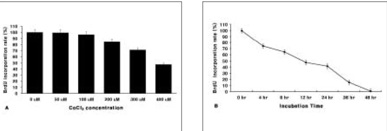
To verify the inhibitory or protective effects of light-emitting diode(LED) irradiation on apoptotic cell death induced by CoCl
2
, human SH-SY5Y cells were treated with CoCl
2
and LED were used to irradiate the cells. In the cell viability assay, cells were died slowly from 50 μM to 250 μM and about 50% of cells died after 12 hours at 400 μM of CoCl
2
. The Diff-Quik staining revealed that cells showed con- densation of DNA and blebbing of the cell membrane. The DNA fragmentation assay revealed the DNA fragmentation, which is anoth- er apoptosis marker, occurred in cells treated with 400 μM CoCl
2
for 16 hours. In the western blot for HIF-1α, HIF-1αwas expressed after 3 hours from induction and peaked maximally at 16 hours. In the cell viability assay of the effects of LED irradiation (at 590 nm for 1 hour 20 minutes), the cells showed more proliferation (about 20%) than the control group. The RPA assay of various apoptosis-related molecules showed that pro-apoptosis molecules such as Bax, Bak, and Bid were upregulated in the CoCl
2
treatment group. This means that the apoptotic cell population was increased. However there was some significant changes in LED irradiated cells. In the CoCl
2
- treated LED irradiation group, those molecules were down-regulated more than in the only CoCl
2
-treated group. These results have shown that CoCl
2
may induce apoptotic cell death in human SH-SY5Y neuroblastoma cells. And LED irradiation has a positive effect on apoptotic cells by down-regulation of pro-apoptotic molecules.
Key words
LED irradiation, Apoptotic cell death, Human SH-SY5Y cells
Abstract