대퇴골 전자부 역사상 골절의 압박고 나사를 이용한 치료
조선대학교 의과대학 정형외과학교실 김동휘∙이상홍∙하상호∙유재원
─ Abstract ─
Treatment of Reverse Oblique Trochanteric Fracture with Compression Hip Screw
Dong-Hui Kim, M.D., Sang-Hong Lee, M.D., Sang-Ho Ha, M.D., Jae-Won You, M.D.
Department of Orthopaedic Surgery, College of Medicine, Chosun University, Gwangju, Korea
Purpose: To investigate the results of treatment of reverse oblique trochanteric fractures with compression hip screw.
Methods: We reviewed the results of 12 cases of reverse oblique trochanteric fracture treated with compres- sion hip screw from January 2000 to December 2006 which could be followed up for more than 1 year. The mean follow up period was 26 months (15~40). The mean age was 48 years old. Injury mechanism was com- posed of 6 cases of traffic accident and 6 cases of fall down. 8 persons were man. We investigated the union time, degree of neck-shaft angle change, amount of sliding of compression hip screw, complications, functional and clinical results.
Results: 10 cases were united and the mean union time were 5 months (3~8). The mean neck-shaft angle change was 3.5 degrees (0~12). The amount of sliding of compression hip screw was 8.9 mm (2~24). There were six coxa vara, six leg due to coxa vara shortening, two nonunion, and one superficial infection.
Unsatisfactory results of Jensen’s social function score and Parker and Palmer’s mobility score were studied.
Conclusion: The results of treatment of reverse oblique trochanteric fractures with compression hip screw were relatively unsatisfied. (J Korean Soc Traumatol 2010;23:1-5)
Key Words: Femur, Trochanteric fracture, Reverse oblique fracture, Compression hip screw
� Address for Correspondence : Sang-Hong Lee, M.D.
Department of Orthopaedic Surgery, Chosun University Hospital 588, Seosuk-dong, Dong-gu, Gwangju 501-717, Korea
Tel : 82-62-220-3147, Fax : 82-62-226-2379, E-mail : [email protected]
접수일: 2010년 4월 23일, 심사일: 2010년 5월 2일, 수정일: 2010년 5월 31일, 승인일: 2010년 6월 7일 이 논문은 2008년도 조선대학교 학술연구비의 지원을 받아 연구되었음.
I. 서 론
대퇴 근위부 골절은 고령에서 발생하는 손상 중 가장 위험한 손상 중 하나로서,(1) 이중 역사상 전자부 골절은
고관절 골절의 2%, 전자부 골절의 5%를 차지하는 드문 골절이고, 일반적인 전자부 골절과는 다른 생역학적 특징 으로 주 골절선이 원위부는 외측, 근위부는 내측으로 진행 하는 양상을 갖는다.(2)
압박고나사는 근위 골절부의 활강을 유도함으로써 골절 선에 압박력을 주고, 안정성을 줌으로써 골유합에 우수한 결과를 갖지만, 역사상 골절에서는 원위 골절부가 내측으 로 전위되며 골절선에 전단력이 작용하여 합병증이 많이 발생하므로,(2,3) 여러 저자들이 골수강내 금속정이 가장 적합한 치료방법으로 보고하였다.(3-6) 그러나 골수강내 금속정 삽입부가 파괴되어있는 경우, 수술 시기가 경과하 고 심한 분쇄가 있어 비관혈적 정복술이 어려운 경우에는 골수강내 금속정의 사용이 어려워, 이러한 경우에 압박고 나사를 이용한 수술적 치료를 시행하였다. 이에 본 저자들 은 A3형인 대퇴골 전자부 역사상 골절에 대하여 압박고 나사를 이용하여 치료한 결과를 후향적으로 분석하고 그 의의에 대하여 살펴보고자 하였다.
II. 대상 및 방법 1. 연구대상
2000년 1월부터 2006년 12월까지 대퇴골 전자부 역사상 골절을 보이는 A3형 골절로 본원에서 수술 받은 환자중 골수강내 금속정이나 인공관절 치환술 등으로 치료한 경 우를 제외하고 압박고 나사를 이용하여 수술을 시행받은 최소 1년 이상 추시 가능한 12예를 대상으로 하였다(Table 1). AO분류상 A3.1은 1예, A3.3은 11예를 보였다. 연령은 21세에서 79세까지 평균 48세였으며, 여자가 4예 남자가 8 예였다. 추시 기간은 15개월에서 40개월로 평균 26개월이 었으며, 수상 원인은 교통사고가 6예, 낙상이 6예였다. 동 반 손상은 하지 골절이 3예로 많았고, 그 외에 상지골절 2 예, 골반내 장기 손상 2예, 뇌 손상 1예를 보였다.
2. 수술방법 및 술후 처치
수술에 있어서 모든 경우에 압박고 나사(Zimmer, Warsaw, USA)를 이용한 내고정을 시행하였으며 부가적 으로 케이블그립 8예, 강선고정 4예, 추가적인 나사못 고정 술 4예를 시행하였다. 골 이식술은 5예를 시행하였는데, 동 종골 3예, 자가골 2예였으며, 수술 후 6주에서 12주 사이에 전 체중부하 보행을 시행하였다.
3. 평가방법
수술 후 기능의 평가는 Jensen의 social function score(7) (Table 2)와 Parker와 Palmer의 Mobility score(8)(Table 3) 를 이용하여 일상 생활능력 및 보행능력을 평가하였고, 수 술 후 방사선상 평가는 수술 후 전후면 및 측면 사진을 시행하였으며 추시 방사선 사진상에서 수술 후 골유합 시 기, 경부 골간각의 변화, 압박고 나사의 활강 정도, 내고정 물의 고정 및 역학적 실패, 불유합 및 지연유합 등의 합병 증을 조사하였고 활강 정도는 최종 추시 전후방 방사선 사진의 비교를 통해 압박고 나사의 내측 끝에서 측면 금 속판의 내측면에 이르는 거리의 차이로 판정하였다. 나이, 성별, 골 이식 유무, 골다공증 유무가 골 유합에 미치는 영 향에 대하여 student t-test를 이용하여 분석하였으며 유의 수준은 0.05 이하에서 의미 있는 것 판단하였다.
III. 결 과
12예중 10예에서 골유합을 얻을 수 있었으며, 골유합까 지의 기간은 평균 5개월(3~8개월)이었다. 방사선학적 이
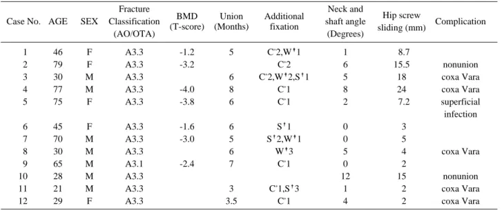
Table 1. Data about the patients Fracture
BMD Union Additional Neck and
Hip screw
Case No. AGE SEX Classification shaft angle Complication
(AO/OTA) (T-score) (Months) fixation
(Degrees) sliding (mm)
01 46 F A3.3 -1.2 5 C*2,W�1 1 08.7
02 79 F A3.3 -3.2 C*2 6 15.5 nonunion
03 30 M A3.3 6 C*2,W�2,S�1 5 18 coxa Vara
04 77 M A3.3 -4.0 8 C*1 8 24 coxa Vara
05 75 F A3.3 -3.8 6 C*1 2 07.2 superficial
infection
06 45 F A3.3 -1.6 6 S�1 0 03
07 70 M A3.3 -3.0 5 S�2,W�1 0 05
08 30 M A3.3 6 W�3 5 04 coxa Vara
09 65 M A3.1 -2.4 7 C*1 0 02
10 28 M A3.3 12 15 nonunion
11 21 M A3.3 3 C*1,S�3 1 02 coxa Vara
12 29 F A3.3 3.5 C*1 4 02 coxa Vara
상 소견으로, 압박고 나사의 활강 정도는 평균 8.9 mm (2~24 mm)였고, 또한 수술 후와 최종 추시때의 대퇴 경 간각을 측정한 결과 0。에서 12。로 평균 3.5。의 변화를 보 였다. 2예에서 불유합을 보였고, 6예에서 내반고를 보였다.
임상적 측면의 합병증으로는 하지 단축이 6예로 1 cm에서 2.5 cm의 평균 1.4 cm의 단축을 보였고, 창상 감염은 1예 를 보였으나 표재성 감염에 국한 되었다. Jensen의 social function score는 1.78±0.56, Parker와 Palmer의 Mobility score는 4.21±0.63을 보여 낮은 보행능력과 관절 운동을 보 였다. Student t-test를 통한 통계학적 분석에서 나이 (P=0.110), 성별(P=0.094), 골이식 유무(P=0.373), 골 다 공증 유무(P=0.179)는 모두 골 유합에 유의성을 보이지 않았다.
IV. 고 찰
대퇴골 전자부 역사상 골절은 전자부 골절의 5%를 차 지하는 골절로써 일반적인 전자부 골절과는 다른 생역학 적인 특징을 갖는다.(2) 대퇴골 전자부 역사상 골절의 치 료에 있어서 압박고 나사의 사용으로 인한 불만족스러운 결과를 보고하였는데,(9) 외측 근위부에서 내측 원위부로 의 일반적인 주골절선을 가진 A1 및 A2형 골절은 축성 부하에 의한 골절부위 압박을 이루지만 A3형 골절은 골절 부위에 전단력이 작용하여 원위 골편의 내측 전위를 유발하
여 불유합 및 고정 실패 등의 합병증을 일으키게 된다.(10) 이러한 골절에서 사용되는 내고정물로는 크게 압박고 나사, 칼날 금속판 등의 금속판군과 골수강내 고나사, 교합 성 골수강내 금속정 또는 아시아형 감마정 등의 골수강내 금속정 군으로 분류할 수 있다.
금속판은 역학적으로 금속정과 달리 더 긴 모멘트 암으 로 인해 많은 굴곡력이 작용하게 되며, 심한 분쇄 골절 또 는 불완전하게 정복된 골절에서 내측 지주가 소실된 경우 장력대로 작용하고 있는 금속판의 한 곳에 모든 응력이 집중되어 기계적 실패가 발생하기 쉬운 단점이 있다.(11) 그러나 관혈적 정복 후 삽입하므로 비교적 해부학적 정복을 할 수 있고, 특히 압박고 나사의 경우 술기가 비교적 쉬운 장점이 있다.(11-13) 그러나 광범위한 절개선에 따른 불유합 의 가능성 및 감염 등의 위험성이 높은 단점이 있다.(14)
골수강내 금속정은 내고정시 골의 혈액 순환이 금속판 보다 좋으며 내고정물이 골수강 내에 위치하여 골절 부위 에 전달되는 부하를 뼈와 공유하고, 뼈의 정상적인 부하 전달을 훼손하지 않는다는 생역학적 장점이 있다.(15) 그 러나 Schatzker와 Waddell 등(11,16)은 전자부 골절에서 근 위 골절편의 길이가 짧고 골수강이 넓어 견고한 내고정을 얻기 어려워 골절 근위부의 내반 변형, 골절 원위부의 외 반 변형이 일어나기 쉬우며 내측 피질골의 분쇄가 있을 경우 예후가 나쁘다고 하였다. 그럼에도 불구하고 많은 저 자들은 대퇴골 전자부 골절의 치료시 골수강내 금속정이 압박고 나사에 비하여 굴곡 모멘트가 작고 근위 골편의 활강을 효과적으로 억제하며, 역사상 골절 및 전자하부 골 절에도 사용할 수 있으며 대전자의 외측 벽에 골절이 있 을 때나 후내측 벽에 골편이 있는 불안정성 골절에도 효 과적으로 사용할 수 있는 점을 들어 우위를 주장하고 있 다.(5,17-19) 저자들은 외과 및 비뇨생식기의 동반 손상으로 인한 3주 이상의 수술지연이나 심한 분쇄를 동반한 역사상 골절(A3.3)로 인하여 폐쇄적 정복술이 불가능하였고 골수 강내 금속정의 삽입부가 심한 분쇄로 인해 파괴되어있는 경우에서 압박고 나사를 이용한 수술적 치료를 시행하였다.
골유합 시기는 평균 5개월 이었으며, 2예의 불유합과 6 예의 내반고 및 하지 단축을 보여 높은 합병증 발생과 다 Table 2. The assessment of social function of Jensen
Score Social function groups Definition 1 Independent Manages everything
Possibly working 2 Slightly dependent Manages household
Meals-on-wheels, home-help
≤4 hours/week
3 Moderately dependent Home-help ≥5 hours/week Possibly district nurse 4 Totally dependent Living in nursing home or
Long-term nursing at home
Table 3. Mobility Score of Parker and Palmer
Alone with With help
Walking ability No from
Not at all difficulty an assistive
another device
person
Able to walk inside house 3 2 1 0
Able to walk outside house 3 2 1 0
Able to go shopping, to a restaurant, or to visit family 3 2 1 0
른 저자들에 비해(20,21) 낮은 골유합율을 보였는데, 대부 분의 골절 유형이 A2형인 반면, 본 연구에서는 A3.3형 골 절이 대부분으로 심한 분쇄를 보이며 역사상 골절만을 대 상으로 하였기 때문일 것으로 사료된다. 골다공증을 보이 는 4예중 3예에서 평균 6.3개월의 골 유합기간을 보였으며, 불유합, 내반고와 창상감염이 각각 1예에서 발생하여 합병 증 발생율이 높을것으로 생각되었으나 통계학적 유의성은 보이지 않았는데, 이것은 증례수가 많지 않고, 고령이며, 전신적인 상태가 좋지 않은 결과인 것으로 사료된다 (Table 1).
견고한 내고정을 얻기 위해서 분쇄가 심한 경우 골이식 과 보조적 나사고정이나 강선 고정을 권유하였다.(11,22,23) 본 연구에서도 8예에서 케이블 그립, 4예에서 강선 고정을 시행하였으며 4예에서 보조적 나사고정을 시행하였다. 그 리고, 분쇄의 정도가 심한 골절에 대해서 2예의 자가골 이 식술, 3예의 동종골 이식술을 시행하였는데, 1예에서 불유 합을 보였으며(Fig. 1), 나머지 4예에서 평균 6.2개월에 골 유합을 얻었다 그러나 이러한 노력에도 불구하고 수술의 결과는 좋지 않았으며, 새로운 치료방법들이 필요하리라 사료된다.
해부학적 정복 후 활강 압박고 나사와 전자부 안정화 금속판을 함께 사용한 불안정성 대퇴전자간 골절 치료에 서 근위 골편의 지지효과(buttress effect)로 불안정한 전자 부의 고정 감소와 골절 부위의 과도한 감입에 의한 각형 성 및 근위 골편의 회전을 막아주고, 원위 골편의 내측 이 동을 막아 하지 단축을 예방하여 대체로 우수한 결과를 보인다고 보고되고 있다.(24,25)
황등(26)과 손등(27)은 대퇴골 전자간부의 불안정성 골 절에서 무시멘트형 인공관절 치환술을 시행한 경우에 임 상적 및 방사선학적으로 비교적 좋은 결과를 보고하고 있 으나 적응에 논란이 많고 과도한 근위부 골 소실과 감염 등의 합병증이 가능성이 높아 주의가 필요하다.
V. 결 론
대퇴골 전자부 역사상 골절의 치료에 있어 압박고 나사 를 이용하여 치료한 결과 골유합이 지연되었으며, 합병증 이 비교적 많았고 수술 후 기능이 매우 저하되었다. 이처 럼 역사상 전자부 골절에서 압박고 나사를 이용한 치료는 불만족스러웠다. 향후 전자부 안정화 금속판의 사용등이 고려되어야 할 것으로 생각된다.
REFERENCES
01) Melton LJ, 3rd, Ilstrup DM, Riggs BL, Beckenbaugh RD. Fifty-year trend in hip fracture incidence. Clin Orthop Relat Res. 1982;162:144-9.
02) George J, Andrew T, Daniel J. Reverse obliquity frac- tures of the intertrochanteric region of the femur. J Bone Joint Surg Am. 2001;83:643-50.
03) Wagner R, Wechbach A, Sellmair U, Blattert T.
Extra-articular proximal femur fracture in the elderly - dynamic hip screw or intramedullary hip screw for fracture management? Langenbecks Arch Chir Suppl Kongressbd. 1996;113:963-6.
04) Hardy DC, Descamps PY, Krallis P, et al. Use of an intramedullary hip-screw compared with a compression hip-screw with a plate for intertrochanteric femoral frac- tures. A prospective, randomized study of one hundred patients. J Bone Joint Surg Am. 1998;80:618-30.
05) Lorich DG, Geller DS, Nielson JH. Osteoporotic pertrochanteric hip fractures: management and current controversies. Instr Course Lect. 2004;53:441-54.
06) Sadowski C, Lubbeke A, Saudan M, Riand N, Stern R, Hoffmeyer P. Treatment of reverse oblique and transverse intertrochanteric fractures with use of an intramedullary nail or a 95 degrees screw-plate: a prospective, randomized study. J Bone Joint Surg Am.
2002;84:372-81.
Fig. 1. (A) Preoperative anteroposterior radiograph of the right hip of a seventy-five-year-old woman who fall down, revealing an A3.3 reverse oblique intertrochanteric fracture. (B) Postoperative radiograph after fixation with a compression hip screw. (C)
A B C
07) Jensen JS. Determining factors for the mortality follow- ing hip fractures. Injury. 1984;15:411-4.
08) Parker MJ, Palmar CR. A new mobility score for pre- dicting mortality after hip fracture. J Bone Joint Surg Br. 1993;75:797-8.
09) Boldin C, Seibert FJ, Fankhauser F, Peicha G, Grechenig W, Szyszkowitz R. The proximal femoral nail (PFN)-a minimal invasive treatment of unstable proximal femoral fractures: a prospective study of 55 patients with a follow-up of 15 months. Acta Orthop Scand. 2003;74:53-8.
10) Baumgaertner MR, Curtin SL, Lindskog DM.
Intramedullary versus extramedullary fixation for the treatment of intertrochanteric hip fractures. Clin Orthop Relat Res. 1998;348:87-94.
11) Fielding JW, Cochran GV, Zickel RE. Biomechanical characteristics and surgical management of sub- trochanteric fractures. Orthop Clin North Am. 1974;
5:629-50.
12) Aune AK, Ekeland A, Odegaard B, Grogaard B, Alho A. Gamma nail vs compression screw for trochanteric femoral fractures. 15 reoperations in a prospective, ran- domized study of 378 patients. Acta Orthop Scand.
1994;65:127-30.
13) Thorngren KG. Optimal treatment of hip fractures.
Acta Orthop Scand. 1991;241:31-4.
14) Jeon TS, Kim WS, Kim SB, Hwang CM, Kim KT, Kim SH. Treatment of communited subtrochanteric fractures of the femur by high energy trauma. J Korean Fracture Soc. 2006;19:135-40.
15) Joachim W, Cirk AH. Pitfalls and complications in the use of the proximal femoral nail. Langenbeck Arc Surg. 2004:196-212.
16) Schatzker J, Waddell JP. Subtrochanteric fractures of the femur. Orthop Clin North Am. 1980;11:539-54.
17) Yoo JH, Yang KH, Park SY, Won JH, Yoon HK. The treatment of unstable reverse oblique intertrochanteric
fractures with proximal femoral nail (PFN). J Korean Orthop Assoc. 2005;40:733-40.
18) Kyle RF, Gustilo RB, Premer RF. Analysis of six hun- dred and twenty-two intertrochanteric hip fractures. J Bone Joint Surg Am. 1979;61:216-21.
19) Haidukewych GJ, Israel TA, Berry DJ. Reverse obliqui- ty fractures of the intertrochanteric region of the femur.
J Bone Joint Surg Am. 2001 May;83-A: 643-50.
20) Herrera A, Domingo LJ, Calvo A, Martinez A, Cuenca J. A comparative study of trochanteric fractures treated with the gamma nail or the proximal femoral nail. Int Orthop. 2002;26:365-9.
21) Domingo LJ, Cecilia D, Herrera A, Resines C.
Trochanteric fractures treated with a proximal femoral nail. Int Orthop. 2001;25:298-301.
22) Velasco RU, Comfort TH. Analysis of treatment prob- lems in subtraochanteric fractures of the femur. J Trauma. 1978;18:513-23.
23) Lee SH. The treatment of unstable intertrochanteric fracture of the femur with compression hip screw. The Korean Society of Traumatology. 1998;11:187-92.
24) Cho JH. The compression hip screw with trochanter stabilizing plate for internal fixation of unstable intertrochanteric fractures. J Korean Fracture Soc.
2005;18-3:221-6.
25) Bridle SH, Patel AD, Bircher M, Calvert PT. Fixation of intertrochanteric fracture of femur randomized prospective comparison of gamma nail and the dynamic hip screw. J Bone Joint Surg Br. 1991;73:330-5.
26) Hwang DS, Kwak SK, Woo SM. Results of cementless hemiarthroplasty for elderly patients with unstable intertrochanteric fractures. J Korean Hip Soc.
2004;16:386-91.
27) Sohn JM, Jahng JH, Ha NK, Cho ST, Choi KY.
Primary bipolar hemiarthroplasty for treatment of unsta- ble intertrochanteric fracture of the femur in elderly patients. J Korean Fracture Soc. 2003;16:37-44.