J. Biomed. Lab. Sci. 10 (2004) 99–105
Effect of Acute Ethanol Intoxication on Hepatic Rhodanese Activity in Rats with Extrahepatic Cholestasis
Ki-Suk Choi, Kyo-Cheol Mun, You-Hee Kim and Chun-Sik Kwak†
Department of Biochemistry, Keimyung University, School of Medicine, Taegu, 700-712, Korea
Liver and serum rhodanese activities were determined in acute ethanol intoxicated rats with extrahepatic cholestasis induced by common bile duct ligation (CBD) to manifest the biochemical background of alcohol drinking hazard under the hepatobiliary disease. Liver cytosolic and microsomal rhodanese activities and these Vmax values in CBD ligated rats with acute ethanol intoxication were found to be decreased much more than that in CBD ligation alone. However, the difference of Km value on above hepatic enzyme was not found between the experimental groups. On the other hand, serum rhodanese activity in CBD ligated rats with acute ethanol intoxication was greater increased more than that in CBD ligation alone. These results indicate that the biosynthesis of the hepatic rhodanese decreases and the serum rhodanese activity increases in cholestasis combined with acute ethanol intoxication, reflecting damage of aggravated hapatocytic membrane. Accordingly, the resulting data supported the fact that alcoholic drinks were enzymologically harmful to the hepatobiliary disease.
Key Words: Common bile duct ligation, Ethanol intoxication, Extrahepatic cholestasis, Rhodanese
서 론
최근 주류의 소비량이 늘어남에 따라 음주로 인해 야기되 는 질병들이 관심의 대상이 되고 있으며, 그 중에서도 주정 대사의 주된 장기인 간에 미치는 주정의 영향이 주목을 받고 있다. 간은 물질 대사의 주된 기관으로서 대단히 복잡하고 다양한 기능을 가진 장기이며 체외로부터 흡수되거나 체내 에서 생성된 물질 특히 유해한 물질을 생체 변환시켜 배설 케 하는 기구를 가짐으로써 생체를 보호하고 있다18,38). 그러 나 이러한 기능을 가진 간이라 하더라도 장기간 많은 양의 음주를 하면 지방간, 간염, 간경변증 등이 초래될 수 있다
14,44)
. 일반적으로 간담도 질환 시 음주는 유해하다고 하며 이 사실은 음주로 인한 간 질환의 유발로 미루어 볼 때 당연하 다고 생각된다. 그러나 아직도 그 생화학적 뒷받침은 충분치 않다. 간은 생체이물 (xenobiotic)들을 생체 변환시키는 주된 장기이므로 생체이물의 생체 변환에 관련된 효소들이 다량 존재하며18) 특히 담즙울체 (cholestasis)로 간 손상이 있을 때 에는 간에서 이 효소들의 활성도가 변동된다17,26,28,30,32)
. 따라
서 간담도 질환 시 음주를 하거나 주정 중독이 야기된다면 간 조직에서 생체이물 생체 변환 (xenobiotic biotransformtaion) 효소의 활성도는 변동이 있을 것으로 생각된다.
Rhodanese (thiosulfate: cyanide sulfurtransferase, EC 2. 8. 1. 1) 는 cyanide, sulfite, organic sulfinate 및 dithiol를 sulfur 포합하 여 배설시키는 생체이물 생체 변환 과정을 촉매하는 효소이 며10,20,41~43)
포유 동물의 간에 주로 분포되어 있다41,42). 이 효 소는 혈중에도 출현되며10) 간세포에서는 세포질, 미토콘드 리아 및 내형질세망에 국재되어 있다16,24,42). 그리고 이 효소 는 흰쥐에서 만성 주정 중독 시 담즙울체를 야기시켰을 때 간 조직에서는 그 활성도가 감소되고 혈청에서는 그 활성도 가 증가되는 것6)으로 밝혀져 있다. 이와 같이 생체이물 생체 변환 효소인 rhodanese는 만성 주정 중독과 담즙울체를 병행 시켰을 때 간 조직과 혈청에서 그 활성도가 변동되고, 또한 담즙울체 시 주정 중독을 시키면 간 손상이 심해진다는 보고
8,14)가 있고 보면 담즙울체 시 급성 주정 중독을 야기시켰을
때도 이 효소의 활성도는 심한 변동이 있을 것으로 생각된다.
그러나 이에 대한 보고는 찾아 볼 수 없었다.
이 연구는 간담도 질환 시 음주의 유해함에 대한 생화학적 배경의 일단을 밝히고자 시행한 실험으로서 담즙울체가 진 행되는 흰쥐에게 급성 주정 중독을 시킨 후 간의 세포질, 미 토콘드리아 및 마이크로솜과 혈청에서 rhodanese의 활성도를 측정하였으며 아울러 총담관을 결찰한 후 14일에 급성 주정 중독을 시킨 흰쥐의 간세포 분획에서 이 효소의 Km 값과
*논 문 접 수: 2004년 3월 25일 수정재접수 : 2004년 5월 11일
†별책 요청 저자: 곽춘식, (우) 700-712 대구광역시 중구 동산동 194번지, 계명대학교 의과대학 생화학교실
Tel: 053-250-7461, Fax: 053-250-7461 e-mail: [email protected]
Vmax 값도 함께 측정하여 이들 성적을 보고하고자 한다.
재료 및 방법
1. 시 약
Potassium cyanide, sodium thiosulfate pentahydrate, formalde- hyde, ferric nitrate, ferric thiocyanate, potassium phosphate mono- basic, rhodanese (thiosulfate sulfurtransferase, type II, from bo- vine liver) 및 단백질 표준액 (10 g/100 ml bovine albumin) 등 은 Sigma사 (미국) 제품을 사용하였으며 그 외 시약들은 시 판하는 특급 또는 일급품을 사용하였다.
2. 동물 및 처치
동물은 4주 이상 같은 조건으로 사육한 체중 280~320 g 되는 Sprague-Dawley 종의 숫흰쥐를 사용하였으며 1군을 5 마리로 하여 다음과 같이 총 6개군으로 나누었다. 즉 정상군 (1군), 총담관 결찰 후 14일에 희생시킨 총담관 결찰군 (1군), Liu 등35)의 방법에 따라 체중 Kg 당 4 g의 에탄올을 투여하 고 각각 1.5시간 및 24시간에 죽인 급성 주정 중독군 (총 2 군), 총담관 결찰 14일 후 체중 Kg 당 4 g의 에탄올을 투여 하고 각각 1.5시간 및 24시간에 죽인 총담관 결찰 후 급성 주정 중독을 시킨 군 (총 2군) 등이다.
각 실험군은 개별 분리 수용하였으며 실험 전 후에 일정한 조건으로 사육하였다. 사료는 삼양유지사료주식회사의 실험 동물 사료를 먹게 하였다. 급성 주정 중독은 흰쥐 체중 Kg 당 4 g의 에탄올이 투여되도록 25% (v/v) 에탄올 용액을 조 제하여 단회 경구 투여하였다. 총담관 결찰술 및 간 적출술 은 효소 활성의 일중 변동을 고려하여 쥐를 일정한 시간에 희생시킬 수 있도록 수술 시간을 조절하였으며 12시간 금식 시킨 후 이터 마취하에서 실시하였다. 총담관 결찰은 간 근 위부와 약 1 cm 아래쪽의 원위부의 총담관을 각각 이중 결 찰한 후 그 중간 부위를 절단하였으며 간 적출 시 담관의 폐 쇄 상태를 확인하였다. 모든 실험군에서 간의 적출은 12시간 금식시킨 후 이터 마취하에서 시행하였으며 복부 대동맥으 로부터 채혈하여 쥐를 실혈사 시켰다. 그리고는 간문맥에 삽 관한 후 4℃의 0.25 M sucrose 액으로 관류하여 간에 남아 있던 혈액을 제거한 다음 간을 적출하였다. 적출한 간은 면 포로 균등히 압박하여 간에 남아 있던 sucrose 액을 가능한 한 제거하였다. 한편 채혈한 혈액은 원심분리하여 혈청을 얻 고 곧 효소 활성도를 측정하였다.
3. 간세포의 분획
간의 세포분획은 적출한 간들을 즉시 2~4℃로 냉각한 후 잘게 썰어서 절편으로 만들고 혼합하여 그 중 약 7 g을 취하 여 9배량의 0.25 M sucrose 액을 넣은 다음 Teflon pestle glass
homogenizer (chamber clearance 0.005~0.007 inches, Thomas사, 미국)로 2~4℃를 유지하면서 400 rpm의 속도로 조심스럽게 5회 왕복 마쇄하여 10% (w/v)의 간 조직 균질액을 만들었다.
이 간 균질액 모두를 취하여 sucrose density gradient 원심분 리법29)으로 세포질, 미토콘드리아 및 마이크로솜 분획을 분 리하였다. 세포 분획법에서 모든 조작은 2~4℃에서 시행하 였으며, 이때 사용한 원심분리기는 Du Pont Sorvall사 (미국) 의 RC-5B refrigerated superspeed centrifuge와 OTD-65B ultra- centrifuge였다. 이때 사용한 rotor는 Du Pont Sorvall사의 SS- 34 및 T865 rotor였고 sucrose linear density gradient 용액의 제조는 gradient former (model 570, ISCO, 미국)를 사용하였다.
4. 효소 시료 조제
Rhodanese 측정용 효소 시료의 조제는 분리한 세포질, 미 토콘드리아 및 마이크로솜 분획을 단백질 양으로 5 mg/ml가 되도록 0.25 M sucrose 액에 현탁시켜 사용하였다.
5. 효소 활성도 측정
혈청과 간의 세포질, 미토콘드리아 및 마이크로솜 분획의 rhodanese의 활성도 측정은 시료와 함께 potassium cyanide와 sodium thiosulfate를 기질로 사용하여 25℃에서 20분간 반응 시키는 동안에 생선된 thiocyanate를 ferric nitrate와 반응시켜 생성된 ferric thiocyanate를 460 nm 파장에서 비색하여 효소 의 활성도를 산출하는 Westley43)의 법에 의하였다. 이 효소 의 활성도 단위는 1분간에 1 ml의 혈청 또는 1 mg의 단백질 이 반응하여 생성한 ferric thiocyanate를 nmol로 나타내었다.
이 실험에서 채택한 효소 활성도 측정법들의 정확도를 높 이기 위하여 Sigma사 (미국)의 정제된 효소를 사용하여 검정 하였으며 같은 시료에 대하여 2회 측정하여 그 평균치를 취 하였다. 이 실험에서 각 효소 활성도 측정에 사용한 분광광 도계는 computer controlled enzyme spectrophotometer (Cary 210, Varian사, 미국)였다.
6. Km 값 및 Vmax 값의 측정
정상쥐, 총담관 결찰술 후 14일 경과한 쥐, 급성 주정 중 독 후 1.5 및 24시간 경과한 쥐 및 총담관 결찰술 후 14일에 급성 주정 중독을 시키고 1.5 및 24시간 경과한 흰쥐의 간세 포 분획 시료들과 rhodanese의 2종 기질 중 sodium thiosul- fate를 선택하여 기질 원액과 기질 희석액을 제조한 후 이 기질액들과 potassium cyanide 기질 원액을 사용하여 rhoda- nese의 활성도를 측정한 후 이들 성적으로부터 1/vi 값을 그리고 기질 농도로부터 1/[S] 값을 계산하여 이중역수도 (double reciprocal plot)를 작도한 다음 이것으로부터 Km 값 과 Vmax 값을 산출하였다.
7. 단백질 정량
효소액 중의 단백질 정량은 0.5 M perchloric acid와 me- thanol-ether 혼합액 (3:1)으로 단백질을 정제하는 Greenberg 와 Rothstein13) 법으로 효소액 중의 단백질을 정제한 다음 biuret 법12)으로 정량하였다.
8. 성적 검정
유의성 검정은 Student's t-test로 하였으며 유의 수준은 0.05 이하로 하였다.
결 과
1. 총담관을 결찰한 흰쥐에서 급성 주정 중독이 간과 혈 청의 rhodanese 활성도에 미치는 영향
쥐 혈청과 간의 세포질, 미토콘드리아 및 마이크로솜 rho- danese 활성도는 급성 주정 중독만 시킨 군 (결과 Table에서 Ethanol 1.5 hrs 및 Ethanol 24 hrs)에서는 통계학적으로 유의 한 변동을 나타내지 않았다 (Table 1).
총담관 결찰 후 14일에 급성 주정 중독을 시키고 1.5시간 및 24시간 후 희생시킨 군 (결과 Table에서 Ethanol 1.5 hrs + CBDL 및 Ethanol 24 hrs + CBDL)의 세포질 rhodanese 활성 도는 대조군인 급성 주정 중독만 시킨 군에 비해 각각 약 74% (P<0.001) 및 약 75% (P<0.001)의 감소를 나타내었으며 미토콘드리아 rhodanese는 각각 약 58% (P<0.01) 및 약 64%
(P<0.001)의 활성도 감소를 나타내었다. 또한 마이크로솜
rhodanese도 각각 약 67% (P<0.001) 및 약 80% (P<0.001)의 활성도 감소를 나타내었다. 그리고 총담관 결찰 후 급성 주 정 중독을 시킨 군들과 대조군인 총담관만 결찰한 군 (결과 Table에서 CBDL 14 days)을 비교했을 때는 총담관 결찰 후 급성 주정 중독을 시키고 1.5시간 및 24시간 후에 희생시킨 군이 대조군인 총담관만 결찰한 군에 비해 세포질 rhodanese 활성도는 각각 약 31% (P<0.05) 및 약 35% (P<0.05)의 감소 를 나타내었으며 마이크로솜 rhodanese는 총담관 결찰 후 급 성 주정 중독을 시키고 24시간 후에 희생시킨 군만이 대조군 인 총담관만 결찰한 군에 비해 약 48% (P<0.05)의 감소를 나타내었다. 그러나 미토콘드리아 rhodanese 활성도는 양군 간에 통계학적으로 유의한 차이가 없었다 (Table 1).
혈청의 rhodanese 활성도는 총담관 결찰 후 14일에 급성 주정 중독을 시키고 1.5시간 및 24시간 후 희생시킨 군은 대조군인 급성 주정 중독만 시킨 군에 비해 각각 약 294%
(P<0.001) 및 약 327% (P<0.001)의 증가를 나타내었다. 또한 혈청의 rhodanese 활성도는 총담관 결찰 후 급성 주정 중독 을 시킨 군들과 총담관만 결찰한 군을 비교했을 때는 총담 관 결찰 후 급성 주정 중독을 시키고 1.5시간 및 24시간에 희생시킨 군이 대조군인 총담관만 결찰한 군에 비해 각각 약 50% (P<0.05) 및 약 69% (P<0.05)의 증가를 나타내었다 (Table 1).
2. 흰쥐에서 총담관을 결찰한 후 14일에 급성 주정 중독 을 시켰을 때 간 rhodanese의 Km 값 및 Vmax 값의 변동 흰쥐에게 총담관을 결찰한 후 14일에 급성 주정 중독을 Table 1. Effect of acute ethanol intoxication on serum and liver cytosolic, mitochondrial and microsomal rhodanese activities in common
bile duct ligated rats
Rhodanese activities
(Liver rhodanese; nmol ferric thiocyanate min-1 mg protein-1, serum rhodanese; nmol ferric thiocyanate min-1 ml-1) Normal CBDL 14 days Ethanol 1.5 hrs Ethanol 1.5 hrs + CBDL Ethanol 24 hrs Ethanol 24 hrs + CBDL (Cytosol)
3,071±277 1,106±285c 2,983±320 766±141c,f,i 2,996±292 753±136c,i,j (Mitochondria)
4,250±852 2,166±718b 3,945±798 1,639±622c,e 4,013±897 1,460±553c,i (Microsome)
235±57 122± 46b 316± 81 105± 44b,f 324± 74 64± 22c,i,j
(serum)
204±47 558±121c 212± 52 836±227c,f,j 221± 60 943±273c,i,j All values are expressed as mean ± SD with 5 rats in each group; CBDL 14 days: The rats were sacrificed at 14th day after common bile duct ligation. Ethanol 1.5 hrs or 24 hrs: The rats were sacrificed at the 1.5 hours or 24 hours after acute ethanol intoxication (16 ml of 25% (v/v) ethanol solution per Kg of body weight was oral administration).
b, P<0.01 vs. Normal; c, P<0.001 vs. Normal; e, P<0.01 vs. Ethanol 1.5 hrs; f, P<0.001 vs. Ethanol 1.5 hrs; i, P<0.001 vs. Ethanol 24 hrs; j, P<0.05 vs. CBDL 14 days
시켰을 때 간의 rhodanese를 sodium thiosulfate를 기질로 사 용하여 측정한 Km 값 및 Vmax 값의 변동은 Table 2, 3 및 4와 같다.
세포질, 미토콘드리아 및 마이크로솜 rhodanese의 Km 값 은 모든 실험군에서 별 변동이 없었다 (Table 2, 3 및 4). 흰 쥐에게 총담관을 결찰한 후 14일에 급성 주정 중독을 시키 고 1.5시간 및 24시간 후 희생시킨 군들에서 세포질 rhoda- nese의 Vmax 값은 그 대조군인 급성 주정 중독만 시킨 군 또는 총담관만 결찰한 군에 비해 현저한 감소를 나타내었다 (Table 2). 그리고 마이크로솜 rhodanese의 Vmax 값도 흰쥐 에게 총담관 결찰 후 14일에 급성 주정 중독을 시키고 24시 간 후 희생시킨 군에서 그 대조군인 급성 주정 중독만 시킨 군 또는 총담관만 결찰한 군에 비해 현저한 감소를 나타내었 다 (Table 4). 그러나 흰쥐에게 총담관 결찰 후 14일에 급성 주정 중독을 시키고 1.5시간 및 24시간 후 희생 시킨 군들에
서 간의 미토콘드리아 rhodanese의 Vmax 값은 그 대조군인 급성 주정 중독만 시킨 군보다는 현저한 감소를 나타내었으 나 총담관만 결찰한 군과 비교했을 때는 유의한 차이가 없었 다 (Table 3).
고 찰
주정 중독 시에 간세포는 심한 형태학적 변화를 일으킨다
4,5,14)
. 주정 중독으로 인한 형태학적 변화는 주로 간세포의 미토콘드리아와 내형질세망에서 관찰되며 미토콘드리아에서 나타나는 형태학적 변화는 종창, 변형 및 능선의 배열 문란
등5,14)이고 내형질세망에서 나타나는 변화는 평활 내형질세
망의 증식4,14)을 들 수 있다. 이 외에도 Mallory 소체의 증식 과 간세포 괴사를 수반하는 형태학적 변화14)도 관찰된다. 음 주로 인한 간세포 손상 시 나타나는 대사성 변화로는 락트산 Table 2. Kinetic parameters of liver cytosolic rhodanese in cholestasis with acute ethanol intoxicated rat determined with sodium thio-
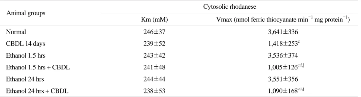
sulfate
Cytosolic rhodanese Animal groups
Km (mM) Vmax (nmol ferric thiocyanate min-1 mg protein-1) Normal 246±37 3,641±336
CBDL 14 days 239±52 1,418±253c
Ethanol 1.5 hrs 243±42 3,536±374
Ethanol 1.5 hrs + CBDL 241±48 1,005±126c,f,j
Ethanol 24 hrs 244±44 3,551±356
Ethanol 24 hrs + CBDL 238±53 1,090±168c,i,j
Michaelis-Menten constants for rhodanese were determined using sodium thiosulfate and potassium cyanide at 25℃ for cytosolic fraction in male rat livers of acute intoxication with ethanol done after 14 days of the common bile duct ligation. The data are expressed as mean ± SD with 5 rats in each group. Animal groups are described in Table 1.
c, P<0.001 vs. Normal; f, P<0.001 vs. Ethanol 1.5 hrs; i, P<0.001 vs. Ethanol 24 hrs; j, P<0.05 vs. CBDL 14 days
Table 3. Kinetic parameters of liver mitichondrial rhodanese in cholestasis with acute ethanol intoxicated rat determined with sodium thiosulfate
Mitochondrial rhodanese Animal groups
Km (mM) Vmax (nmol ferric thiocyanate min-1 ml protein-1) Normal 263±49 4,672± 974
CBDL 14 days 255±57 2,425± 722b
Ethanol 1.5 hrs 261±51 4,337± 916
Ethanol 1.5 hrs + CBDL 257±55 1,844± 645c,e
Ethanol 24 hrs 259±47 4,412±1,162
Ethanol 24 hrs + CBDL 254±53 1,665± 576c,h
Michaelis-Menten constants for rhodanese were determined using sodium thiosulfate and potassium cyanide at 25℃ for mitochondrial fraction in male rat livers of acute intoxication with ethanol done after 14 days of the common bile duct ligation. The data are expressed as mean ± SD with 5 rats in each group. Animal groups are described in Table 1.
b, P<0.01 VS. Normal; c, P<0.001 vs. Normal; e, P<0.01 vs. Ethanol 1.5 hrs; h, P<0.01 vs. Ethanol 24 hrs
생산의 증가, 파이브르산 생성의 감소, 지방산 합성의 촉진, 시트르산 회로 활성의 저하 및 지방산 산화의 감소 등11,14)을 들 수 있다.
간 조직에 담즙울체가 야기되는 경우들은 원발성 담즙성 간경변증, 담관염, 담즙울체형 간염, 선천성 담도 폐쇄, 종양 이나 담석에 의한 담도 폐쇄 등15)이며 이와 같은 간담도 질 환으로 간에 담즙울체가 야기되면 간 조직은 괴사, 담도증식, 섬유화 및 경화성 변화 등9)이 나타날 뿐만 아니라 심한 간 기능의 장애도 나타난다15,38).
흰쥐의 총담관을 결찰하면 간에 담즙울체가 야기되며 시 간이 경과함에 따라 담즙울체간은 괴사, 담도증식, 섬유화 및 경화성 변화가 연속적으로 나타나며21,25) 동시에 간 기능 도 장애가 초래되는 것19,37,40)으로 알려져 있다. 간의 배설 기 능에 장애가 오면 간에는 담즙울체가 야기되며38) 이때 담즙 울체간과 혈청에서는 각종 효소들의 활성도가 증감되는 것 으로 알려져 있다. 특히 생체이물 생체 변환 효소의 일종인 rhodanese는 담즙울체간에서 그 활성도가 변동된다16).
체내에 흡수된 주정은 간에서 대부분 대사 되며 이 대사 과정은 에탄올이 아세트 알데하이드로 산화되고 다시 아세 트산으로 산화되어 이용3,33)되는 것이다. 이러한 대사 과정에 서 생성된 아세트 알데하이드는 간세포의 괴사를 초래하는 물질39)로 알려져 있고 또한 주정 중독 시 심한 형태학적 변 화가 초래4,5)되는 만큼 담즙울체와 주정 중독이 병행된다면 간 손상의 정도는 더욱 심해질 것으로 생각된다. 특히 흰쥐 에서는 급성 주정 중독과 담즙울체가 병행되었을 때는 생체 이물 생체 변환 효소들의 활성도 변동이 심하다고 한다. 즉 흰쥐에서 급성 주정 중독 시 담즙울체가 야기되면 담즙울체 만 있을 때보다 간에서 그 활성도가 증가되는 생체이물 생체 변환 효소들은 xanthine oxidase7), catalase36), 세포질 glutath-
ione S-transferase27), 세포질 glutathione peroxidase27), 미토콘드 리아 monoamine oxidase8) 등을 들 수 있으며 간에서 그 활 성도가 감소되는 생체이물 생체 변환 효소들은 마이크로솜 glutathione S-transferase27), arylesterase2) 및 carboxylesterase1) 를 들 수 있다. 또한 급성 주정 중독 시 담즙울체가 야기되 면 담즙울체만 있을 때보다 혈청에서 그 활성도가 증가되는 생체이물 생체 변환 효소들은 aryl sulfotransferase31), alcohol dehydrogenase36) 및 xanthine oxidase7)이며 그 활성도가 감소 되는 생체이물 생체 변환 효소는 arylesterase2) 및 carboxyle- sterase1)를 들 수 있다. 따라서 이 실험에서 측정한 rhodanese 는 간에서 그 합성이 활발할 뿐만 아니라 생체이물 생체 변 환 효소로서 담즙울체 시 간에서 그 활성도가 감소되는16) 만 큼 주정 중독 시 담즙울체가 야기되면 그 활성도의 변동은 더욱 심해질 것으로 생각된다.
이 실험 결과에서 총담관 결찰 후 14일에 급성 주정 중독 을 시킨 군과 총담관만 결찰한 군간에 간의 rhodanese 활성 도를 비교했을 때 간의 세포질과 마이크로솜 분획에서는 총 담관 결찰 후 14일에 급성 주정 중독을 시킨 군이 총담관만 결찰한 군보다 이 효소의 활성도는 더 현저한 감소를 나타내 었다. 이 결과를 볼 때 쥐 간의 세포질 및 마이크로솜 rho- danese는 담즙울체 시 급성 주정 중독이 야기되면 담즙울체 만 있을 때보다 그 활성도가 감소되는 효소라 생각된다. 한 편 이 실험 결과에서 총담관 결찰 후 14일에 급성 주정 중독 을 시킨 군에서 간의 세포질, 미토콘드리아 및 마이크로솜 rhodanese의 Km 값를 총담관만 결찰한 군의 Km 값과 비교 했을 때는 상호간에 별 차이가 없었다. 그러나 총담관 결찰 후 14일에 급성 주정 중독을 시킨 군에서 간의 세포질 및 마이크로솜 rhodanese의 Vmax 값은 총담관만 결찰한 군의 Vmax 값보다 유의하게 감소된 값을 나타내었다. 이와 같이 Table 4. Kinetic parameters of liver microsomal rhodanese in cholestasis with acute ethanol intoxicated rat determined with sodium
thiosulfate
Microsomal rhodanese Animal groups
Km (mM) Vmax (nmol ferric thiocyanate min-1 mg protein-1) Normal 604±121 276±60
CBDL 14 days 592±133 148±56b
Ethanol 1.5 hrs 599±126 368±83
Ethanol 1.5 hrs + CBDL 594±131 128±52b,f
Ethanol 24 hrs 597±128 381±78
Ethanol 24 hrs + CBDL 595±134 77±27c,i,j
Michaelis-Menten constants for rhodanese were determined using sodium thiosulfate and potassium cyanide at 25℃ for microsomal fraction in male rat livers of acute intoxication with ethanol done after 14 days of the common bile duct ligation. The data are expressed as mean ± SD with 5 rats in each group. Animal groups are described in Table 1.
b, P<0.01 VS. Normal; c, P<0.001 vs. Normal; f, P<0.001 vs. Ethanol 1.5 hrs; i, P<0.001 vs. Ethanol 24 hrs; j, P<0.05 vs. CBDL 14 days
담즙울체 시 급성 주정 중독을 야기시키면 이 효소의 Km 값이 변동이 없으면서도 담즙울체만 시켰을 때보다 그 활성 도가 감소되고 또한 Vmax 값이 감소된 것은 이 효소의 활 성도 감소가 촉매 효율의 감소라 보기는 어렵다. 따라서 담 즙울체 시 급성 주정 중독이 야기되면 이 생체이물 생체 변환 효소는 담즙울체만 있을 때보다 그 합성이 감소되는 것 으로 생각된다.
이 실험에서 혈청의 rhodanese 활성도는 총담관 결찰 후 14일에 급성 주정 중독을 시킨 군이 총담관만 결찰한 군보 다 더 현저한 증가를 나타내었다. 이 결과는 담즙울체 시 급 성 주정 중독을 야기시키면 간 손상이 심해져서 이 효소의 혈중 누출이 심해진다는 것을 나타낸 결과라 할 수 있다. 담 즙울체간과 주정 중독 간은 괴사가 진행되는 간이며14,21,25) 간의 괴사가 진행될 때는 간에서 각종 효소들의 누출이 심해
진다23,28,34)고 한다. 또한 담즙울체 시 간에서 rhodanese의 누
출이 심해져서 혈중에 그 활성도가 증가된다는 보고16)가 있 고 보면 이 실험에서 흰쥐에게 담즙울체와 급성 주정 중독을 병행시켰을 때 혈청에서 이 효소의 활성도가 담즙울체만 시 켰을 때보다 증가된 것은 이 효소의 혈중 누출이 담즙울체만 시켰을 때보다 더욱 심해진 것이라 생각된다.
이상 이 실험 성적과 문헌상의 지견으로 보아 간의 rhoda- nese는 담즙울체 시 급성 주정 중독이 야기되면 그 합성이 담즙울체만 있을 때보다 감소되는 효소로 생각되며, 또한 담 즙울체 시 급성 주정 중독이 야기되면 담즙울체만 있을 때 보다 간 손상이 심해져서 간에서 혈중으로 rhodanese의 누출 이 더욱 증가되는 것으로 생각된다. 따라서 이들 성적은 담 즙울체로 간 손상이 있을때 음주를 하면 간 손상이 더욱 심해질 것이라는 것을 시사해 준다.
참 고 문 헌
1) Ahn KW and Kim YH (1999): Effects of common bile duct ligation on serum and hepatic carboxylesterase activity in ethanol-intoxicated rats. J Biochem Mol Biol, 32(4): 331-338.
2) Ahn KW and Kim YH (1999): Effects of common bile duct ligation on serum and hepatic arylesterase activity in ethanol intoxicated rats. Keimyung Med J, 18(3): 371-386.
3) Bosron WF and Li TK (1980): Alcohol dehydrogenase, pp.
213-244. In Jakoby WB (ed.), "Enzymatic Basis of Dectoxi- cation", Vol. 1. Academic Press, New York.
4) Chang ES (1985): Light and electron microscopic studies of liver biopsies on alcoholic patients. J Clin Electron Micro- socopy, 18(4): 331-347.
5) Chang ES (1987): Ultrastructural morphogenesis of mitoch- ondria in alcoholic liver. Acta Pathol Jpn, 37(2): 213-224.
6) Choi KS, Mun KC, Kim YH and Kwak CS (2002): Effect of extrahepatic cholestasis on hepatic rhodanese activity in chronic ethanol intoxicated rats. Keimyung Med J, 21(4):
218-228.
7) Chung SK, Kim YH and Kwak CS (1994): Effect of common bile duct ligation on liver xanthine oxidase activity in ethanol intoxicated rats. The Keimyung Univ Med J, 13(1): 64-72.
8) Chung SK and Kwak CS (1992): Effect of common bile duct ligation on the liver monoamine oxidase activity in ethanol intoxicated rats. Korean Biochem J, 25(3): 210-218.
9) Desmet VJ (1994): Cholestasis; extrahepatic obstruction and secondary billiary cirrhosis, pp.425-474. In MacSween RNM, Anthony PP, Scheuer PJ, Burt AD, Portman BC (eds.),
"Pathology of the Liver", 3rd Ed., Churchill Livingstion, New York.
10) Drawbaugh RB and Marrs TC (1987): Interspecies differences in rhodanese (thiosulfate sulfurtransferase, EC 2. 8. 1. 1) acti- vity in liver, kidney and plasma. Comp Biochem Physiol, 86(2): 307-310.
11) Ellenhorn MJ and Barceloux DG (1988): Medical Toxico- logy Diagnosis and Treatment of Human Poisoning, pp.782 -796. Elsevier Science Publishing, New York.
12) Gornall AG, Bardawill CJ and David MM (1949): Determi- nation of serum protein by means of biuret reaction. J Biol Chem, 177(3): 751-766.
13) Greenberg DM and Rothstein M (1957): Method for isolation and degradation of labelled compounds, pp.708-731. In Colo- wick SP, Kaplan NO (eds.), "Method in Enzymology", Aca- demic Press, New York.
14) Hall PM (1994): Alcoholic liver disease, pp.317-348. In MacSween RNM, Anthony PP, Scheuer PJ Burt AD, Portman BC (eds.), "Pathology of the Liver", 3rd Ed., Churchill Livingstion, New York.
15) Halsted JA (1976): The Laboratory in Clinical Medicine. In- terpretation and Application, pp.426-429. Sannders, London.
16) Ihm JS and Kim YH (1997): Thiosulfate sulfurtransferase and UDP-glucuronosyltransferase activities in cholestatic rat liver induced by common bile duct ligation. Exp Mol Med, 29(4):
197-201.
17) Ihm JS, Kim YH and Kwak CS (1995): Aryl sulfotransferase activity in cholestatic rat liver induced by common bile duct ligation. Korean J Biochem, 27(3): 141-147.
18) Jakoby WB, Bend JR and Caldwell J (1982): Metabolic Basis of Detoxication, Metabolism of Functional Groups, pp.5-317.
Academic Press, New York.
19) Kaplan MM and Righetti ABB (1970): Induction of rat liver alkaline phosphatase: The mechanism of the serum elevation in bile duct obstruction. J Clin Invest, 49(3): 508-516.
20) Kim BK (1984): Enzyme Nomencleature, IUB, pp.264-265.
Academic Press, New York.
21) Kim HS, Park JY, Kim EY, Kwak KS, Choi YH and Chung JM (1989): Morphologic change of hepatocytes induced by common bile duct ligation. Korean J Intern Med, 36(4): 459 -470.
22) Kim SS, Park SD and Kwak CS (1993): Effect of common bile ligation on liver cathepsins B, D, H and acid phosphatase activities in ethanol intoxicated rats. Kor J Gastroenterol, 25(4): 696-708.
23) Kim YH, Kwak CS and Chung SK (1990): Effect of common bile duct ligation on the serum and liver aspartate amino- transferase activities in ethanol intoxicated rats. The Keimyung Univ Med J, 9(1): 87-95.
24) Koj A, Frendo J and Woitczak L (1975): Subcellular distribu- tion and intramitochondrial localization of three sulfurtrans- ferase in rat liver. FEBS Lett, 57(1): 42-46.
25) Kountouras J, Billing BH and Scheuer PJ (1984): Prolonged bile duct obstruction: a new experimental model for cirrhosis in the rats. Br J Exp Pathol, 65(3): 305-311.
26) Kwak CS (1985): Xanthine oxidase activity in the cholestatic rat liver. The Keimyung Univ Med J, 4(2): 125-130.
27) Kwak CS, Kim YH and Jo JS (1990): Effect of common bile duct ligation on the liver glutathione S-transferase, glutathione peroxidase and glutathion reductase activities in ethanol in- toxicated rats. Korean Biochem J, 23(2): 251-262.
28) Kwak CS, Kim YH and Mun KC (1988): Activities of alcohol metabolizing enzymes in the cholestatic rat liver. The Kei- myung Univ Med J, 7(1): 64-75.
29) Kwak CS and Kwak JS (1986): Cell fractionation method of the rat liver. I. Isolation of mitochondria and microsomes. The Keimyung Univ Med J, 5(1): 45-53.
30) Kwak CS and Lee SH (1992): Carboxylesterase, arylesterase and cholinesterase activities in cholestatic rat liver induced by common bile duct ligation. Korean Biochem J, 25(3): 251-261.
31) Kwon SO and Kim YH (1997): Effect of common bile duct ligation on liver aryl sulfotransferase and UDP-glucuronosyl- transferase activities in acute ethanol intoxicated rats. The
Keimyung Med J, 16(2): 182-194.
32) Kwon YC, Mun KC and Kwak CS (1990): Glutathione S -transferase, glutathione reductase and glutathion perexidase activities in cholestatic rat liver. The Keimyung Univ Med J, 9(2): 159-170.
33) Lieber CS (1985): Alcohol metabolism, pp.3-40. In Hall P (ed.),
"Alcoholic liver Disease, Pathobiology, Epidemiology and Clinical Aspects", Edward Arnold, London.
34) Lind S (1958): A comparison between the patterns (GOT, GPT, LD) in serum and tissue extracts in cardiac hepatic disease.
Scand J Clin Lab Invest, 10(2): 303-307.
35) Liu SJ, Ramsey RK and Fallon HJ (1975): Effect of ethanol on hepatic microsomal drug metabolizing enzymes in the rat.
Biochem Pharmacol, 24(3): 369-378.
36) Mun KC, Kwak CS and Jo YH (1999): Activities of hepatic enzymes for ethanol metabolism in cholestatic rats with acute ethanol intoxication. Kor J Gastroenterol, 34(1): 50-55.
37) Righetti ABB and Kaplan MM (1971): Effects of actinomycin D on rat liver alkaline phosphatase. Proc Soc Exp Biol Med, 136(2): 491-495.
38) Sherlock S and Dooley J (2002): Disease of the Liver and Biliary System, 11th Ed., pp.1-17. Blackwell Science, Oxford.
39) Sherlock S and Dooley J (2002): Disease of the Liver and Biliary System, 11th Ed., pp.381-398. Blackwell Science, Oxford.
40) Toda G, Ikeda Y, Kako M, Oka H and Oda T (1980): Mech- anism of elevation of serum alkaline phophatase activity in biliary obstruction: An experimental study. Clin Chim Acta, 107(1-2): 85-96.
41) Westley J (1973): Rhodanese. Adv Enzymol Relat Areas Mol Biol, 39: 327-368.
42) Westley J (1980): Rhodanese and the sulfane pool, pp.245 -262. In Jakoby WB (ed.), "Enzymatic Basis of Detoxication", Vol. II. Acade- mic Press, New York.
43) Westley J (1981): Thiosulfate: cyanide sulfurtransferase (Rho- danese), pp.285-291. In Jakoby WB (ed.), "Methods in Enzy- mology", Vol. 77. Academic Press, New York.
44) Wooddell WJ (1980): Liver disease in alcohol addicted pa- tients, pp.125-134. In Davidson SV (ed.), "Alcoholism and Health", Aspen System, Century Boulevard.