• 검색 결과가 없습니다.

KASL clinical practice guidelines : management of hepatitis C

N/A
N/A
Protected

Academic year: 2021

Share "KASL clinical practice guidelines : management of hepatitis C"

Copied!
64
0
0

로드 중.... (전체 텍스트 보기)

전체 글

(1)

http://dx.doi.org/10.3350/cmh.2016.22.1.76 Clinical and Molecular Hepatology 2016;22:76-139

Review

Corresponding author : KASL (Committee Chair: Sook-Hyang Jeong) Room A1210 MapoTrapalace, 53 Mapo-daero, Mapo-gu, Seoul 04158, Korea Tel: +82-2-703-0051, Fax: +82-2-703-0071

E-mail: kasl@kams.or.kr

* Clinical Practice Guidelines Committee of KASL for the Management of Hepatitis C

Sook-Hyang Jeong (Committee Chair, Seoul National University College of Medicine), Jung Il Lee (Yonsei University College of Medicine), Young Seok Kim (SoonChunHyang University Bucheon Hospital), Kyung-Ah Kim (Inje Unversity Ilsan Paik Hospital), Sang Hoon Park (Hallym University Kangnam Sacred Heart Hospital), Byung Seok Lee (Chungnam National University College of Medicine), Jun Yong Park (Yonsei University College of Medicine), Ki Tae Yoon (Pusan National University School of Medicine), Young Kul Jung (Korea University College of Medicine), Chang Wook Kim (Catholic University College of Medicine), Eun Young Cho (Wonkwang University School of Medicine), Geum-Youn Gwak (Sungkyunkwan University School of Medicine), Woo Jin Chung (Keimyung University School of Medicine), Jeong Han Kim (Konkuk University School of Medicine)

Abbreviations:

3TC, lamivudine; AAR, AST/ALT ratio; ABC, abacavir; AGREE, Appraisal of guidelines for research and evaluation; ALT, alanine transaminase; anti-HCV, antibody to hepatitis C; APRI, aminotransferase-platelet ration index; ARFI, acoustic radiation force; AST, aspartate aminotransferase; ATV, atazanavir; /r, /ritonavir; /c, /cobicistat;

Cat, catalyzed by human cathepsin; CEA, carboxylesterase; cEVR, complete EVR;

CLIA, chemiluminescent immunoassay; CrCl, creatinine clearance; CTP, Child-Pugh Turcotte; DAA, direct-acting antivirals; DDI, drug-drug interactions; DRV, darunavir;

DTG, dolutegravir; DVR, delayed virologic response; ECLIA, electrochemiluminescence;

EFV, efavirenz; EIA, enzyme immunoassay; ESRD, end-stage renal disease; ETR, end-of treatment response; ETR, etravirine; EVG, elvitegravir; EVR, early virologic response; FIB- 4, fibrosis-4 score; FPV, fosamprenavir; FTC, emtricitabine; G-CSF, granulocyte colony stimulating factor; GFR, glomerular filtration rate; GRADE, grading of recommendations, assessment, development and evaluation; HAV, hepatitis A virus; HBV, hepatitis B virus;

HCC, hepatocellular carcinoma; HCV, hepatitis C virus; HINT, histidine triad nucleotide- binding protein; HIRA, Health Insurance Review and Assessment Service; HIV, human immunodeficiency virus; IL 28B, interleukin 28B; IVDU, intravenous drug user; KASL, Korean Association for the Study of the Liver; KCDC, Korea Centers for Disease Control and Prevention; KHF, Korea Hemophilia Foundation; LC, liver cirrhosis; LPV, lopinavir;

MELD, Model for End-Stage Liver Disease; MR, magnetic resonance; MVC, maraviroc;

ddi, didanosine; NNRTIs, non-nucleoside reverse transcriptase inhibitors; NS, non- structural; NVP, nevirapine; OATP, organic anion-transporting polypeptide; Opr+D, ombitasvir/paritaprevir/ritonavir and dasabuvir; PCR, polymerase chain reaction;

PegIFN, peginterferon; pEVR, partial EVR; PI, protease inhibitor; PR, pegylated interferon- α+ribavirin therapy; PWID, people who inject drugs; R*, ribavirin started from 600 mg/

d; R, weight-based ribavirin; RAL, raltegravir; RAV, resistance-associated variants; RFMP, restriction fragment mass polymorphism; RIBA, recombinant immunoblot assays;

RPV, rilpivirine; RVR, rapid virologic response; SNP, single nucleotide polymorphism;

SQV, saquinavir; SVR, sustained virologic response; TDF, tenofovirdisoproxil fumarate;

TMA, transcription-mediated amplification; TPV, tipranavir; TSH, Thyroid-stimulating

hormone; UTR, untranslated region; ZDV, zidovudine Received : Feb. 15, 2016 / Accepted : Feb. 22, 2016

PREAMBLE Aims

The Korean Association for the Study of the Liver (KASL) Prac- tice Guidelines for Management of Hepatitis C were first estab- lished in 2004, and revised in 2013, when direct-acting antivirals (DAA) were not approved in Korea. Since then, numerous studies of the efficacy, adverse effects and drug-drug interactions (DDI) of interferon-free DAA combination therapy have been published.

With all oral DAA therapy showing a sustained virologic response

(SVR) rate of 80-90% with minimal adverse events, hepatitis C vi- rus (HCV) eradication is a realistic goal of treatment. Therefore, a screening strategy for HCV-infected populations according to each country’s epidemiology and disease burden is required.

DAA combination therapeutics were approved and adapted to practice in Korea in 2015, and KASL revised the guidelines based on a systematic approach that reflects evidence-based medicine and expert opinions.

The clinical practice guidelines for the management of hepatitis C have been revised to be useful for treatment, research and edu- cation. These recommendations are not absolute standards of

KASL clinical practice guidelines: management of hepatitis C

The Korean Association for the Study of the Liver (KASL)*

Keywords: Hepatitis C; Management; Guidelines; KASL

(2)

care, and adoption of the guidelines in clinical practice may differ among individual patients.

Target population

The target groups of these guidelines are newly or previously diagnosed patients with hepatitis C virus (HCV) infection, includ- ing not only chronic hepatitis C and cirrhosis but also acute hepa- titis C patients, hepatitis C patients with chronic kidney diseases, and those patients coinfected with human immunodeficiency virus (HIV) or hepatitis B virus (HBV).

Intended users

The guidelines are intended to provide useful information and guidance to physicians and healthcare providers involved in the diagnosis and treatment of hepatitis C, and resident physicians, practitioners, and trainers.

Development, funding, and revision process

The Clinical Practice Guidelines Committee for the Management of Hepatitis C (Committee) comprising 14 hepatologists, was or- ganized according to the proposal and approval of the KASL Board of Executives. Funding for the revision was provided by KASL. Each committee member collected and analyzed the source data in his or her own field, and the members then wrote the manuscript together.

Literature review for evidence collection

The committee systematically collected and reviewed the inter-

national and domestic literature published in PubMed, MEDLINE, KoreaMed, and other databases. The key words used were ‘hepa- titis C virus’, ‘hepatitis C’, ‘liver cirrhosis’, ‘liver cancer’ and other related specific key words.

Levels of evidence and grades of recommendations

The quality of evidence was classified according to the GRADE (Grading of Recommendations, Assessment, Development, and Evaluation) system (Table 1). 1 Based on the types of study, ran- domized control studies were approached from a high level of evi- dence, while observational studies were approached from a low level of evidence. Then, the level of evidence was adjusted by ac- counting for the factors influencing the quality of the studies.

Through follow-up studies, the level of evidence was defined as follows: A, the highest level of evidence with the smallest possi- bility of changes in the conclusion; B, a moderate level of poten- tial changes; and C, the lowest level of evidence with the greatest possibility of changes.

The strength of a recommendation was also classified according to the GRADE system. Each study was classified as strong recom- mendation (1) or weak recommendation (2) based on the quality of evidence, the balance between the desirable and undesirable effect of an intervention, and socioeconomic aspects including cost or availability. A strong recommendation indicated that the interventions could be applied in most patients with strong cer- tainty and that there was a greater possibility of desirable effects, high-quality evidence, and presumed patient-important outcomes, cost-effectiveness, preference, and compliance. A weak recom- mendation indicated a suggestion made with less certainty but that could be considered favorable for many patients, based on

Table 1. Grading of Recommendations, Assessment, Development, and Evaluation (GRADE) Quality of evidence Criteria

High (A) Further research is very unlikely to change our confidence in the estimate of effect.

Moderate (B) F urther research is likely to have an important impact on our confidence in the estimate of effect and may change the estimate.

Low (C) F urther research is very likely to have an important impact on our confidence in the estimate of effect and is likely to change the estimate. Any change of estimate is uncertain.

Strength of Recommendation Criteria

Strong (1) F actors influencing the strength of the recommendation included the quality of the evidence, presumed patient-important outcomes, and cost.

Weak (2) V ariability in preference and values, or more uncertainty. Recommendation is made with less certainty, higher cost or resource consumption.

Note: Of the quality levels of evidence, we excluded “very low quality (D)” in our guideline for convenience, which was originally included in the GRADE system.

(3)

the level of evidence, the cost or preferences of the patients or medical practitioners.

List of key questions

The revision committee considered the following clinical ques- tions as the key components to be covered in these guidelines.

1. What is the epidemiology, natural history and prevention strategy of hepatitis C in Korea?

2. How should diagnosis and evaluation of severity of chronic hepatitis C be made?

3. What is the goal of treatment and who are the targets for an- tiviral treatment of hepatitis C?

4. How to define the treatment response and what are the pre- dictors of the response?

5. How to treat patients with genotype 1 chronic hepatitis C and compensated cirrhosis?

6. How to treat patients with genotype 2 chronic hepatitis C and compensated cirrhosis?

7. How to treat patients with genotype 3 chronic hepatitis C and compensated cirrhosis?

8. How to treat patients with genotype 4 chronic hepatitis C and compensated cirrhosis?

9. How to treat patients with genotypes 5 and 6 chronic hepati- tis C and compensated cirrhosis?

10. How to treat patients with decompensated cirrhosis?

11. How to treat patients who underwent liver or extrahepatic or- gan transplantation?

12. How to treat patients with acute hepatitis C?

13. How to monitor the patients and the adverse effects of antivi- ral drugs during and after antiviral treatment?

14. How to treat patients with special conditions (people who in- ject drugs, chronic kidney diseases, coinfection with HIV or HBV, hemophilia or thalassemia, immunosuppressive therapy or cytotoxic chemotherapy, and pediatric patients)?

Review of the manuscript and approval process

Each manuscript written by committee members was reviewed, agreed, and approved through meetings of the committee. The quality of the manuscript was evaluated based on the standards suggested by AGREE II (Appraisal of Guidelines for Research and Evaluation II) along with the academic integrity of the contents.

The guidelines were reviewed after counsel from an infection spe-

cialist, at a meeting of an external review board composed of 14 KASL members, and were further modified following opinions aired at a public hearing, and a symposium open to all KASL members. The final manuscript was approved by the KASL Board of Executives.

Release of the guidelines and plan for updates

The Korean version of the KASL Clinical Practice Guidelines for the Management of Hepatitis C was released in November 2015 at a KASL meeting and published in January 2016 on the KASL website (http://www.kasl.org). Future plans for revision will be conducted under the judgment that the revision is necessary for promotion of health in South Korea with accumulation of research on the management of hepatitis C. In addition, use of DAA is to be allowed in South Korea in the near future, so that updating or partial revision of the guidelines, as appropriate, is warranted.

EPIDEMIOLOGY

HCV is one of the main causes of acute and chronic hepatitis, cirrhosis, and hepatocellular carcinoma. 2 Hepatitis C is on the list of National Notifiable Infectious Disease in South Korea and has

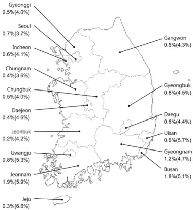
Figure 1. Map of South Korea showing age and sex-adjusted anti-HCV

seroprevalence in each area. 11

(4)

been under surveillance since 2000. An effective HCV vaccine is yet to be discovered, so that understanding of national epidemiol- ogy and a preventive strategy to block routes of HCV infection is important for public health.

Prevalence of HCV infection

The worldwide prevalence of HCV infection was 2.8% in 2005 and 1.6% in 2014, equating to about 115 million persons positive for antibody to hepatitis C (anti-HCV) and 1.1% equating to about 80 million persons positive for HCV RNA. 3 The prevalence varies among geographic regions; regions with a high prevalence of over 3.5% include Central Asia (including Mongolia and China), South- East Asia (including Pakistan and Thailand), and North Africa (in- cluding Egypt). Low prevalence (below 1.5%) regions are Asia (in- cluding South Korea and Japan), North America (including the United States), and South America. 4

1. Prevalence in adult health check examinees

The prevalace of HCV infection in adult health check examinees was reported to be 1.7% using a first-generation enzyme immu- noassay (EIA) in the early 1990s soon after HCV was discovered. 5 The estimated age-standardized prevalence of anti-HCV in adult health check examinees in > 40 years of age was reported to be 1.29% (95% confidence interval, 1.12-1.48) and was over 193,000 persons in a collective study of health checkup examin- ees from Seoul, Ulsan, Jeollanam-do, and Daegu between 1995 and 2000. 6-10

In 2009, the anti-HCV prevalence in health examinations of 291,314 adults ≥20 years of age from 29 health examination cen- ters was 0.78% using third-generation EIA after adjusting for age, sex, and area. 11 The anti-HCV prevalence was higher in females (0.83%) than in males (0.75%) and increased with age (20-29 years: 0.34%, 30-39 years: 0.41%, 40-49 years: 0.60%, 50-59 years: 0.80%, 60-69 years: 1.53%, ≥70 years: 2.31%). In addi- tion, the anti-HCV prevalence varied geographically; in compari- son with the prevalence of 0.50-1.20% in most regions, including Seoul and Gyeonggi-do, the prevalence in Pusan and Jeollanam- do were 1.53% and 2.07%, respectively, while the Jeju Special Self-Governing Province had the lowest rate of 0.23% (Fig. 1).

And also, there was a significant geographic difference in the po- tential risk factors for HCV infection in Korea. 12 An update of the national HCV infection prevalence in South Korea is expected to be released at the end of 2015, and the Korea National Health and Nutrition Examination Survey has included anti-HCV testing

since 2012.

2. Prevalence of anti-HCV in blood donors, pregnant women, and children

The anti-HCV prevalence in 2,040,151 blood donors in 1997 in South Korea was 0.34%, as determined by third-generation EIA. 13 From 2005 to 2009, the anti-HCV prevalence in 11,064,532 blood donors was 0.16%, and the HCV RNA-positive rate was 8.4 (0.0084%) of 100,000 donors, among whom 81% were young people aged 10-30 years. 14 In South Korea, the risk of blood transfusion-related HCV infection decreased from 1 in 81,431 in 2000/2001 to 1 in 2,984,415 after implementation of nucleic acid testing for HCV screening of donated blood in February 2005. 15

The anti-HCV prevalence in pregnant women was reported to be 0.49-1.7%, 16-18 and a domestic report investigating over 5,000 pregnant women reported rates of 0.42-0.44%. 19,20 Among anti- HCV-positive pregnant women, 57-60% of the patients positive for HCV RNA. 19,20

Domestic studies on anti-HCV prevalence in children and ado- lescents are insufficient. A 0.82% anti-HCV-positive rate tested by third-generation EIA in 2,080 children between 6 and 11 years of age living in Seoul was reported. 21 However there have been no other reports or studies on children and adolescents, hindering the accurate assessment of the HCV infection prevalence in the pediatric population of South Korea.

3. Prevalence of anti-HCV in high-risk groups

The high-risk groups for HCV infection include people who in- ject drugs (PWID), patients under hemodialysis, and those with HIV infection, hemophilia, and leprosy. However, the HCV preva- lences in this group have been reported mostly before 2000; few studies were performed thereafter.

The domestic anti-HCV prevalence in the intravenous drug user (IVDU) group was 48.4-79.2%. 22-25 Among anti-HCV-positive per- sons, 98.1% were HCV RNA-positive. 24 The anti-HCV prevalence in those who share cocaine suction pipes was similar to that in the IVDU group. 26

The anti-HCV prevalence was 5.9-14.7% in previous studies of

>200 patients with chronic kidney diseases, 27,28 2.2% in the 2014 report of the Korean Society of Nephrology 29 and was significantly correlated with hemodialysis duration.

The HCV coinfection rate was high in those infected with HIV;

~25% of westerners and 5.0-6.3% of HIV-infected individuals in South Korea were coinfected with HCV. 30-32

The anti-HCV prevalence in 104 hemophilia patients tested by

(5)

third-generation EIA was 42.3% in 2002, and the risk of infection was correlated with age and severity of hemophilia. 33 In their 2012 annual report, the Korea Hemophilia Foundation (KHF) re- ported that 430 of 2,148 (20.0%) hemophilia patients were anti- HCV-positive and 118 of 2,148 (5.5%) were HCV RNA-positive. 34

Leprosy patients can be considered at high risk of HCV infection due to their skin lesions and long-term cohabitation in limited ar- eas. The anti-HCV prevalence of 96 leprosy patients tested by second-generation EIA was 67.7% in 1997; 82% of these individ- uals were immunoblot-positive. 35

HCV incidence rate

Studies on HCV infection incidence rate are rare, since only 20- 30% of those with acute HCV infection develop symptoms. HCV incidence rates are decreasing in Western countries. 36 In the US the incidence rate decreased from 7.4 of 100,000 people from 1982-1989 to 0.7 of 100,000 people from 1994-2006, 37 and in It- aly, it decreased from 2.02 of 100,000 people in 1996 to 0.55 of 100,000 people in 2006. 38

The HCV infection incidence rate in South Korean blood donors was reported to be 13.8 per 100,000 according to a survey con- ducted on those who donated blood at least twice from 1994- 1996. 39 The recent HCV infection incidence rates among blood donors who donated at least twice in 2 years between 2000 and 2010 were estimated to be 6.80 in 2001, 3.19 in 2003, 2.69 in 2005, 1.83 in 2007, and 0.80 in 2009 per 100,000 person-years, showing significant decrease in the incidence of HCV in this popu- lation in South Korea. 15 According to surveillance sample data from the Korea Centers for Disease Control and Prevention (KCDC), the number of reported hepatitis C cases was 1,927 in 2002, 6,407 in 2008, and 4,280 in 2012. Future studies should evaluate the nationwide incidence of HCV in Korea.

Distribution of HCV genotypes

Globally, HCV genotypes 1, 2, and 3 are common and geno- types 4, 5, and 6 are localized to limited regions. 40,41 Genotype 1a is the most common in Northern Europe and North America, and 1b is the most common in Far East Asia and Europe. Genotype 2 is less common than genotype 1. Genotype 3 is common in Southeast Asia and genotype 4 is common in the Middle East, Egypt, and Central Africa. Genotype 5 is commonly found in South Africa and genotype 6 is common in Hong Kong, Macau, and Vietnam. Common HCV genotypes in South Korea are geno- type 1b (45- 59%) and 2a (26-51%); types 1a, 2b, 3, 4, and 6 are

rare in South Korea. 42,43

Whether genotype 1 HCV infection provokes faster progression of hepatic disease than the other genotypes is controversial. A re- cent meta-analysis reported that genotype 1b patients showed a 1.78-fold higher risk of developing HCC (95% CI, 1.36-2.32) com- pared to non-1 genotype patients. 44 Nevertheless, the HCV geno- type is the most crucial factor in determining the efficacy of anti- viral therapy. 45

PREVENTION Route of transmission

HCV transmission occurs by parenteral exposure. The main routes of transmission include transfusion of contaminated blood or blood products, organ transplantation, PWID, unsafe injection or medical procedures, stabs by contaminated syringe or needle, sexual contact with HCV-infected person, or perinatal transmis- sion from an infected mother to her newborn.

Transmission via transfusion was a main route of infection until 1991, but the possibility has become extremely low since intro- duction of a screening test for blood donors. 46-48 Recently, in de- veloped regions such as the US and Europe which have low HCV prevalence, the most common route of HCV transmission has been the use of illicit drugs, 49 and anti-HCV prevalence in the PWID group was reported to be up to 50-90%. 50 Meanwhile, un- safe injection with multiple-use medication vials or reused syring- es, or unsanitary medical procedures including surgery, endosco- py, and dental treatment without proper disinfection are the main causes of HCV transmission in developing countries. 51-53 In addition, meta-analyses have reported that risk factors for HCV transmission include piercing, acupuncture, or tattooing without proper disinfec- tion. 54-56 The risk of HCV infection due to percutaneous exposure to a small dose, such as needle sticks, is 1.8% (0-7%) 57-60 in other countries and 0.92% in South Korea. 61 Heterosexual persons with chronic HCV infection in long-term monogamous relationships with a partner had little evidence of sexual transmission of HCV.

However, the risk becomes higher with multiple sex partners, and unsafe sex including anal sex, sex accompanying wounds, sex with carriers of other sexually transmitted diseases such as HIV, or in homosexuals. 62,63 The percentage of perinatal transmission was 1-6.2%. 64,65 It was reported to be 1.7% when the mothers were positive for anti-HCV regardless of HCV RNA-positivity, and 4.3%

(3.9-7.1%) in the case of HCV RNA-positive mothers. 65,66 The risk

(6)

of perinatal transmission increased in female infants, HIV-positive mothers, and mothers with high blood HCV RNA levels. 67 Cesare- an section reportedly does not prevent HCV transmission, 67,68 and the frequency of transmission via nursing was very low. Thus, it is not necessary to limit breast-feeding unless nipples are injured or bleeding. 69 Reports of horizontal transmission between siblings or family members of HCV-infected persons are based on a low level of evidence. 70

A comparative study of 1,173 HCV patients and 534 controls in five university hospitals between 2007 and 2011 in South Korea reported several independent risk factors of infection, including use of illicit drugs, needle-stick injury, transfusion before 1995, tattoo, and age. 71

Counseling for prevention

Since an effective vaccine has not been developed, the main strategy for prevention is to educate people on the risk factors for HCV infection and to maintain strict sanitation standards in all lo- cations performing percutaneous procedures.

HCV-infected persons should be counseled not to donate blood, organs, tissues, or semen, and not to share any instrument pene- trating skin. They should use their own instruments including toothbrushes, oral hygiene devices, razors, or nail clippers so oth- er people are not exposed to his/her blood. Finger-stabbing nee- dles commonly used for Korean home remedies should not be shared. IVDU should be persuaded to stop drug abuse and they should not reuse syringes, needles, injection solution, cotton swab, or alcohol sponges. They must be reminded that other peo- ple can be infected via recklessly disposed needles. Since the risk of infection among monogamous couples is very low, use of barri- er protection by these couples is not necessarily recommended.

Nevertheless, if the partner of the infected individual requests, or if the infected person has multiple sex partners, it is recommend- ed to use condoms. Routine screening for HCV is not recommend- ed for all pregnant women. However, for those with a risk factor, prenatal testing for HCV is required. HCV infection does not mean a restriction of breast-feeding or recommendation for a specific means of delivery, such as Cesarean section. Healthcare facilities should take precautions to prevent HCV transmission. Proper dis- infection, cleaning, and management of materials and instru- ments are essential in medical and invasive procedures including tattooing, piercing, and acupuncture.

[Recommendations]

1. HCV-infected persons should not donate blood, organs, tis- sues, or semen (A1). HCV-infected persons should avoid sharing toothbrushes, oral hygiene devices, razors, nail clip- pers, or any instrument penetrating skin, so as not to ex- pose other people to his/her blood (C1).

2. People who inject drugs should be counseled to stop abuse of illicit drugs (A1). They should be educated about routes of infection and tested regularly for HCV infection (B1).

3. Proper disinfection, cleaning, and management of materials and instruments are essential in medical and invasive proce- dures including tattooing, piercing, and acupuncture (B1).

4. As the risk of infection among monogamous sexual partners is very low, use of barrier protection is not advised in these couples (B1). However, for those with multiple sex partners, it is recommended to use condoms (B1).

5. For pregnant women, if a risk factor for HCV infection is detected or HCV infection is suspected otherwise, prena- tal testing for HCV infection is recommended (B1). HCV in- fection does not mean a restriction of breast-feeding or a recommendation of specific delivery, such as Cesarean section (B2).

NATURAL HISTORY Acute HCV infection

After 1-3 weeks of HCV infection, HCV RNA becomes detectable in blood and the level rapidly increases. 72,73 The serum alanine transaminase (ALT) level increases due to hepatocyte damage after 4-12 weeks of infection. Most infections are asymptomatic (70- 80%), but symptoms including flu-like symptoms, fatigue, vomit- ing, nausea, right upper quadrant pain, muscle pain, or pruritus may develop within 2-12 weeks. About 20% of acute infection accompanies jaundice with serum bilirubin level <3-8 mg/dL, and acute liver failure occurs in <1% of cases.

Acute hepatitis progressed to chronic infection in 54-85% of

patients, and 20-50% of patients recovered spontaneously within

3-4 months. 74-76 The spontaneous recovery rate differs according

to route of infection; the spontaneous recovery rate in post-trans-

fusion cases was 12%, while in the cases not related to transfu-

sion it was 29-52%. 76-78 Factors related to spontaneous recovery

are hepatitis accompanying jaundice, female gender, low viral

(7)

load, and genotype 3. 77-79 A Korean study reported that among 18 acute hepatitis C patients (17 patients showed symptoms), 12 pa- tients spontaneously recovered and 6 patients progressed to chronic hepatitis. 80 Another study including 47 acute hepatitis C patients with a mean age of 45.8 years in seven Korean institu- tions reported that 21 of 47 (44.7%) patients recovered sponta- neously, and 16 patients received antiviral therapy. All 12 patients who were treated and followed-up achieved a sustained virologi- cal response (SVR). Ten patients who did not receive antiviral therapy progressed to chronic hepatitis. 81

A single nucleotide polymorphism (SNP) of the interleukin 28B (IL28B) gene is strongly related to spontaneous recovery from acute hepatitis C infection. 82-84 IL28B is located on chromosome 19 and encodes interferon-lamda-3. One study reported a sponta- neous recovery rate of 53% in cases with genotype CC of IL28B SNP rs12979860 and of 28% in genotype CT or TT (Odds ratio (OR)=0.33, P<10-12). 85 However, future studies are needed as there has been no Korean study of the role of IL28B SNP in acute HCV infection.

Chronic HCV infection

About 50-80% of HCV-infected patients progress to chronic in- fection. Chronic hepatitis can cause persistent liver injury without spontaneous recovery, leading to cirrhosis and HCC. Most (60- 80%) patients with chronic hepatitis show no symptoms, but some can experience abdominal discomfort, fatigue, nausea, muscle pain, arthritis, or weight loss. About 60-70% of chronic HCV-infected patients show chronic hepatitis accompanying steady or intermittent elevation of serum ALT. About 15-56% of chronic hepatitis may progress to cirrhosis through a period of 20- 25 years. 73,86-88 Among patients with liver cirrhosis, the annual in- cidence of HCC is reported as 1-4.9%, 89-91 that of decompensated liver cirrhosis is 3-6%, 73,90-92 and the overall annual mortality rate is 2-4%. 73,90-92 An observational study including 1,137 Korean chronic HCV infected patients for an average follow-up of 55.2 months reported a 14.2% rate of disease progression, defined as development of HCC, spontaneous bacterial peritonitis, variceal bleeding, hepatic encephalopathy, and death due to hepatic dis- eases, and the overall annual mortality rate was 2.0-2.5%. 93 The cumulative probability of disease progression was 6.3%, 12.9%, and 26.1% at 1, 2, and 3 years, respectively. Among 1,137 pa- tients, 490 (43.0%) received antiviral treatment and 60.4%

showed an SVR. Chronic infection without antiviral treatment showed a significantly higher risk of disease progression com-

pared to chronic infection with antiviral treatment (37.4% vs.

10.7%, respectively, P<0.05). The 5-year cumulative probability of disease progression was higher in a non-SVR group compared to a group with SVR (13.0% vs. 3.7%, respectively, P<0.05). Ac- cording to prospective cohort data from 196 patients with HCV- related cirrhosis in Korea, during a mean follow-up period of 39.2 months, 31 (15.8%) patients developed HCC, and 33 (16.8%) pa- tients died or underwent liver transplantation. The estimated HCC incidence was 5.8 per 100 person-years, and the independent factors for HCC were absence of anti-HBV surface antibody (HBs hazard ratio [HR], 5.018; 95% confidence interval [CI], 1.710- 14.726; P=0.003) and serum albumin < 3.8 g/dL (HR, 3.051; 95%

CI, 1.318-7.067; P=0.009). The overall mortality rate was 5.1 per 100 person-years, and the related independent factors were the presence of ascites (HR, 2.448; 95% CI, 1.142-5.210; P=0.022), serum albumin < 3.8 g/dL (HR, 3.067; 95% CI, 1.254-8.139, P =0.014), and nonachievement of SVR (HR, 0.066; 95% CI,

0.001-0.484, P=0.002). 94

Factors affecting disease progression include duration of infec- tion, age at the time of infection (≥40 years of age), male gender, alcohol intake, coinfection with other viruses (HBV or HIV), insulin resistance, obesity, immune-depressed patients, organ transplant- ees, elevation of ALT, and genetic factors such as IL28B. 73 Exces- sive alcohol intake by chronic hepatitis C patients is strongly relat- ed to occurrence of cirrhosis, and increases the risk of HCC. 88,95-98 Fatty liver, insulin resistance, and obesity increase the risks of he- patic fibrosis and HCC development in chronic hepatitis C pa- tients. 99-102 Coinfection with HIV or HBV accelerates progression of liver diseases and increases the risk of HCC compared to HCV sin- gle infection. 103-105 In addition, coinfection with hepatitis A virus (HAV) in chronic hepatitis C increases the risk of hepatic failure. 106 The pathologic stage of hepatic fibrosis at the time of chronic hepatitis C diagnosis is the most important predictor of progres- sion to cirrhosis (refer to the Diagnosis section of this paper). 88,103 Stage 1 hepatic fibrosis has a 10-30% incidence of cirrhosis over a period of 15 years, while most cases of stage 3 hepatic fibrosis are expected to progress to cirrhosis within 15 years. Therefore, patients diagnosed as having hepatic fibrosis over stage 2 must be considered for active antiviral treatment.

[Recommendations]

1. Continuous management and surveillance for development

of cirrhosis and HCC is necessary in chronic hepatitis C pa-

tients (A1).

(8)

2. Chronic hepatitis C patients should abstain from alcohol or drink in moderation, and maintain suitable body weight through physical exercise and dietary control, since disease progression is related to alcohol, obesity, and insulin resis- tance (B1).

3. Patients with chronic HCV infection without antibodies against HAV and HBV should be vaccinated for HAV and HBV (C1).

Screening test for HCV infection

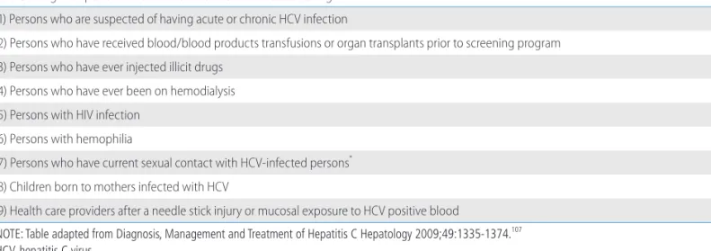
Routine screening for HCV infection is recommended in popula- tions at risk, such as those with a history of blood transfusions or organ transplantation prior to 1992; persons who have injected il- licit drugs; persons with HIV infection, hemophilia, or Hansen’s disease; persons who have been on hemodialysis; children born to

mothers infected with HCV; and health care providers after a needle stick injury or mucosal exposure to HCV-positive blood (Table 2). 107 In 2012, the US Centers for Disease Control and Prevention expand- ed the screening population to the birth cohort born between 1945-1965 and recommended screening for HCV once in a life- time, based on cost effectiveness. 108-110 A Japanese study revealed that hepatitis C screening appears cost-effective in the general population as well as in high-risk groups, 111 but a European study showed that screening of the general population is cost-effective only in HCV prevalent areas. 112 Therefore, since the epidemiologic characteristics and healthcare systems differ among nations, fur- ther research on cost effectiveness is needed to determine the op- timal screening strategy for Korea.

According to anti-HCV prevalence rates in Korea, the estimated number of anti-HCV infected persons over 20 years old was about 320,000 in 2009. However, the number of patients who were Table 3. Interpretation of HCV assays

Anti-HCV HCV RNA Interpretation Further evaluation

Positive Positive Acute hepatitis C Chronic hepatitis C

Positive Negative Resolution of HCV infection Recheck anti-HCV & HCV RNA, 3-6 months later Acute HCV infection during period of low-level viremia

False positive anti-HCV test False negative HCV RNA test

Negative Positive Early acute HCV infection Recheck anti HCV & HCV RNA, 3-6 months later Chronic HCV infection in setting of immunosuppressed state

False positive HCV RNA test

NOTE: Table adapted from Diagnosis, Management and Treatment of Hepatitis C Hepatology 2009;49:1335-1374. 107 HCV, hepatitis C virus.

Table 2. High-risk persons recommended for HCV infection screening 1) Persons who are suspected of having acute or chronic HCV infection

2) Persons who have received blood/blood products transfusions or organ transplants prior to screening program 3) Persons who have ever injected illicit drugs

4) Persons who have ever been on hemodialysis 5) Persons with HIV infection

6) Persons with hemophilia

7) Persons who have current sexual contact with HCV-infected persons * 8) Children born to mothers infected with HCV

9) Health care providers after a needle stick injury or mucosal exposure to HCV positive blood

NOTE: Table adapted from Diagnosis, Management and Treatment of Hepatitis C Hepatology 2009;49:1335-1374. 107 HCV, hepatitis C virus.

* The prevalence of infection is low.

(9)

treated under coverage of Health Insurance Review and Assess- ment Service (HIRA) due to liver disease associated with HCV in- fection was 64,501 in 2009. 113 These data suggest the possibility that only about 20% of the patients were diagnosed and treated.

An online survey in 2013 about awareness of HCV infection showed that about 90% of the general population had not been tested for hepatitis C or did not know whether they had been tested for hepatitis C, which implies that awareness of HCV infec- tion in the general population was low. According to HIRA data, the direct annual medical cost per patient in South Korea was markedly higher for liver cirrhosis (1,522 USD), hepatocellular car- cinoma (6046 USD) or liver transplantation (57,940 USD) com- pared to in chronic hepatitis (842 USD). 113 Such results demon- strate that every effort to intervene disease progression should be exerted to reduce the HCV disease burden. The high accuracy rate and low cost of testing for anti-HCV as a screening tool, and the

>90% cure rate of DAA in early stage liver disease, should also be taken into consideration. One-time screening for HCV infection in the South Korean population aged 40-70 years is likely to be more cost-effective compared to current practice. 114 Therefore, a nationwide screening and therapeutic strategy to diagnose and cure hepatitis C patients prior to progression to advanced liver disease would not only decrease the disease burden but also be a successful model for HCV elimination.

Diagnosis of HCV infection

Biochemical tests, serologic assays, and HCV RNA testing are needed to confirm HCV infection. Physical examination and histo- ry taking should be performed to understand the routes of trans- mission and prevent further reinfection. HCV genotyping is essen- tial for treatment, and radiologic examination, liver biopsy, or noninvasive evaluation of hepatic fibrosis can be performed to determine the necessity of treatment, and to assess liver disease severity. Interpretation of serological and virological test results is summarized in Table 3.

DIAGNOSIS

Serologic assays: Anti-HCV test

Detection of anti-HCV in serum or plasma is used for screening of high-risk groups and for diagnosis of acute or chronic hepatitis C. 115 The third-generation EIA uses recombinant core, NS3, NS4,

and NS5 HCV proteins, and its sensitivity and specificity are 97.2- 99% and 99.8-100%, respectively, when tested in immune-com- petent individuals. 116-118 If signal/cutoff (S/CO) ratios of third-gen- eration EIA exceed 3.8, a positive result will be apparent in 95%

of recombinant immunoblot assays (RIBA). 119-121 However, the cut- off S/CO ratio can differ according to the type of equipment, so that high S/CO ratios do not always mean true positive. 122 Recent- ly, use of enhanced chemiluminescent immunoassay (CLIA) or electrochemiluminescence immunoassay (ECLIA) is increasing since those assays detect antigen-antibody reaction more sensi- tively compared to the third-generation EIAs. Meanwhile, point- of-care tests using saliva or fingerstick blood that produce results within 20 minutes can also be employed. 123,124

Average time between HCV infection and seroconversion is 8-9 weeks, and anti-HCV is detectable in >97% of patients with HCV infection within 6 months. 107,125 Anti-HCV is not a neutralizing an- tibody and persists indefinitely in chronic hepatitis C patients and after recovery. Therefore, the differentiation of current from past infection after recovery is impossible using anti-HCV positivity.

Anti-HCV can be tested repeatedly in high-risk persons because hepatitis C virus can reinfect after recovery. Negative result for anti-HCV in combination with a positive result for HCV RNA may represent an early stage of acute infection or chronic infection in the setting of severe immunosuppression, such as patients on he- modialysis, HIV coinfection, solid organ transplantation recipients, hypo-/ a-gammaglobulinemia, and patients with HCV-associated essential mixed cryoglobulinemia. 126-128 In these patients, HCV RNA testing is necessary for diagnosis of HCV infection. In con- trast, false-positive result for anti-HCV and negative result for HCV RNA can occur in patients with autoimmune diseases. 129

Virological assays

1. HCV RNA assays

HCV RNA assays are classified as quantitative and qualitative.

Since the detection cutoff of qualitative assays is 50 IU/mL, and

these are more sensitive than previous-generation quantitative

assays, HCV RNA qualitative assays have been used for diagnostic

confirmation of HCV infection, and HCV RNA quantification is

used for pretreatment assessment and monitoring of a virological

response during and after antiviral therapy. 115,121,130 However, re-

cently available quantitative HCV RNA assays use real-time poly-

merase chain reaction (PCR) and transcription-mediated amplifi-

cation (TMA), and are highly sensitive with lower detection limits

of 12-15 IU/mL, while they have a broad measuring range with an

(10)

upper limit of 7-8 log IU/mL and 98-99% diagnostic specificity ir- respective of HCV genotype. 115,131-135 Therefore, quantitative HCV RNA tests are now widely used for both diagnosis and evaluation of the treatment response. 127,136

In 1997, the World Health Organization established an interna- tional standard for HCV RNA quantification unit, IU, rather than HCV copy number. 137,138 However, since viral quantification results can differ among laboratories, 139 it is recommended to use the same laboratory test before, during, and after-treatment for moni- toring, if possible. 107,127

Blood HCV RNA is detectable as early as 2 weeks after infec- tion, 76 rapidly increases to reach a plateau, and decreases along with ALT after ALT has peaked. 140 HCV RNA levels remain steady in patients with chronic hepatitis C. 140,141 HCV RNA levels are not significantly correlated with the severity of hepatic inflammation or fibrosis, and change little during chronic infection without anti- viral treatment. 142,143

2. Genotyping/subgenotyping assays

HCV genotyping is useful for epidemiologic studies as well as for predicting treatment response. Therefore, HCV genotype should be assessed before treatment to determine the optimal therapeutic duration and dose of ribavirin. 144 HCV is classified into six major genotypes (1-6) and is subdivided into subtypes identi- fied by lower-case letters, such as 1a or 1b. Differences of 31- 33% at the nucleotide level differentiate the genotypes, com- pared with 20-25% for the subtypes. 145 HCV genotype does not change within a person unless reinfected.

HCV genotypes and subtypes can be determined by direct se- quence analysis, reverse hybridization, or restriction fragment mass polymorphism (RFMP). 146 Most genotyping assays analyze both the 5’-untranslated region (UTR) and HCV core regions, the nucleotide sequences of which are highly conserved. 147-150 Subtyp- ing is not necessary in antiviral therapy using interferon alpha and

ribavirin, but in treatment that includes DAAs, subtypes may need to be confirmed since DAAs act differently according to subgeno- type. 148-153 Genotyping is not possible in <5% of patients, because of low HCV RNA levels, problems with PCR amplification, or the high nucleotide variability of the HCV genome. 154

3. HCV drug-resistance mutation tests

As various DAAs began to be used for therapy, amino acid se- quence variants associated with resistance to DAA have been found (Table 4). 155-166 HCV resistance-associated variants (RAV) can be selected during the HCV life cycle, or during DAA therapy.

Naturally occurring RAV has been found more commonly in HCV 1a than in HCV 1b. 167 RAV to protease inhibitors (PI) were found in 9-48% of patients infected with HCV 1a and 0.5-4.9% of those with HCV 1b, 168-170 while RAV to daclatasvir, an NS5A inhibi- tor, are found in 11.2% of patients infected with HCV 1b. RAVs to both NA5A inhibitor and NS3/4 PI are found in only 0.4% of pa- tients. 170 Asunaprevir-resistant substitutions found during therapy can disappear within a few weeks, but daclatasvir-resistant sub- stitutions can persist up to 48 weeks post-treatment. 160 Therefore, patients who failed the first DAA therapy should be retreated with drugs showing no cross-resistance with the drugs already admin- istered. 171 RAVs can be detected using several methods, including population sequencing, clonal sequencing and deep sequencing, by which variants with frequencies less than approximately 25%, 5% and 0.5% respectively, cannot be detected. 172 HCV RAV test- ing prior to first-line therapy is not generally required. However, the Q80K polymorphism should be tested in HCV 1a-infected pa- tients who were previously treated with boceprevir or telaprevir, and will be treated with a simeprevir-containing regimen. In pa- tients with HCV genotype 1a who have the Q80K variant, simeprevir is not recommended. In HCV 1b patients with baseline NS5A polymorphisms such as L31 or Y93H, daclatasvir plus asu- naprevir combination therapy showed a significantly lower SVR Table 4. HCV drug resistance-associated variants

Target enzyme Drug Mutation

NS3/4 Protease inhibitor 156-164 Boceprevir V36, T54, V55, Q80, S122, R155, A156, D168 Telaprevir

Simeprevia Asunaprevir

NS5A inhibitors 156-159,164-166 Daclatasvir L28, Q30, L31, Y93

Ledispasvir

NS5B Polymerase inhibitor 164 Sofosbuvir S282, M289, C316, L320

(11)

than that in those without NS5A RAVs. Therefore, HCV NS5A RAV testing should be performed prior to treatment of HCV patients with daclatasvir plus asunaprevir. Otherwise, HCV RAV testing is not recommended during DAA therapy. 173

Diagnosis in case of accidental exposure

The average incidence of anti-HCV seroconversion in healthcare providers after accidental percutaneous exposure to HCV-infected blood is 1.8% (0-7%) in various countries, 59,60,174-178 and 0.92% in South Korea. 61 When a person is exposed to an HCV-positive source, baseline testing for anti-HCV and serum ALT level should be performed. If anti-HCV is negative, HCV RNA assay should be performed 4-6 weeks after exposure for early diagnosis. Even if all baseline tests for HCV infection are negative, follow-up testing for anti-HCV and serum ALT level should be performed 4-6 months after exposure. 127,175 If anti-HCV is positive, a confirmative test is needed.

Assessment of liver disease severity

To decide the treatment for HCV-infected patients, the severity of liver disease must be evaluated through liver biopsy and/or noninvasive tests. It is important to confirm whether the patient has liver cirrhosis or not before treatment since the existence of liver cirrhosis can affect the treatment response, prognosis, and necessity of surveillance for HCC. Liver cirrhosis can be diagnosed using clinical characteristics, histologic findings and/or noninva-

sive tests for evaluation of liver fibrosis.

Liver biopsy

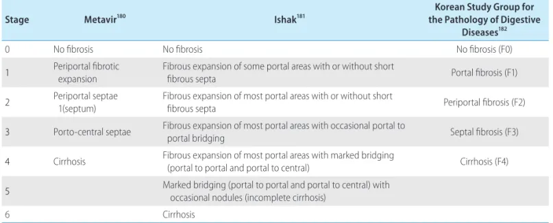
Liver biopsy is performed to assess the grade and stage of the hepatic injury. 107,179 The Metavir 180 and Ishak 181 scoring systems are most widely used, and the scoring system proposed by the South Korean Study Group for the Pathology of Digestive Diseases 182 is used in South Korea (Table 5). Although liver biopsy is not man- datory prior to treatment, it can help to determine when to start treatment and to provide information regarding the treatment re- sponse and prognosis. Considering the natural history of the dis- ease, the cost of treatment and its possible adverse effects, treat- ment can be postponed if liver histopathology shows minimal to moderate fibrosis, state <2 (Metavir stage 2 or periportal fibrosis of the South Korean Study Group for the Pathology of Digestive Diseases, F2). 76,183,184 In this case, a liver biopsy should be repeated 4-5 years later to reassess the necessity of treatment according to the progression of liver disease. 185 About 5-30% of patients with genotype 1 with consistently normal serum ALT may have severe fi- brosis 186-188 and liver biopsy would be useful to determine treatment initiation in this group. 88,189,190 Although hepatic steatosis 179,191,192 and liver iron load overload 193 might impede the treatment response, these findings are not contraindications for treatment. 194-196 If a liver biopsy is not conducted and treatment is not undertaken, continu- ous monitoring is needed. Liver biopsy and treatment initiation should be considered when there is elevation of the serum ALT lev- el and evidence of liver disease progression. 107

Table 5. Comparison of scoring systems for histological stage

Stage Metavir 180 Ishak 181 Korean Study Group for

the Pathology of Digestive Diseases 182

0 No fibrosis No fibrosis No fibrosis (F0)

1 P eriportal fibrotic expansion

F ibrous expansion of some portal areas with or without short

fibrous septa Portal fibrosis (F1)

2 P eriportal septae

1(septum) F ibrous expansion of most portal areas with or without short

fibrous septa Periportal fibrosis (F2)

3 Porto-central septae F ibrous expansion of most portal areas with occasional portal to

portal bridging Septal fibrosis (F3)

4 Cirrhosis F ibrous expansion of most portal areas with marked bridging

(portal to portal and portal to central) Cirrhosis (F4)

5 M arked bridging (portal to portal and portal to central) with occasional nodules (incomplete cirrhosis)

6 Cirrhosis

NOTE: Table adapted from Diagnosis, Management and Treatment of Hepatitis C Hepatology 2009;49:1335-1374. 107

(12)

Noninvasive tests for evaluation of liver fibrosis

Although liver biopsy is accepted as the gold standard test for evaluation of liver fibrosis, 197,198 it is associated with serious com- plications, 199,200 sampling errors, 201 high costs, and interobserver variations. Therefore, various blood marker panels have been de- veloped—including aspartate aminotransferase (AST)-platelet ra- tio index (APRI) and AST/ALT ratio (AAR), and Fibrosis-4 score (FIB-4)—that use combinations of AST, ALT, and platelet count.

FibroTest, Hepascore, FibroMeter, Fibrospect II, and Enhanced Liv- er Fibrosis tests can also be used. 202-213

APRI is calculated by the formula (AST/upper limit of normal for AST) × 100/platelet count (×10 9 /L) (www.kasl.org). This formula is accurate for predicting both significant fibrosis (Ishak score ≥3) defined as APRI >1.5 (AUROC=0.8) and cirrhosis defined as APRI>2 (AUROC=0.89). 214 A normal value of AAR is <0.8, but it increases with hepatic fibrosis progression, so that an AAR value

>1.0 has a 73.7-100% positive predictive value for diagno- sis. 207,215-217 FIB-4 is calculated using the formula: age (yr) × AST (IU/

L)/platelet count (10 9 /L × [ALT (IU/L) 1/2 ] (www.kasl.org). A FIB-4 score <1.45 has a negative predictive value of 90% for advanced fibrosis (Ishak fibrosis score 4-6). In contrast, a FIB-4 score of >3.25 has a positive predictive value of 65% for advanced fibrosis. 218

Liver stiffness measurement using transient elastography can be used to assess hepatic fibrosis. 219-222 However, transient elastogra- phy cannot totally replace liver biopsy, because it often cannot produce reliable measurements in obese patients, and tends to give falsely high results in cases of acute hepatitis with severe in- flammation and necrosis with mild fibrosis. 223,224 In the case of chron- ic hepatitis C, cutoff values determining significant fibrosis (≥F2) vary among studies, ranging from 7.1 to 8.8 kPa, with an AUROC of 0.79- 0.83. 225 The AUROC for diagnosis of liver cirrhosis ranged from 0.95- 0.97, with cutoff values of 12.5-14.6 kPa (77-78% positive predictive value, 95-97% negative predictive value). 202,209,219,225-228

Other newly developed noninvasive tests include acoustic radia- tion force impulse (ARFI) imaging, real-time elastography, mag- netic resonance (MR) elastography, diffusion-weighted MR image, and MR spectroscopy. However, their effectiveness remains to be validated. 229-231

[Recommendations]

1. Screening for HCV infection could be considered in popula- tions at risk as well as those over 40 years old with increas-

ing prevalence of HCV infection (C1).

2. Anti-HCV should be tested in patients suspected of having acute or chronic HCV infection (A1).

3. HCV RNA should be tested in patients with a positive anti- HCV test to confirm the diagnosis (A1).

4. Even if anti-HCV is negative, HCV RNA testing is required when acute HCV infection is suspected or in the presence of unexplained liver disease in immunosuppressed patients (B1).

5. HCV RNA quantitative assay and genotyping/subgenotyping (1a/1b) should be performed prior to antiviral treatment (A1).

6. Immediately following exposure to infected blood or body fluids, anti-HCV and serum ALT level testing should be per- formed. If anti-HCV is negative, an HCV RNA assay should be conducted 4-6 weeks after exposure for early diagnosis.

If all baseline tests are negative, follow-up testing for anti- HCV and serum ALT level should be performed 4-6 months after the exposure (B2).

7. Assessment of liver disease severity is essential prior to an- tiviral treatment (A1).

8. Liver biopsy and/or noninvasive tests for assessment of he- patic fibrosis can be performed to make treatment decision and predict prognosis (B1).

TREATMENT GOALS

The goals of hepatitis C treatment are to eradicate HCV and to prevent complications of liver cirrhosis, hepatocellular carcinoma, extrahepatic manifestations of HCV infection and death. It is diffi- cult to evaluate the treatment goal in a short period of time due to the gradual progression of chronic hepatitis C over several de- cades. Therefore, the short-term goal of hepatitis C treatment is to achieve an SVR, defined as undetectable serum HCV RNA by a sensitive assay 12 or 24 weeks after the end of treatment. SVR was determined 24 weeks after the end of treatment using an as- say with a lower limit of detection of <50 IU/mL in studies of combination therapy with Peginterferon (PegIFN)-α and ribavi- rin, 232 whereas it was determined 12 weeks after the end of treat- ment using an assay with a lower limit of detection of 10-25 IU/

mL in studies on DAA therapy. 233,234 Since HCV does not reappear

in 99% of patients who achieve an SVR, 232 the SVR is considered

as eradication of HCV. Histological hepatic fibrosis improves or

does not get worse, 235,236 complications of cirrhosis significantly

decrease, 237 occurrence of hepatocellular carcinoma decreas-

es, 238,239 and survival rate improves. 240,241 SVR also improves the

(13)

extrahepatic manifestations of HCV infection, such as mixed cryo- globulinemia and glomerulonephritis. 242,243

[Recommendations]

1. The goals of hepatitis C treatment are to eradicate HCV and to prevent complications of liver cirrhosis, hepatocellular carcinoma, extrahepatic manifestations of HCV infection and death (A1).

2. A short-term goal of hepatitis C treatment is to achieve an SVR, defined as an undetectable serum HCV RNA using a sensitive assay at 12 or 24 weeks after the end of treat- ment (A1).

INDICATIONS FOR TREATMENT

All hepatitis C patients who have no contraindications to treat- ment can be considered for antiviral treatment. However, treat- ment would be applicable in cases in which the benefits of treat- ment outweigh the risks. Generally, treatment is strongly recommended for patients with significant hepatic fibrosis (≥

stage F2). Treatment for patients with advanced fibrosis (stage F3-4) who are at high risk of cirrhotic complications and hepato- cellular carcinoma should be prioritized. 90 Treatment of patients in the pre- and post-liver transplant setting should be a priority as successful eradication of HCV increases patient and graft surviv- al. 244,245 Treatment of patients with HCV-related mixed cryoglobu- linemia and glomerulonephritis should also be a priority since HCV eradication can improve the prognosis of those patients. 242,243 With advent of DAA, patients with decompensated liver cirrhosis (LC), which is a contraindication to the combination of PegIFN-α and ribavirin are candidates for antiviral treatment. HCV eradica- tion can reduce the recurrence of hepatocellular carcinoma (HCC) and improve survival in patients with HCC. 246,247 Therefore, pa- tients with HCC can be treated using the rules applicable to those without HCC, taking into consideration the prognosis of HCC and life expectancy.

In cases of mild hepatic fibrosis, treatment can be determined after considering patients’ age, willingness to undergo treatment, and perspectives regarding new drugs.

Contraindications of DAA are limited due to the few adverse ef- fects. Sofosbuvir is contraindicated in patients with several renal impairment (Glomerular filtration rate [GFR] <30 mL/min) in

whom the serum sofosbuvir concentration is markedly increased.

Safety of daclatasvir, ledipasvir, and asunaprevir has not been ful- ly evaluated in patients with severe renal impairment (GFR <30 mL/min). Paritaprevir, dasabuvir, and asunaprevir are contraindi- cated in patients with decompensated cirrhosis in whom the se- rum concentrations of these drugs are markedly increased. Abso- lute contraindications to the combination of PegIFN-α and ribavirin include uncontrolled depression or psychiatric illness, un- controlled autoimmune diseases, transplantation of solid organs except the liver, untreated thyroid illness, pregnancy or unwilling- ness to comply with adequate contraception, severe concurrent medical illness such as poorly controlled hypertension, heart fail- ure, significant coronary heart disease, poorly controlled diabetes mellitus, and chronic obstructive pulmonary disease, age ≤2 years, and hypersensitivity to PegIFN-α or ribavirin.

[Recommendations]

1. All HCV-infected patients with no contraindication to treat- ment should be considered for treatment (A1).

2. Patients with advanced fibrosis ≥F3 (including compensat- ed and decompensated cirrhosis) should be given priority for treatment (A1).

3. Treatment should be prioritized in the pre- and post-liver transplant setting (A1).

4. Treatment should be prioritized for patients with severe ex- trahepatic manifestations, including HCV-related mixed cryoglobulinemia and glomerulonephritis (A1).

5. Treatment should be individualized taking into consider- ation the severity of liver disease, probability of treat- ment success, risks of severe adverse effects, accompany- ing diseases, and patients’ willingness to undergo treatment (B1).

6. HCV treatment is not recommended in patients with limit- ed life expectancy due to extrahepatic diseases (B1).

DEFINITION OF TREATMENT RESPONSE

An end-of-treatment response (ETR) is defined as undetectable

HCV RNA at the end of treatment using a sensitive assay. An SVR

is defined as undetectable HCV RNA by a sensitive assay at 12 or

24 weeks after completion of treatment. The concordance of SVR

12 and SVR 24 is 98% irrespective of treatment regimen. 248 Viral

breakthrough refers to the reappearance of HCV RNA during treat-

(14)

ment after a virological response, and relapse is defined as the re- appearance of HCV RNA after treatment has been completed.

The combination of PegIFN-α and ribavirin is associated with considerable cost and adverse effects. The likelihood of SVR in- creases as the time of HCV RNA disappearance is shorter. 249 Re- sponse-guided therapy is a strategy to modify the duration of treatment based on the time of HCV RNA disappearance by mea- suring serum HCV RNA at weeks 4, 12, and 24 of treatment.

However, treatment with DAA does not comply with this strategy.

A rapid virological response (RVR) is defined as undetectable HCV RNA using a sensitive assay with a lower limit of detection of <50 IU/mL at week 4 of treatment. The SVR rate is expected to be 87.5-100% in HCV genotype 1 patients with a RVR and 33.3- 63.8% in those without a RVR. 250-252 The SVR rate is expected to be 85-86.5% in HCV genotypes 2 and 3 patients with a RVR and 54-58.3% in those without a RVR. 251,253 An early virological re- sponse (EVR) is defined as undetectable HCV RNA using a sensi- tive assay with a lower limit of detection of <50 IU/mL or a ≥ 2 log reduction in HCV RNA compared with the baseline level. The SVR rate is as low as 3% in HCV genotype 1 patients without an EVR. 254-256 Therefore, medical cost and adverse effects can be re- duced by discontinuing therapy in cases without an EVR. An EVR is classified as a complete EVR (cEVR), which is defined as unde-

tectable HCV RNA, and a partial EVR (pEVR), which is defined as an EVR with detectable HCV RNA at week 12. A delayed virologi- cal response (DVR) is defined as a pEVR that results in undetect- able HCV RNA at week 24. 257,258 A null response is defined as a

<2 log reduction in HCV RNA level from baseline at week 12 of therapy, whereas a partial nonresponse is defined as a ≥ 2 log re- duction in HCV RNA level from baseline but detectable HCV RNA at week 12 and 24 (Table 6).

PREDICTORS OF TREATMENT RESPONSES

HCV genotype 3, 259 liver cirrhosis, 259 previous treatment fail- ure, 259 and RAV 233,260 are predictive factors for decreased SVR when treated with DAAs.

In cases of combination treatment of PegIFN-α and ribavirin, the strongest pretreatment predictors of an SVR include HCV gen- otype, 254,261,262 degree of hepatic fibrosis, 263 and IL28B genetic polymorphism. 263,264 The SVR rates are 40-60% in HCV genotype 1 patients and 70-80% in HCV genotypes 2 and 3 patients. 254,265 Patients with F0-F2 fibrosis have a 2.7-fold higher SVR rate than those with F3-F4 fibrosis. 263 The SVR rates are 2.4-2.7-fold higher in patients with a viral load of <400,000-800,000 IU/mL com- Table 6. Definitions of virological responses during the combination therapy of PegIFN- α and ribavirin

Virological response Definition Clinical implication

Rapid virological response (RVR) Undetectable HCV RNA (<50 IU/mL) at week 4 of therapy M ay allow shortening of course for genotypes 2&3 and possibly genotype 1 with low viral load Early virological response (EVR) ≥ 2 log reduction of HCV RNA level from baseline at week 12 of therapy Negative predictor of SVR

Complete EVR (c-EVR) Undetectable HCV RNA at week 12 of therapy Partial EVR (p-EVR) EVR but detectable HCV RNA at week 12 of therapy D elayed virological response

(DVR) ≥ 2 log reduction of HCV RNA level from baseline but detectable HCV RNA at week 12 and undetectable HCV RNA at week 24

End of treatment response (ETR) Undetectable HCV RNA at the end of 24 or 48 weeks of treatment S ustained virological response

(SVR)

Undetectable HCV RNA (<50 IU/mL) at 12 or 24 weeks after treatment Be st predictor of a long-term response to treatment Null response (NR) <2 log reduction of HCV RNA level from baseline at week 12 of therapy

Partial nonresponse ≥ 2 log reduction of HCV RNA level from baseline but detectable HCV RNA at week 12 and 24

Breakthrough R eappearance of HCV RNA in serum during treatment after virological response

Relapse Reappearance of HCV RNA after treatment is discontinued

HCV, hepatitis C virus.

(15)

pared to those with a viral load of >800,000 IU/mL. 250,263,266 SVR rates are lower in older patients (>40 years), 254 African-Ameri- cans, 267 body weight >70 kg, 254,261 and insulin resistance. 268,269 SVR rates vary depending on SNP of IL28B, in other words, a C or T allele at the rs12979869 locus. 270 The SVR rates of HCV geno- type 1 Caucasian patients are 69%, 33%, and 27% in CC homo- zygotes, CT heterozygotes, and TT homozygotes, respectively. The SVR rates of HCV genotype 1 African-American patients are 48%, 15%, and 13%. 263,271 The SVR rates of HCV genotype 1 Korean patients are 73-88% in CC homozygotes and 0-40% in CT het- erozygotes. 272-274 The prevalence of the IL28B genetic polymor- phism varies among ethnicities. CC homozygotes in Korea account for 88-89%, 272-274 compared to 17% in African-Americans, and 37% in Caucasians. 263 Therefore, the usefulness of the IL28B ge- netic polymorphism as a predictive factor is limited as over 90%

of the population of Korea are CC homozygotes.

During combination treatment with PegIFN-α and ribavirin, a RVR is the strongest on-treatment predictor for an SVR, 250-252 and an SVR rate increases ninefold with a RVR. 263 Meanwhile, an EVR is a strong negative predictor for an SVR. Without EVR, the SVR rate is only 3%. 255 In addition, SVR rates increase when medica- tion adherence is higher than 80%, 275 so assessment and mainte- nance of medication adherence can increase the SVR rate.

NEW DRUGS, DIRECT-ACTING ANTIVIRALS (DAA)

DAA acts at a specific step of the viral life cycle. DAAs include NS3/4A protease inhibitors (PI), NS5A inhibitors, and NS5B poly- merase inhibitors. NS3/4A PI is a first-generation DAA and blocks the polyprotein processing essential for HCV replication. To date, available drugs are boceprevir, telaprevir, simeprevir, asunaprevir, and paritaprevir. NS5A inhibitors, such as daclatasvir, ledipasvir or ombitasvir, affect various HCV genotypes and show a synergistic effect in combination with other DAAs. The NS5B polymerase in- hibitors are divided into nucleoside polymerase inhibitors (sofos- buvir) and non-nucleoside polymerase inhibitors (dasabuvir, be- clabuvir). Because the basic characteristics of each DDA differ, selection and use of the appropriate drugs should take into con- sideration hepatic and renal function. DAA regimens may have a risk of interactions with other medications used by patients. Prior to starting treatment, patients should be evaluated for potential drug-drug interactions with selected DAAs. A more comprehen- sive list of drug-drug interactions is available at several websites,

such as www.hep-druginteractions.org.

[Recommendations]

1. The characteristics of the DAAs should be understood, and the appropriate drugs selected taking into consideration hepatic and renal function (A1).

2. The potential for drug-drug interactions must be consid- ered before and during treatment with DAAs. The full pre- scribing information must be consulted prior to use of DAAs due to the potential for drug-drug interactions (A1).

Simeprevir

Simeprevir is a HCV NS3/4A protease inhibitor.

1. Dosage and administration

One 150 mg capsule is taken orally once daily with food.

2. Pharmacokinetics

Simeprevir primarily undergoes oxidative metabolism by the he- patic CYP3A system. Elimination of simeprevir occurs via biliary excretion. Renal clearance plays an insignificant role in its elimina- tion. Therefore, no dose adjustment of simeprevir is needed in pa- tients with mild, moderate or severe renal impairment. Compared to HCV-uninfected subjects with normal hepatic function, the mean steady-state AUC of simeprevir was higher in HCV-uninfect- ed subjects with moderate hepatic impairment (Child-Pugh Class B) and severe hepatic impairment (Child-Pugh Class C). Simeprevir has not been extensively studied in such patients, but has been used in real-life settings.

3. Drug-drug interactions

Co-administration of simeprevir with substances that are mod-

erate or strong inducers or inhibitors of CYP3A4 is not recom-

mended as this may lead to significantly lower or higher exposure

to simeprevir, respectively. A number of compounds are contrain-

dicated in patients receiving simeprevir, including anticonvulsants

(carbamazepine, phenobarbital, phenytoin), antibiotics (erythro-

mycin, clarithromycin), antimycobacterials (rifampin, rifabutin, ri-

fapentine), antifungals (itraconazole, ketoconazole, fluconazole,

voriconazole), dexamethasone, cisapride, herbal products (milk

thistle, St John’s wort) and antiretroviral drugs (cobicistat-based

regimens, efavirenz, etravirine, nevirapine, ritonavir). Simeprevir

does not require dose changes in combination with the immuno-

수치

Table 1. Grading of Recommendations, Assessment, Development, and Evaluation (GRADE) Quality of evidence Criteria
Figure 1. Map of South Korea showing age and sex-adjusted anti-HCV  seroprevalence in each area
Table 2. High-risk persons recommended for HCV infection screening 1) Persons who are suspected of having acute or chronic HCV infection
Table 5. Comparison of scoring systems for histological stage
+6

참조

관련 문서

Prioritize and invest in inclusive programs that promote a culture of peace, social cohesion, global citizenship principles, environmental sustainability,

3) ACC/AHA/ESC 2006 Guidelines for the Management of Patients with Atrial Fibrillation: a report of the American College of Cardiology/American Heart

&lt; 0.1 mg/L Penicillin G or ampicillin Cefotaxime, ceftriaxone, or chloramphenicol 0.1-1.0 mg/L Cefotaxime or ceftriaxone Cefepime, meropenem.. ≥ 2.0

Severe disease activity and cy- tomegalovirus colitis are predictive of a nonresponse to infliximab in patients with ulcerative colitis. Guidelines for the management of

Especially, Mg-Al system alloys are inexpensive and have standard mechanical properties microstructure and deformation behaviorsat high temperature were investigated..

– Land: Food, Urbanization, Fibre, Water, Conservation – Energy: Utility and cost as a delivered energy form.. • US has land

Consequently, the present study was designed to investigate the effect of adding 60 mg of raloxifene hydrochloride daily to women treated with GnRH-a for severe endometriosis

Harmonics generated by Control Element Drive Mechanism Control System(CEDMCS) affected the MG Set operation such as increased voltage waveform distortion and... MG Set