Abstract. Background: The applicability of the melanoma antigen gene (MAGE) to the molecular diagnosis of cancer, by detecting MAGE mRNA in the sputa of head and neck cancer patients, was investigated. Materials and Methods: Nested reverse transcription-polymerase chain reaction (RT-PCR) with the MAGE common primers designed to detect MAGE A1-6 genes was conducted with sputa of 17 head and neck squamous cell carcinoma patients. Results: Of the 17 sputa specimens from patients with squamous cell carcinoma, MAGE mRNA was detected in 13 cases, while the detection rate was 2 out of 40 in the normal control group, showing significant correlation (p<0.05) between head and neck squamous cell carcinoma and MAGE mRNA expression in sputa. Conclusion: MAGE may be a useful tumor marker of head and neck cancer.
One of the objectives of cancer research is to discover cancer-specific molecules and to use them in the diagnosis and management of cancer. To achieve these objectives, this study addressed whether the expression of the melanoma antigen gene (MAGE) is specific to head and neck cancer.
The MAGE gene is identified from melanoma cell lines and is expressed specifically in cancer cells (1). As this gene is recognized by cytotoxic T-lymphocytes, it has been considered as a potential target for cancer immunotherapy (2). The MAGE gene is classified as MAGE-A, -B, -C and - D based on its locus on the chromosome (2). MAGE-A, consisting of at least 12 genes, is located on chromosome Xq28 (3) and is expressed in most cancers, including cancers of the head and neck (4-6), stomach (7), esophagus (8),
colon (9), lung (10), breast (11), ovary (12) and liver (13), as well as melanoma, but is not expressed in normal tissues with the exception of the testis. Currently, the mainstream investigation of the MAGE gene is to examine whether it is useful in cancer immunotherapy. However, studies on the clinical feasibility of the MAGE gene in cancer diagnosis, based on cancer-specific expression of the gene, have rarely been reported. Recently, the possibility of the molecular diagnosis of cancer through detection of MAGE from blood or sputum samples of patients with stomach, liver and lung cancer has been reported (14-17).
In the head and neck, MAGE-A is known to be expressed specifically in cancer tissues. Eura et al. (4) and Lee et al.
(5) have reported that more than one subtype of MAGE-A mRNA are expressed in 71-75% of squamous and non- squamous cell carcinoma of the head and neck. Moreover, the same group, using monoclonal antibody 57B, confirmed that the MAGE gene encodes the protein specific to cancer (6). These studies supported that mRNA and protein encoded by MAGE are specific to cancer. Recently, Park et al. (17) designed a common MAGE primer which can detect all subtypes of MAGE A1-A6 genes by a single examination.
They performed nested reverse transcription - polymerase chain reaction (RT-PCR) with a common MAGE primer to evaluate the MAGE-A expression in cancer, benign tumor and normal tissues of the head and neck. MAGE A1-A6 were detected in 70.4% of the squamous cell carcinomas with this primer, but not in benign tumor and normal tissues, indicating that the common MAGE primer can be useful in detecting MAGE-A expression.
All the previous studies were conducted on tissue samples collected from patients with head and neck cancer. As a next step, we sought to examine the MAGE expression in body fluids. We hypothesized that MAGE could be a tumor marker of head and neck cancer if specifically expressed in the sputa of cancer patients. In this study, the common MAGE primers we designed were assessed for their ability to detect MAGE-A mRNA in the sputum of cancer patients, Correspondence to: Kang Dae Lee, 34 Amnam-Dong, Suh-Gu,
Busan, 602-702, South Korea. Tel: 82-51-990-6470/6137, Fax: 82- 51-245-8539, e-mail: [email protected]
Key Words: MAGE gene, sputum, tumor marker, head and neck cancer.
Expression of MAGE A 1-6 mRNA in Sputa of Head and Neck Cancer Patients - A Preliminary Report
KANG DAE LEE1, HWAN HO LEE1, HO BUM JOO1, HYOUNG SHIN LEE1, TAI HYUN YU1, HEE KYUNG CHANG2, CHANG HO JEON3and JONG WOOK PARK4
Departments of 1Otolaryngology and 2Pathology, College of Medicine, Kosin University, Busan;
3Department of Clinical Pathology, School of Medicine, Catholic University of Daegu, Daegu;
4Department of Immunology, School of Medicine, Keimyung University, Daegu, Korea
and the clinical feasibility of a molecular diagnosis of head and neck cancer with the MAGE gene was evaluated.
Materials and Methods
Materials. Seventeen patients with squamous cell carcinoma of the head and neck, who had been confirmatively diagnosed at Kosin University Gospel Hospital, Korea, between April 2001 and December 2002, were enrolled in the study, and 40 normal controls were recruited from healthy volunteers. After obtaining informed consent, sputum was collected from both groups (Table I).
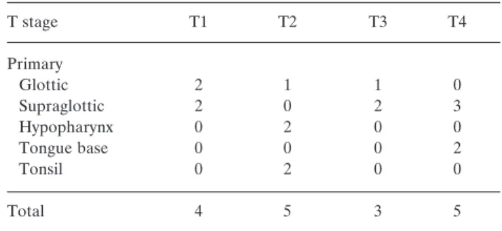
In the patient group, the mean age was 62.5 years and the male- to-female ratio was 16:1. In the control group, the mean age was 31.5 years and the male-to-female ratio was 9:1. The ratio of smokers to non- smokers was 16:1 in the patient group and 1:1 in the control group. The primary sites of the 17 squamous cell carcinomas were 11 laryngeal cancer, including glottic cancer (4 cases) and supraglottic cancer (7 cases), hypopharyngeal cancer (2 cases), base of tongue cancer (2 cases) and tonsillar cancer (2 cases). According to the T stage, 4 cases were T1, 5 cases were T2, 3 cases were T3 and 5 cases were T4 (Table II). The primary therapies for all cases were laser surgery (1 case), radiation therapy only (3 cases) and combined chemotherapy and radiation therapy (13 cases).
Collection of sputum and isolation of mRNA. To detect the expression of MAGE mRNA from sputum, a test was performed once prior to the treatment. During the follow-up period after treatment, including surgery, radiotherapy and chemotherapy, the test was performed more than once. This test has been approved by the Korean Ministry of Health and Welfare. To collect sputum from the patients, each patient was induced to cough deeply and spit the sputum into a 50 mL conical tube so that as many cells as possible might be exfoliated from the larynx and pharynx. We developed a method of sputum management to simultaneously perform the liquefaction of sputum, cytolysis and RNA preservation. In brief, about 3-5 mL sputum digester (iC&G Co., Korea) was applied to the sputum accumulated in the 50 mL conical tube and was stirred for complete sputum liquefaction. A magnetic bead (ABgene, Epsom, UK) of 150 ÌL (7.5 mg) was mixed with the liquefied sputum at room temperature for 30 min, and then the conical tube was centrifuged at 1,100 rpm for 2 min.
The supernatant was removed and the bead was suspended using rinsing solution to transfer to a 1.5 mL test tube. The magnetic bead, washed with rinsing buffer, was separated on a magnetic stand and the supernatants were removed completely. This step
was repeated 4 times. The magnetic bead was then suspended with elution buffer and incubated for 10 min at 70ÆC. After transferring the elution buffer to a new test tube, 3 ÌL biotin- labelled oligo dT (10 pmol/ÌL) and strepavidin magnetic particle (SMP, 10 mg/mL, Roche Diagnostics, Mannheim, Germany) were added and the mixture was incubated at 37ÆC for 10 min. SMP washed with SMP rinsing buffer was separated on a magnetic stand and the supernatants were completely removed. This step was repeated 4 times. Finally, SMP was re-suspended with 20 ÌL RNase-free distilled water and incubated at 70ÆC for 2 min to elute mRNA.
RT-PCR and nested PCR
a) cDNA synthesis by reverse transcription. First, the RT-mixture was prepared by adding 5 x RT buffer of 3 ÌL, 10 mM dNTP mix of 1.5 ÌL, MMLV reverse transcriptase (200 U/ÌL, Promega, USA) 0.5 ÌL, RNase inhibitor (40 U/ÌL, Promega) 0.5 ÌL and 100 pM oligo dT primer of 1.5 ÌL to a PCR tube. After adding 13 ÌL mRNA solution to the RT-mixture, a drop of mineral oil was applied and the PCR tube was kept at room temperature for 10 min. This PCR tube was heated at 42ÆC for 60 min resulting in cDNA synthesis. The cDNA was used for primary PCR.
b) Primary PCR using C1/C2 primer to MAGE 1-6. PCR for MAGE A1-A6 was performed after confirming mRNA isolation with GAPDH (sense: CGTCTTCACCACCATGGAGA, antisense:
CGGCCATCACGCCACAGTTT). The PCR mixture was prepared by adding 3 ÌL of 10 x PCR buffer, 1.8 ÌL of 25 mM MgCl2, 0.3 ÌL of 10 mM dATP, 0.3 ÌL of 10 mM dGTP , 0.3 ÌL of 10 mM dTTP, 0.3 ÌL of 10 mM dCTP, 0.25 ÌL of 50 ÌM sense/antisense primer and 0.25 ÌL of Taq polymerase (5 U/ÌL, Promega) and applying distilled water to reach a final volume of 25 ÌL per PCR tube. Five microliters of reverse transcripts were applied to the PCR tube and were mixed. After applying a drop of mineral oil to the PCR tube and transferring to the PCR system (Cetus 480, Perkin Elmer Co., USA), a primary PCR was performed using C1/C2 common MAGE primer according to the following conditions (Table III) : after heating at 94ÆC for 5 min, cDNA was amplified by reacting in 35 cycles with a cycle of 94ÆC for 30 sec, 57ÆC for 45 sec and 72ÆC for 45 sec. The PCR was completed with a 5-min treatment at 72ÆC.
c) Nested PCR using C3/C4 primer to MAGE A1-A6. The primary PCR product, diluted 10-fold with distilled water, was used for nested PCR. Three microliters of distilled water were added to 2 ÌL of the primary PCR product described above. After applying a drop Table I. Primary site of squamous cell carcinoma of the head and neck.
Primary site No. of patients
Larynx (4 glottic, 7 supraglottic) 11
Hypopharynx 2
Base of tongue 2
Tonsil 2
Total 17
Table II. T stage according to the primary site.
T stage T1 T2 T3 T4
Primary
Glottic 2 1 1 0
Supraglottic 2 0 2 3
Hypopharynx 0 2 0 0
Tongue base 0 0 0 2
Tonsil 0 2 0 0
Total 4 5 3 5
of mineral oil to the test tube with PCR reagents and transferring to the PCR system, nested PCR was performed using a C3/C4 primer according to the following conditions (Table III). After heating at 94ÆC for 5 min, the cDNA was amplified by reacting in 35 cycles with a cycle of 94ÆC for 30 sec, 57ÆC for 45 sec and 72ÆC for 45 sec.
The PCR was completed with a 5-min treatment at 72ÆC, and the PCR product was loaded to 1% agarose gel for electrophoresis, followed by observation using an ultraviolet transilluminator.
Statistical analysis. To evaluate the statistical significance of MAGE expression in each group, the ¯2-test was used. Statistical significance was determined at values of p<0.05.
Results
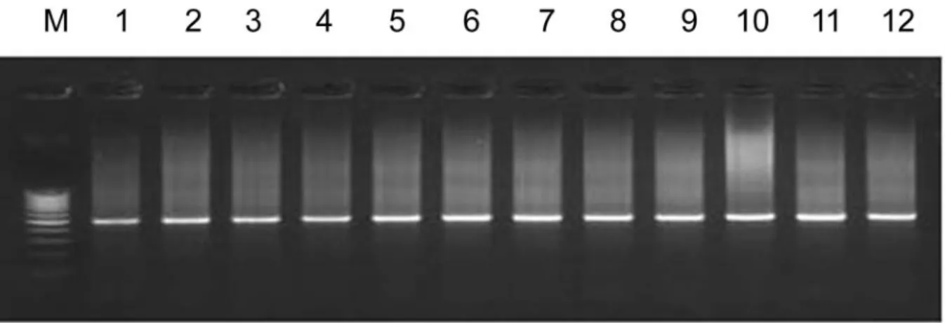
Expression of MAGE A1-A6 mRNA in sputa of the patient group. MAGE mRNA was expressed in 13 out of 17 patients with squamous cell carcinoma (Figure 1). According to the primary site, MAGE was detected in 8 out of 11 of the laryngeal cancer cases, 1 out of 2 of the tongue base cancers, all of the hypopharyngeal cancers, and all of the tonsillar cancer cases (Table IV). MAGE was detected in 2 out of 4 of the cases at T1, all at T2, 2 out of 3 at T3 and 4 out of 5 at T4 (Table V).
Expression of MAGE A1-A6 mRNA during the follow-up.
Among the 17 cases of squamous cell carcinoma, 11 cases achieved complete remission while 6 cases had recurrences.
Six out of the 11 complete remission cases were evaluated and showed negative MAGE expression in the sputum.
However, 3 out of the 6 cases of recurrence after induction chemotherapy and radiotherapy (2 cases of supraglottic cancer and 1 case of hypopharyngeal cancer) were positive for MAGE expression during the follow-up.
Expression of MAGE A1-A6 mRNA in the normal controls.
MAGE A1-A6 mRNA was detected in 2 out of 40 of the normal controls (p<0.001) (Figure 2). These 2 positive cases both had a long-term history of smoking.
Discussion
Although many therapeutic regimens have been developed to manage head and neck cancer, the survival rate has not been improved in advanced disease. Thus, the development of methods for the early diagnosis of cancer are critical to improving the survival rate. The development of a new tumor marker would be one means for the early detection Table III. Common primer sequences for the detection of MAGE A1-A6 genes.
Primer Type PCR Sequence Size (bp)
C1 S RT-PCR CTGAAGGAGAAGATCTGCC 831-855
C2 AS RT-PCR CTCCAGGTAGTTTTCCTGCAC
C3 S Nested PCR CTGAAGGAGAAGATCTGCCWGTG 469-493
C4 AS Nested PCR CCAGCATTTCTGCCTTTGTGA
S: sense primer.
AS: antisense primer.
W: A or T.
Table IV. Expression of MAGE A1-A6 genes detected by nested RT-PCR with common primer in squamous cell carcinoma of the head and neck.
Primary site % of positive expression
Larynx 72.7% (8/11)
Hypopharynx 100% (2/2)
Base of tongue 50% (1/2)
Tonsil 100% (2/2)
Total (n=17) 76.5% (13/17)
Table V. Expression of MAGE A1-A6 genes detected by nested RT-PCR with common primer according to T stage in squamous cell carcinoma of the head and neck.
T stage T1 T2 T3 T4
Primary
Glottic 1/2 1/1 1/1 0
Supraglottic 1(2) 0 1(2) 3(3)
Hypopharynx 0 2(2) 0 0
Tongue base 0 0 0 1(2)
Tonsil 0 2(2) 0 0
Total 50% 100% 66.6% 80%
(2/4) (5/5) (2/3) (4/5)
of cancer. An ideal tumor marker should be cancer-specific and expressed in a variety of cancers with a high sensitivity.
To date, no satisfactory tumor marker has been found for head and neck cancer.
Eura et al. (4) and Lee et al. (5, 6) reported that MAGE- A was detected with relatively high specificity in cancer tissue.
Moreover, Lee et al. (18) and Park et al. (17) performed nested RT-PCR using a common MAGE primer to simultaneously detect MAGE A1-A6 mRNA in malignant tumor, benign tumor and normal tissue of the head and neck. MAGE was detected in 70.4% of squamous cell carcinomas, but not in the benign tumor or normal tissue, indicating that MAGE could be a tumor marker for this disease. Unlike previous studies on surgically removed tumor tissue, this study used the sputum from patients with head and neck cancer. The result was interesting in that the expression rate of MAGE A1-A6 mRNA was 76.5% (13/17).
In contrast to our initial assumption, the detection rate of the gene in sputum was slightly higher than that of cancer tissue. This may have been due to the small size of the
patient group. Park et al. (17) applied a similar technique to lung cancer and reported that 74.2% (23/31) of the sputum samples from lung cancer patients showed MAGE expression. These findings suggest that the MAGE A1-A6 assay may be used to detect a small number of cancer cells in the sputum for early diagnosis of head and neck cancer as well as to detect lung metastasis. Matsuda et al. (19) examined whether it was possible to diagnose head and neck cancers using sputum cytology. According to their results, laryngeal and hypopharyngeal cancer were diagnosed at the rate of 63.5% and 77.4%, respectively, through sputum cytology. In terms of stages, laryngeal cancer was diagnosed at T1 at the rate of 29.4%, T2 at the rate of 63.3%, T3 at the rate of 69.7% and T4 at the rate of 79.2%. The diagnostic rate of T1 laryngeal cancer was low due to the small number of exfoliated cells in the sputum. Although sputum cytology is a non- invasive and simplified diagnostic technique, it may have little clinical significance if the detection rate of early cancer is low. This is also because advanced head and neck cancer can be easily diagnosed Figure 1. Detection of MAGE mRNA in the sputum of the head and neck cancer patients. M : size marker. Lanes 1-12 : squamous cell carcinoma of the head and neck.
Figure 2. Detection of MAGE mRNA in the sputum of the normal controls. The expression of MAGE is noted in lane 11.
using other diagnostic tools. A new diagnostic method for early head and neck cancer should indicate the presence of cancer cells which conventional diagnostic methods cannot detect. This study showed a relatively high detection rate in early stages (50% in T1, 100% in T2).
Park et al. (17) performed an experiment adding various numbers of SNU484 cells, a MAGE-positive cell line, to 107 cells of SNU638, a MAGE-negative cell line, to indirectly determine the minimal number of MAGE-positive cells for cancer detection in body fluid. The results indicated that 20 PCR cycles of nested PCR were enough to detect MAGE messages of 1 to 10 SNU484 cells in a background of 107 SNU638 cells. The results also showed that the detection of MAGE gene expression was possible in 6 ml of normal whole blood samples containing 5 SNU484 cells and in normal sputum samples containing 1-2 SNU484 cells. Thus a molecular diagnosis of cancer is possible as long as the cancer cells express the MAGE gene.
In the present study, the false-positive rate was 5% (2 cases), which is markedly lower than that of conventional tumor markers, including the alpha-fetoprotein (20) and carcinoembryonic antigen (21). However, considering our reports on the cancer-specific expression of the MAGE gene in tissue, the 5% false-positive rate in sputum is not consistent with our results. We assume that the false- positive results were due to factors such as the high sensitivity of the nested RT-PCR, RNA contamination during the test, early carcinogenic changes of cells in the respiratory tract and the expression of MAGE in non- cancerous tissue. Park et al. (17) reported that 2.1% (4/192) of the sputum samples from non-lung cancer patients expressed the MAGE gene. Jang et al. (22) reported that MAGE transcripts were detected in cells under early carcinogenic change as well as in cancer tissues. Lee et al.
(5) noted that MAGE was detected in laryngeal leukoplakia, although only in 1 case.
The false-positive results in this study could also be attributed to various factors as in the cases described above.
However, the normal controls with MAGE-A-positive expression may represent early carcinogenic changes considering the subjects’ history of smoking. The false- positive group should be followed-up because of the risk of carcinogenesis. One case of laryngeal leukoplakia proven as dysplasia was MAGE-positive in the sputum. The change from dysplasia to invasive squamous cell carcinoma was later confirmed in this patient and curative therapy was possible. These findings indicate that MAGE expression was effective in assessing the malignant potential of cells in dysplasia; thus, long-term follow-up is necessary in MAGE false-positive groups.
Radiotherapy for laryngeal or hypopharyngeal cancer causes edema or necrosis in the larynx or hypopharynx and makes early diagnosis of residual or recurrent cancer
difficult. Among the 6 patients with recurrence after radiotherapy, recurrent or residual cancer could be identified in 3 patients (2 supraglottic cancers, 1 hypopharyngeal cancer) on the basis of MAGE expression in the sputum. Since recurrent supraglottic cancers were diagnosed at an early stage by molecular diagnosis using the MAGE gene, these patients were treated with minimal salvage surgery. Therefore, MAGE expression can be effective in identifying recurrent or residual cancer caused by necrosis or edema after radiotherapy. In conclusion, the MAGE-A gene may be a tumor marker for the early diagnosis and detection of primary, residual and recurrent disease of the head and neck.
References
1 van der Bruggen P, Traversari C, Chomez P, Lurquin C, De Plaen E, Van den Eynde B, Knuth A and Boon T: A gene encoding an antigen recognized by cytolytic T lymphocytes on a human melanoma. Science 254: 1643-1647, 1991.
2 Zammatteo N, Lockman L, Brasseur F, De Plaen E, Lurquin C, Lobert PE, Hamels S, Boon T and Remacle J: DNA microarray to monitor the expression of MAGE-A genes. Clin Chem 48: 25-34, 2002.
3 De Plaen E, Arden K, Traversari C, Gaforio JJ, Szikora JP, De Smet C, Brasseur F, van der Bruggen P, Lethe B and Lurquin C: Structure, chromosomal localization, and expression of 12 genes of the MAGE family. Immunogenetics 40: 360-369, 1994.
4 Eura M, Ogi K, Chikamatsu K, Lee KD, Nakano K, Masuyama K, Itoh K and Ishikawa T: Expression of the MAGE gene family in human head-and-neck squamous-cell carcinomas. Int J Cancer 64: 304-308, 1995.
5 Lee KD, Eura M, Ogi K, Nakano K, Chikamatsu K, Masuyama K and Ishikawa T: Expression of the MAGE-1, -2, -3, -4, and -6 genes in non-squamous cell carcinoma lesions of the head and neck. ActaOtolaryngol 116: 633-639, 1996.
6 Lee KD, Chang HK, Jo YK, Kim BS, Lee BH, Lee YW, Lee HK, Huh MH, Min YG, Spagnoli GC and Yu TH: Expression of MAGE 3 gene product in squamous cell carcinomas of the head and neck. Anticancer Res 19: 5037-5042, 1999.
7 Inoue H, Li J, Honda M, Nakashima H, Shibuta K, Arinaga S, Ueo H, Mori M and Akiyoshi T: MAGE-1 mRNA expression in gastric carcinoma. Gastroenterol 109: 1522-1525, 1995.
8 Inoue H, Mori M, Li J, Mimori K, Honda M, Nakashima H, Mafune K, Tanaka Y and Akiyoshi T: Human esophageal carcinomas frequently express the tumor-rejection antigens of MAGE genes. Int J Cancer 63: 523-526, 1995.
9 Mori M, Inoue H, Mimori K, Shibuta K, Baba K, Nakashima H, Haraguchi M, Tsuji K, Ueo H, Barnard GF and Akiyoshi T:
Expression of MAGE genes in human colorectal carcinoma.
Ann Surg 224: 183-188, 1996.
10 Weynants P, Lethe B, Brasseur F, Marchand M and Boon T:
Expression of MAGE genes by non-small cell lung carcinomas.
Int J Cancer 56: 826-829, 1994.
11 Russo V, Traversari C, Verrecchia A, Mottolese M, Natali PG and Bordignon C: Expression of MAGE gene family in primary and metastatic human breast cancer: implications for tumor- specific immunotherapy. Int J Cancer 64: 216-221, 1995.
12 Russo V, Dalerba P, Ricci A, Bonazzi C, Leone BE, Mangioni C, Allavena P, Bordignon C and Traversari C: MAGE, BAGE and GAGE genes expression in fresh epithelial ovarian carcinomas. Int J Cancer 67: 457-460, 1996.
13 Yamashita N, Ishibashi H, Hayashida K, Kudo J, Takenaka K, Itoh K and Niho Y: High frequency of the MAGE-1 gene expression in hepatocellular carcinoma. Hepatology 24: 1437- 1440, 1996.
14 Mori M, Mimori K, Tanaka F, Ueo H, Sugimachi K and Akiyoshi T: Molecular diagnosis of circulating cancer cells using MAGE gene assays. JAMA 278: 476-477, 1997.
15 Mou DC, Cai SL, Peng JR, Wang Y, Chen HS, Pang XW, Leng XS and Chen WF: Evaluation of MAGE-1 and MAGE-3 as tumour-specific markers to detect blood dissemination of hepatocellular carcinoma cells. Br J Cancer 86: 110-116, 2002.
16 Miyamoto A, Fujiwara Y, Sakon M, Nagano H, Sugita Y, Kondo M, Eguchi H, Dono K, Umeshita K, Nakamori S and Monden M: Development of a multiple-marker RT-PCR assay for detection of micrometastases of hepatocellular carcinoma.
Dig Dis Sci 45: 1376-1382, 2000.
17 Park JW, Kwon TK, Kim IH, Sohn SS, Kim YS, Kim CI, Bae OS, Lee KS, Lee KD, Lee CS, Chang HK, Choe BK, Ahn SY and Jeon CH: A new strategy for the diagnosis of MAGE- expressing cancers. J Immunol Methods 266: 79-86, 2002.
18 Lee KD, Lee CS and Lee HH: Experimental studies on the significance of new MAGE common primers detecting MAGE 1-6 mRNA in head and neck cancers. Korean J Otolaryngol 44:
736-743, 2001.
19 Matsuda M, Nagumo S, Horai T and Yoshino K: Cytologic diagnosis of laryngeal and hypopharyngeal squamous cell carcinoma in sputum. Acta Cytol 32: 655-657, 1988.
20 Adamiak G, Ligezinski A, Jurkiewicz D, Hermanowski M, Konieczna M. Adamiak G, Ligezinski A, Jurkiewicz D, Hermanowski M and Konieczna M: Alpha-fetoprotein – diagnostic utility in squamous cell carcinoma of the head and neck. Pol Merkuriusz Lek 11: 320-322, 2001.
21 Rosati G, Riccardi F and Tucci A: Use of tumor markers in the management of head and neck cancer. Int J Biol Markers 15:
179-183, 2000.
22 Jang SJ, Soria JC, Wang L, Hassan KA, Morice RC, Walsh GL, Hong WK and Mao L: Activation of melanoma antigen tumor antigens occurs early in lung carcinogenesis. Cancer Res 61:
7959-7963, 2001.
Received October 18, 2005 Revised January 3, 2006 Accepted January 13, 2006