Tuberc Respir Dis 2011;70:315-322
CopyrightⒸ2011. The Korean Academy of Tuberculosis and Respiratory Diseases. All rights reserved.
폐암환자의 객담 및 암 조직에서 MAGE A 1-6와 SSX 1-9 유전자의 발현
1
이연재맥의원, 영남대학교 의과대학
2흉부외과학교실,
3내과학교실
이연재1, 이장훈2, 이정철2, 이관호3Expression of MAGE A 1-6 and SSX 1-9 Genes in the Sputum and Cancer Tissue of the Lung Cancer Patients
Yeun Jae Lee, M.D., Ph.D.
1, Jang Hoon Lee, M.D., Ph.D.
2, Jung Cheul Lee, M.D., Ph.D.
2, Kwan Ho Lee, M.D., Ph.D.
31
Leeyeunjae Meck Clinic, Departments of
2Thoracic and Cardiovascular Surgery,
3Internal Medicine, Yeungnam University College of Medicine, Daegu, Korea
Background: A variety of diagnostic modalities for lung cancer have been developed. To achieve efficient and early detection of lung cancer, we tried to measure the expression rates of the melanoma associated gene (MAGE) and synovial sarcoma on X chromosome (SSX) genes.
Methods: We designed primers for the SSX gene. In addition to the pre-developed MAGE A primer, using an SSX gene primer was attempted to increase the detection rate. We obtained cancer tissues and cancer-free lung tissues from resected lung, sputum from lung cancer patients who had not undergone surgery, and sputum from healthy people and patients with benign intrathoracic diseases.
Results: The sensitivity of the MAGE or SSX gene RT-PCR to identifying cancer tissue of the 69 lung cancer patients was 95.2% for squamous cell carcinoma (scc), 87.0% for adenocarcinoma, and 100% for small cell carcinoma. The mean sensitivity value was 94.2% (p=0.001). For adenocarcinoma, the additional use of the SSX gene resulted in a higher expression rate than MAGE alone (87% vs. 69.6%). The expression rate for the cancer-free lung tissue was 14.3% in scc, 17.4% in adenocarcinoma, and 25.0% in small cell carcinoma. In the induced sputum of 49 lung cancer patients who had not undergone surgery, the expression rate for one of the two genes was 65.5%.
The expression rate for the sputum of healthy people and benign intrathoracic diseases by MAGE or SSX gene reverse transcription polymerase chain reaction (RT-PCR) was 3.8% and 17.7%.
Conclusion: Detecting lung cancer using the expression of MAGE and SSX genes in lung cancer tissue has high sensitivity.
Key Words: Lung Neoplasms; Genes
Address for correspondence: Jung Cheul Lee, M.D.
Department of Thoracic and Cardiovascular Surgery, Yeungnam University Medical Center, 317-1, Daemyung- dong, Daegu 705-717, Korea
Phone: 82-53-620-3882, Fax: 82-53-626-8660 E-mail: [email protected]
Received: Jan. 5, 2011 Accepted: Apr. 4, 2011
서 론
우리나라에서 폐암의 발생률은 위암에 이어서 2위이지 만 암사망률은 2005년 통계에서 28.4%로 수위를 차지하
고 있으며 2002년도의 20.0%에 비해서 증가율이 매우 높 다1. 그 동안 폐암에 대한 수많은 진단법과 치료법이 개발 되었으나 장기 생존율의 향상은 만족스럽지 않으며 이의 가장 주요한 원인은 조기 진단의 실패에 있다. 폐암의 조 기 진단을 위해서 영상의학적 발전이 지속되어 초기 암의 검진력은 향상되었지만, 양성결절 및 유사질환의 검출이 함께 증가하여2-4 특이도가 높지 못한 실정이다. 형광 기관 지 내시경 검사3,4는 전산화 단층촬영에서 진단하기 어려 운 초기 중앙부 폐암에 효과적인 것으로 알려져 있으나 이는 침습적인 방법으로 상용적 사용이 어렵다. 한편 바
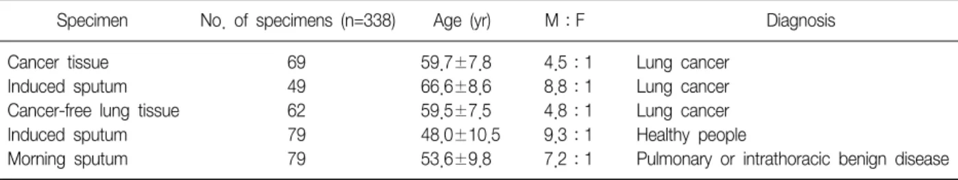
Table 1. Specimens used for MAGE and SSX gene expression
Specimen No. of specimens (n=338) Age (yr) M:F Diagnosis
Cancer tissue 69 59.7±7.8 4.5:1 Lung cancer
Induced sputum 49 66.6±8.6 8.8:1 Lung cancer
Cancer-free lung tissue 62 59.5±7.5 4.8:1 Lung cancer
Induced sputum 79 48.0±10.5 9.3:1 Healthy people
Morning sputum 79 53.6±9.8 7.2:1 Pulmonary or intrathoracic benign disease
*Values are presented as mean±SD.
MAGE: melanoma associated gene; SSX: synovial sarcoma on X chromosome; M: male; F: female; SD: standard deviation.
이오 표지자의 발굴도 지속되어 methylation specific PCR5,6, volatile organic compounds (VOC)7,8를 이용한 조 기 폐암의 진단법들이 개발되었다. 특히 VOC를 이용한 진단법은 날숨에 함유된 VOC를 분석하여 폐암 여부를 확 인하는 것으로 원발성 폐암 환자에서 민감도 85.1%, 특이 도 80.5%를 보였으나6 좀 더 연구가 필요한 실정이다. 최 근 melanoma associated gene (MAGE)을 이용한 폐암 검 출법이 발표되고 있으며9-12 이 중에 MAGE A 1에서 A 6까 지 동시에 증폭이 가능한 공통 시발체를 사용한 이중 중합 효소 연쇄반응법이 소개되었다13. 이 검사법은 폐암조직 에서 민감도 83.3%, 특이도 91.1%를 나타내었고14, 유도 객담과 기관지 세척액 등에서도 70% 이상의 민감도와 98%정도의 특이도를 나타내어6,14 매우 우수한 검사법으 로 보고되었다. MAGE A 1-6 공통 시발체를 개발한 연구 자들은 종양 검출률을 높이기 위하여 추가적인 표지자를 연구하였고 synovial sarcoma on X chromosome (SSX)으 로부터 SSX 1-9 유전자를 동시에 증폭하는 시발체를 고안 하였다. SSX 유전자는 cancer testis antigen (CTA) 유전자 중의 한 가지로서 아홉 종류의 아형을 가지고 있으며 종양 세포, 고환세포 및 갑상선세포 등에서만 표현되는 것으로 알려져 있다15-17. 따라서 정상 조직에서는 발현되지 않는 다고 알려진 두 가지 유전자를 이용하여 폐암조직 및 객담 에서의 발현 정도를 파악하여, 폐암의 조기 진단율을 높이 고자 본 연구를 시행하였다.
대상 및 방법
1. 대상
2002년부터 2004년까지 폐암으로 진단되어 폐암 절제 수술을 시행한 환자 69예, 수술을 하지 않은 폐암환자 49 예, 폐암 조기검진을 실시하여 건강인으로 판정된 79예 그리고 폐 및 흉강의 양성질환자 79예, 총 276예를 대상으
로 338 검체를 채취하였다(Table 1). 수술로 적출한 폐를 절개하여 종양의 가장 중심부위에서 암조직을 채취하고, 종양에서 되도록 멀리 떨어진 폐구역에서 가장 정상으로 보이는 폐조직을 채취하여 각각 1 mL의 Trizol (Invitro- gen, Calsbad, CA, USA)이 들어있는 시험관에 주입하여 검사 당일까지 영하 70oC에서 보관하였다. 건강검진을 시 행하러 내원한 건강인과 폐암진단을 받은 환자들은 대체 적으로 객담량이 많지 않았으며 이들을 대상으로 다음과 같은 방법으로 유도객담을 채취하였다. 먼저 물로 입안과 목안을 헹구어 낸 다음 β-2 agonist (FenoterolR; Boeh- ringer Ingelheim, Ridgefield, CT, USA)를 2회 흡입하게 한 후 약 10분 동안 기다렸다. 3% 생리식염수 16 mL를 초음파 분무기로 연무시켜 환자에게 흡입시켰다. 환자가 연무를 흡입한 후 객담을 뱉을 느낌이 있으면 50 mL 시험 관에 3 mL 정도를 모았다. 채취한 검체에 즉시 3배의 RNA 보존제(iC&G, Daegu, Korea)를 넣은 후 진탕하여 잘 혼합한 다음 검사 당일까지 영하 70oC에서 보관하였 다. 아침 객담은 주로 객담 분비가 많은 환자들을 대상으 로 미생물 배양 검사를 실시하기 위해 채취하였으며, 배양 검사 후 남은 잔여 검체를 즉시 수거한 뒤 3배의 RNA 보 존제를 주입하여 보관하였다.
2. RNA 추출
조직에서의 RNA 추출은 Trizol사에서 제공하는 추출법 을 사용하였다. 먼저 분쇄기를 사용하여 조직을 잘 간 다 음 실온에서 5분간 방치하였다. Chloroform 0.2 mL를 첨 가하여 마개를 잘 닫고 15초간 손으로 강하게 혼합한 다 음 12,000 g, 4oC에서 15분간 원심분리하였다. 무색의 상 층액을 새 시험관에 옮기고 동량의 isopropyl alcohol을 주입하고 혼합한 다음, 실온에서 10분간 방치하고 12,000 g, 4oC에서 10분간 원심분리 하였다. 상층액을 제거하고 75% ethanol 1 mL를 가하여 진탕 혼합한 다음 7,500 g,
Figure 1. Electrophoresis of MAGE (A), SSX gene (B) and GAPD (C) RT-PCR products in ten sputa of lung cancer patients. MAGE: melanoma associated gene; SSX: syno- vial sarcoma on X chromosome; GAPD: glyceraldehyde phosphate dehydrogenase; RT-PCR: reverse transcrip- tion polymerase chain reaction; SM: size markers.
4oC에서 5분간 원심분리 하였다. 다시 상층액을 제거하고 RNA 침사층을 5∼10분 동안 공기 중에서 건조시키고 RNase가 없는 증류수 30μL를 가하여 혼합한 다음 60oC 에서 10분간 방치하였다. 객담에서의 추출은 RNA 추출 시약(iC&G, Daegu, Korea)으로 시행하였다. 먼저 검체를 녹인 후 1,500 RPM에서 5분간 원심하여 상층을 15 mL 시험관으로 옮겼다. Oligo dT가 결합된 작은 자석 구슬로 이루어진 용액 50μL를 가하고 실온에서 30분간 roller 혼 합기에서 반응시켰다. 시험관을 자석 분리대에 설치한 다 음 10분간 방치하였다. 구슬이 자석에 완전히 붙은 것을 확인한 후 보존제를 제거하고 세척액 1 mL를 첨가하여 구슬을 부유시키며 세척하였다. 소시험관으로 옮겨 자석 분리대에 방치하여 구슬을 결합시킨 다음 세척액을 제거 하였다. 세척액을 교환하면서 이 과정을 2회 추가적으로 반복하고 세척액을 자석 분리대에서 제거하였다. RNase 가 없는 증류수 30μL를 가하여 혼합한 다음 70oC에서 8분 동안 방치하여 RNA를 용출시키고 다시 자석 분리대 에 붙였다. RNA 용출액을 새 시험관에 옮겨 4oC에서 보관 하였다.
3. 역전사 반응
추출한 RNA를 분광광도계(Beckman Coulter, Fullerton, CA, USA)를 이용하여 RNA 농도와 비를 측정하였으며 RNA 1μg을 역전사 반응에 사용하였다. 반응액은 완충액 4μL, 25 mM dNTP 0.4μL, 100 pmol oligo-dT 0.5μL, 25 mM MgCl2 2.4μL, 40 U/μL RNase inhibitor 0.5μL, RT enzyme (Promega, Madison, WI) 1μL로 구성된 반 응액에 추출한 RNA를 주입하여 총 20μL를 사용하였다.
MasterCycler (Eppendorf, Hamburg, Germany)를 이용하 여 25oC 10분, 42oC 60분 및 70oC 15분간 역전사 반응을 진행하였다.
4. 중합효소 연쇄반응
역전사 반응 산물 2μL를 취하여 GAPD (glyceralde- hyde phosphate dehydrogenase), MAGE 및 SSX 유전자 를 각각 증폭하였다(Figure 1). 중합효소 연쇄반응 시약은 MgCl2 1.5 mM, dNTP 0.2 mM, primer 20 pmol, PCR en- zyme 1.5 U 및 PCR 완충액 5μL로 구성된 총 50μL를 사용하였다. MAGE 유전자의 반응 조건은 94oC에서 2분 간 반응하여 효소를 활성화한 다음, 95oC 30초, 60oC 45 초, 72oC 45초로 30회 증폭하고 최종 증폭주기에서는 95oC 30초, 62oC 45초, 72oC 5분으로 1회 반응시켰다. SSX
유전자는 결합온도만 59oC 및 58oC로 각각 시행하였다.
MAGE 및 SSX 유전자는 이중 중합효소 연쇄반응을 시행 하였으며 GAPD 유전자는 한번의 중합효소 연쇄반응을 시행하였다.
5. MAGE 및 SSX 유전자의 양성률 분석
총 338 검체를 대상으로 임상진단에 따라 MAGE 및 SSX 유전자의 발현율 및 특이도 분석을 시행하였다. 또한 폐암의 세포형태에 따른 양성률과 폐암 환자의 객담에서 폐암의 T (종양) 병기에 따른 양성률을 각각 구하였다.
6. 실험성적의 통계분석
MAGE, SSX 및 두 가지 방법에 의한 검출률의 차이는 SPSS version 14.0 (SPSS Inc., Chicago, IL, USA)을 이용 하여 χ2 검정으로 실시했으며 p<0.05를 기준으로 하였 다.
결 과
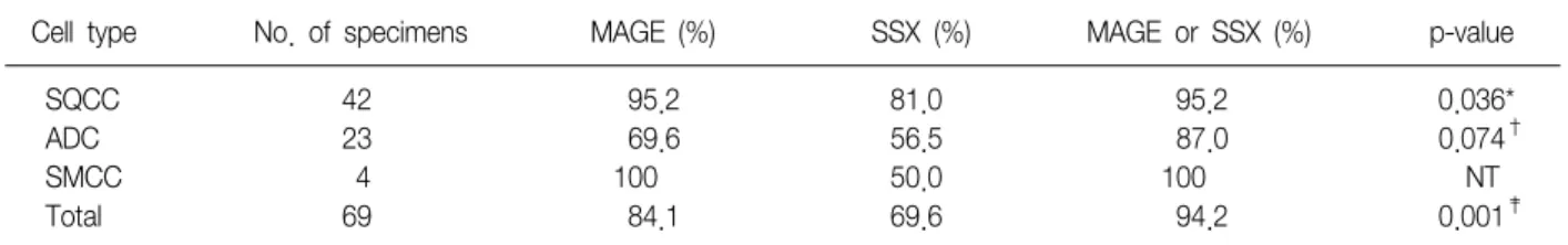
1. 폐암조직에서 MAGE 및 SSX 유전자의 발현율
폐암조직 69 검체를 대상으로 시행한 검사에서 평균적 으로 MAGE (84.1%)가 SSX 유전자(69.6%)에 비하여 발현 율이 높았으며, MAGE 및 SSX 유전자(이하 조합유전자)를 동시에 사용하였을 때는 편평상피암 95.2%, 선암 87.0%, 소세포암 100%로서 평균 94.2%의 양성률을 보였다 (p=0.001). 특히 선암에 있어서는 MAGE 단독(69.6%)보 다는 SSX 유전자를 동반했을 때 발현율이 87%로 증가되 었으나 통계적 유의성은 없었다(Table 2).Table 2. Expression rates of MAGE and SSX gene RT-PCR for lung cancer tissue
Cell type No. of specimens MAGE (%) SSX (%) MAGE or SSX (%) p-value
SQCC 42 95.2 81.0 95.2 0.036*
ADC 23 69.6 56.5 87.0 0.074
†SMCC 4 100 50.0 100 NT
Total 69 84.1 69.6 94.2 0.001
‡p-values were obtained by χ
2test.
*There is a higher expression rate in MAGE or SSX than SSX alone and it has a statistically significance in squamous cell carcinoma,
†
There is higher expression rate in MAGE or SSX than SSX alone but it has not statistically significance in adenocarcinoma,
‡There is a higher expression rate in MAGE or SSX than SSX alone and it has a statistically significance in lung cancer tissue.
MAGE: melanoma associated gene; SSX: synovial sarcoma on X chromosome; SQCC: squamous cell cancer; ADC: adenocarcinoma;
SMCC: small cell carcinoma; RT-PCR: reverse transcription polymerase chain reaction; NT: not tested.
Table 3. Expression rates of MAGE and SSX gene RT-PCR for cancer-free lung tissue
Diagnosis No. of
specimens MAGE (%) SSX (%) MAGE or SSX (%)
SQCC 35 8.6 11.4 14.3
ADC 23 8.7 17.4 17.4
SMCC 4 0.0 25.0 25.0
Total 62 8.1 14.5 17.7
MAGE: melanoma associated gene; SSX: synovial sarcoma on X chromosome; SQCC: squamous cell cancer; ADC: ad- enocarcinoma; RT-PCR: reverse transcription polymerase chain reaction.
2. 정상조직에서 MAGE 및 SSX 유전자의 발현율
MAGE 및 SSX 유전자의 특이도를 관찰하기 위하여 수 술환자에서 채취한 정상 폐조직 62 검체에서 발현율을 조 사하였다. 정상 조직에서의 발현율은 SSX 유전자가 MAGE에 비해서 평균적으로 높았으며(14.5% vs. 8.1%), 조합유전자를 사용하였을 때는 17.7%의 양성률을 보였 다. 따라서 조합유전자의 폐암조직에 대한 진단특이도는 편평상피암 85.7%, 선암 82.6%, 소세포암 75.0%, 그리고 평균 82.3%였다(Table 3).3. 폐암환자의 유도객담에서 MAGE 및 SSX 유전자의 발현율
MAGE 및 SSX 유전자가 조기폐암의 검출표지자로서 임상적 활용성이 있는지를 검증하기 위하여 폐암환자의 유도객담에서 각 유전자들의 발현양상을 관찰하였다 (Table 4). 총 49예의 객담에서 MAGE 및 SSX 유전자는 각각 51.0%, 42.9%, 조합유전자는 65.3%의 양성률을 나
타내었다. 암의 T병기에 따른 분류에서는 대체로 높은 T 병기에서 발현율이 높게 나타났으며, 조합 유전자의 경우 T1 33.3%, T2 72.2%, T3 80.0% 그리고 T4에서 66.7%의 발현율을 각각 나타내었다(Table 4).
4. 정상인과 폐 및 흉강 내 양성질환자의 객담에서 MAGE 및 SSX 유전자의 발현율
MAGE 및 SSX 유전자의 폐암환자의 객담에 대한 특이 도를 관찰하기 위하여 건강인의 유도객담 및 양성질환자 의 아침객담을 대상으로 하였다. 유도객담에 대한 MAGE, SSX 유전자 그리고 조합유전자의 발현율은 각각 2.5%, 2.5% 그리고 3.8%였으며 아침객담에서는 각각 2.5%, 15.2% 그리고 17.7%였다. 따라서 폐암환자의 객담에 대 한 조합유전자의 진단 특이도는 82.3∼96.2%로 나타났다 (Table 5).
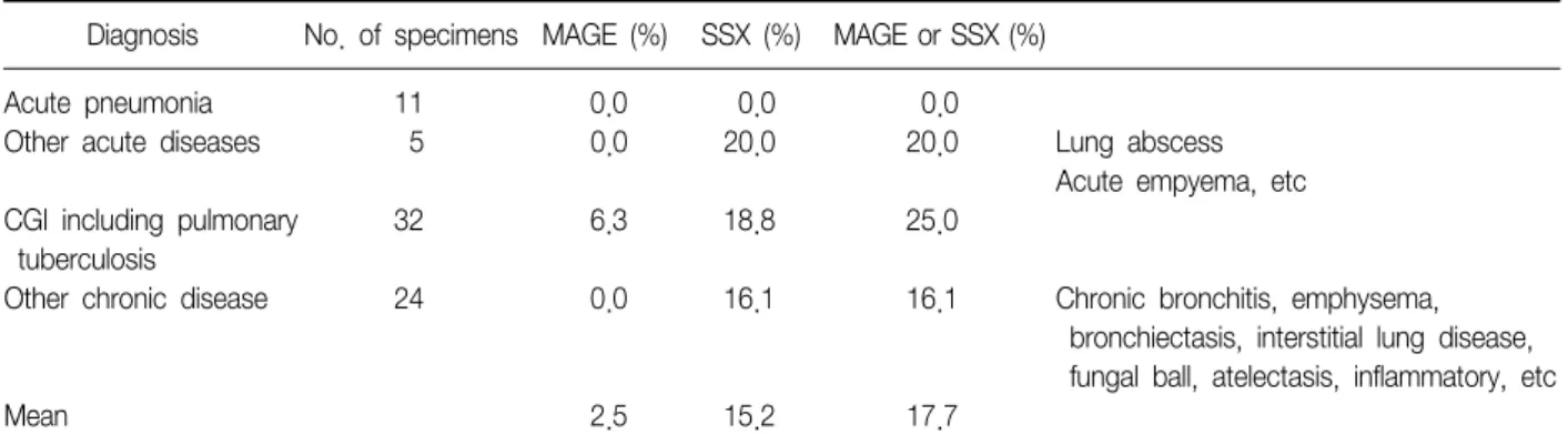
5. 폐 및 흉강 내 양성질환자들의 객담에서 MAGE 및 SSX 유전자의 발현율
임상적 진단에 따른 객담에서의 발현율을 분석해 보았 다. 전체적으로 SSX 유전자가 MAGE에 비해서 발현율이 높았다(15.2% vs. 2.5%). 그리고 조합유전자의 발현은 폐 렴환자의 전 예에서 음성이었으며 폐결핵을 포함한 만성 육아종성 염증에서 25.0%, 다른 만성 폐질환에서도 16.1%
로 높게 나타났다(Table 6).
고 찰
MAGE 유전자는 A, B, C, D, E, F, G, H 등 다양한 아형이 존재하며 A 아형은 다시 A 1부터 A 12로 나누어
진다18-20. 이 중 MAGE C4를 제외한 A, B, C 유전자가 암
Table 4. Expression rates of MAGE and SSX gene RT-PCR for the induced sputum of lung cancer patients
Stage No. of specimens MAGE (%) SSX (%) MAGE or SSX (%) p-value
T1 9 22.2 22.2 33.3
T2 18 61.4 50.0 72.2
T3 10 70.0 50.0 80.0
T4 12 41.7 41.7 66.7
Total 49 51.0 42.9 65.3 0.074*
p-values were obtained by χ
2test.
*There is a higher expression rate increasing T-stage but it has not statistically significance.
MAGE: melanoma associated gene; SSX: synovial sarcoma on X chromosome; RT-PCR: reverse transcription polymerase chain reaction.
Table 5. Expression rates of MAGE and SSX gene RT-PCR for the sputum of healthy people and benign diseases
Specimen Diagnosis No. of specimens MAGE (%) SSX (%) MAGE or SSX (%)
Induced sputum Healthy people 79 2.5 2.5 3.8
Morning sputum Pulmonary or intrathoracic benign diseases 79 2.5 15.2 17.7 MAGE: melanoma associated gene; SSX: synovial sarcoma on X chromosome; RT-PCR: reverse transcription polymerase chain reaction.
Table 6. Expression rates of MAGE and SSX gene RT-PCR in benign diseases
Diagnosis No. of specimens MAGE (%) SSX (%) MAGE or SSX (%)
Acute pneumonia 11 0.0 0.0 0.0
Other acute diseases 5 0.0 20.0 20.0 Lung abscess
Acute empyema, etc
CGI including pulmonary 32 6.3 18.8 25.0
tuberculosis
Other chronic disease 24 0.0 16.1 16.1 Chronic bronchitis, emphysema,
bronchiectasis, interstitial lung disease, fungal ball, atelectasis, inflammatory, etc
Mean 2.5 15.2 17.7
CGI: chronic granulomatous inflammation; MAGE: melanoma associated gene; SSX: synovial sarcoma on X chromosome; RT-PCR:
reverse transcription polymerase chain reaction.
세포에 특이적으로 발현하며 MAGE D, E, F, G, H 등은 정상세포에도 발현된다20. MAGE A 유전자는 암세포 검출 및 표적 치료제의 항원 등으로 많이 연구되고 있다21,22. MAGE 유전자는 3번, 15번, X 염색체 등에 다양하게 존재 하며20, 그 동안 이 유전자의 기능에 대해서 잘 알려지지 않았으나 최근 Yang 등23에 의하여 그 기능이 연구되었다.
암세포에서 발현되는 MAGE 단백은 KAP1 단백이 p53 단 백과 결합하여 암세포의 자멸사를 억제하는데 중요한 조 효소로 작용한다23. 즉 MAGE 유전자는 p53에 의한 종양 세포의 사멸을 억제하여 암세포로 이행하는데 매우 중요
한 역할을 담당한다. 세포에서 MAGE 유전자의 발현은 그 세포가 암으로 이행하는 과정 중에 있음을 암시하여 앞으 로 MAGE 유전자를 대상으로 한 연구가 더욱 활발해질 것으로 예상된다. SSX 유전자는 1∼9까지의 아형이 있으 며 이 아형이 모두 X 염색체에 존재한다17. Synovial sar- coma에서는 SSX 유전자와 SYT (synovial sarcoma trans- located to X chromosome) 유전자 간의 전위, 즉 t (X;18) (p11.2;q11.2)가 일어나 융합유전자인 SYT/SSX 유전자가 합성된다24. SSX 유전자 중 SSX 1, 2, 4 등이 종양에서 주 로 발현되며16, 특히 폐암조직에서는 SSX 1 28%, SSX 2
11% 그리고 SSX4는 35%가 발현되는 것으로 보고되었 다19. SSX 1, 2, 4, 6, 8 및 9는 정상 조직에서는 표현되지 않고 종양에서만 발현된다25,26. 반면에 SSX3와 SSX5는 종 양에서 잘 표현되지 않으며16 SSX 6-9는 종양과의 관계에 대한 연구가 미비하다. 악성 종양의 검출을 위해서 SSX 유전자 단독보다는 주로 다른 CTA (cancer testis antigen) 유전자와 조합하여 이용한다. Tajima 등10은 폐암조직에 서 MAGE A1-6 및 SSX 1, 2 및 4 중 어느 한 가지라도 양성인 경우가 94%라고 하였는데, 이는 MAGE와 SSX 유 전자를 이용한 폐암 검출이 우수한 민감도를 갖고 있다는 것을 시사한다. 하지만 SSX 유전자 아형 별로 독립적인 중합효소 연쇄반응을 시행하는 것은 매우 번거롭고 비효 율적이다. 따라서 본 연구에서는 SSX 아형간의 유전자 동 질성이 88∼95%에 달한다는 점15에 착안하여, SSX 1에서 SSX 9까지 동시에 증폭할 수 있는 공통시발체를 고안하여 기존의 MAGE A 1-6 공통시발체와 함께 사용하여 종양의 검출률을 높이고자 하였다. 본 연구 결과, 폐암조직에서 SSX 유전자의 발현은 편평상피암 및 소세포암에서 MAGE 의 발현과 중복되는 경우가 많아 종양검출률이 증가되지 는 않았다. 하지만 선암인 경우 SSX 유전자는 MAGE와 독립적으로 발현되는 경우가 많아서 두 가지 유전자를 동 시에 사용하는 경우 검출률이 MAGE 단독보다 17% 정도 증가하여 87%의 양성률을 보였다. 따라서 두 가지 유전자 를 동시에 사용하는 경우 전체 폐암에 있어서 양성률이 94.2%에 달하여 폐암 검출의 표적으로 매우 유용하였다.
이러한 성적은 Tajima 등10의 연구와 비슷한 양성률이지 만, Türeci16, Sahin 등25의 연구에서는 폐암에서 SSX 유전 자의 양성률이 21%에 불과하여 다소 차이를 보이고 있다.
SSX 유전자의 동양인과 서양인간의 발현율의 차이 여부 가 보고된 자료가 부족하여 파악하기 힘든 실정이다.
MAGE 및 SSX 유전자의 특이도를 파악하기 위하여 종양 인근 부위의 육안상 정상 폐조직, 비종양성 질환자의 아침 객담, 폐암 검진자들의 유도 객담을 대상으로 유전자 발현 율을 살펴보았다. 정상 폐조직에서의 MAGE 및 SSX 유전 자의 발현율은 17.7%로 나타나서 폐암에 대한 특이도는 82.3%였으며, 이는 MAGE 단독인 경우에 비해서 약 10%
정도 특이도가 감소하였다. MAGE는 정상 조직에서 객담 에 비하여 특이도가 다소 높게 나타났는데 이는 기존의 만성염증에 기인하거나 가능성은 적지만 종양세포의 구 역화(field cancerization)에 의한 미세한 종양세포의 존재 가능성27에 기인할 수 있을 것이다. 객담에서 MAGE 혹은 SSX 유전자 검사의 특이도를 분석한 결과 MAGE가 SSX
유전자에 비하여 우수한 특이도를 나타내었다(97.5% vs.
84.8%). MAGE는 결핵환자들에서만 위양성이 관찰되었 으나 SSX 유전자는 폐렴을 제외한 대부분의 비종양성 질 환에서 16.1∼20.0%의 위양성률이 관찰되었다. 두 유전 자 모두 폐렴 등의 급성 염증성 질환보다는 결핵 및 만성 염증성 질환에서 양성률이 증가함으로써, 이런 만성 폐질 환이 폐암 진단의 특이도를 감소시키는 주요 원인으로 작 용됨을 시사한다. 결핵환자에서 MAGE 및 SSX 유전자의 발현율 증가는 결핵 감염이 활성화되기 위해서 오랜 기간 의 발병과정이 소요되므로 만성화에 따른 세포 내 유전자 의 변화가 동반된 것으로 유추된다. 결핵환자의 폐암 발 생율이 결핵병변이 없는 사람보다 높다는 연구 결과28,29에 의거하면 결핵병변에서 만성염증성 자극에 의한 발암과 정을 배제할 수 없는 실정이다. 더우기 MAGE의 역할이 종양의 사멸을 방지하는 것이므로 MAGE 양성인 결핵환 자들은 폐암 발생 여부를 확인하기 위한 보다 적극적인 조기검진이 요망된다. 만성염증성 병변에서 SSX 유전자 의 양성률이 증가하는 것도 동일한 맥락에서 해석할 수 있을 것이다. 하지만 SSX 유전자는 아직 기능이 밝혀지지 않았고, 위양성률이 MAGE보다 높기 때문에 이의 양성률 에 대해서는 신중한 해석이 필요할 것이다. 폐암환자의 유도객담을 대상으로 한 MAGE와 SSX 유전자의 병용사용 시 65.3%의 발현율을 나타내었으며 MAGE 단독 보다 약 14%의 양성률이 증가됨을 보여 주었다. T병기에 따른 분 석에서는 T1병기에서 33.3%로 T2, 3, 4 병기보다는 낮게 발현되었으나 기존의 객담세포 검사(sputum cytology)의 양성률 10∼28%보다는 높게 나왔다30-32. 결론적으로 폐 암조직에 있어서 MAGE 또는 SSX 유전자의 발현율은 평 균 94.2%로 높았으며, 객담에서는 조직보다는 다소 낮았 지만 65.3%의 민감도와 82.3∼96.2%의 특이도를 보여주 었다. 특히 선암에 있어서는 MAZE단독보다는 SSX 유전 자를 동반했을 때 발현율이 증가하였다(69.6% vs. 87%).
따라서 향후 이 방법이 보완되고 개선된다면 기관지경 검 사와 같은 침습적인 검사를 시행하지 않고 환자의 유도객 담을 이용하여 폐암을 screening하여 폐암의 조기 검출에 매우 효과적인 검사법으로 활용될 수 있을 것이다.
참 고 문 헌
1. Korea Central Cancer Registry. Republic of Korea. 2002 Annual report of the Korea central cancer registry:
based on registered data from 139 hospitals. Seoul:
Ministry of Health and Welfare; 2005.
2. Kawahara M. Screening for lung cancer. Curr Opin Oncol 2004;16:141-5.
3. McWilliams A, Lam S. Lung cancer screening. Curr Opin Pulm Med 2005;11:272-7.
4. Lee P, Sutedja TG. Lung cancer screening: has there been any progress? Computed tomography and auto- fluorescence bronchoscopy. Curr Opin Pulm Med 2007;13:243-8.
5. Greenberg AK, Lee MS. Biomarkers for lung cancer:
clinical uses. Curr Opin Pulm Med 2007;13:249-55.
6. Lee KH, Shin KC, Lee CH, Jheon SH, Jeon CH.
Detection of lung cancer using MAGE A1-6 and SSX4 RT-PCR expression profiles in the bronchial wash fluid.
Cancer Res Treat 2007;39:69-73.
7. Phillips M, Cataneo RN, Cummin AR, Gagliardi AJ, Gleeson K, Greenberg J, et al. Detection of lung cancer with volatile markers in the breath. Chest 2003;123:
2115-23.
8. Poli D, Carbognani P, Corradi M, Goldoni M, Acampa O, Balbi B, et al. Exhaled volatile organic compounds in patients with non-small cell lung cancer: cross sec- tional and nested short-term follow-up study. Respir Res 2005;6:71.
9. Mecklenburg I, Stratakis DF, Huber RM, Häussinger K, Morresi-Hauf A, Riethmüller G, et al. Detection of mel- anoma antigen-A expression in sputum and bronchial lavage fluid of patients with lung cancer. Chest 2004;
125 Suppl 5:164S-6S.
10. Tajima K, Obata Y, Tamaki H, Yoshida M, Chen YT, Scanlan MJ, et al. Expression of cancer/testis (CT) anti- gens in lung cancer. Lung Cancer 2003;42:23-33.
11. Sugita M, Geraci M, Gao B, Powell RL, Hirsch FR, Johnson G, et al. Combined use of oligonucleotide and tissue microarrays identifies cancer/testis antigens as bi- omarkers in lung carcinoma. Cancer Res 2002;62:
3971-9.
12. Wu Y, Li Y, Lin Y. Expression of MAGE-12 in lung cancer. Lung Cancer 2002;35:95.
13. Park JW, Kwon TK, Kim IH, Sohn SS, Kim YS, Kim CI, et al. A new strategy for the diagnosis of MAGE-ex- pressing cancers. J Immunol Methods 2002;266:79-86.
14. Jheon S, Hyun DS, Lee SC, Yoon GS, Jeon CH, Park JW, et al. Lung cancer detection by a RT-nested PCR using MAGE A1--6 common primers. Lung Cancer 2004;43:29-37.
15. Gure AO, Türeci O, Sahin U, Tsang S, Scanlan MJ, Jäger E, et al. SSX: a multigene family with several members transcribed in normal testis and human cancer. Int J
Cancer 1997;72:965-71.
16. Türeci O, Chen YT, Sahin U, Güre AO, Zwick C, Villena C, et al. Expression of SSX genes in human tumors. Int J Cancer 1998;77:19-23.
17. Güre AO, Wei IJ, Old LJ, Chen YT. The SSX gene fam- ily: characterization of 9 complete genes. Int J Cancer 2002;101:448-53.
18. van der Bruggen P, Traversari C, Chomez P, Lurquin C, De Plaen E, Van den Eynde B, et al. A gene encod- ing an antigen recognized by cytolytic T lymphocytes on a human melanoma. Science 1991;254:1643-7.
19. De Plaen E, Arden K, Traversari C, Gaforio JJ, Szikora JP, De Smet C, et al. Structure, chromosomal local- ization, and expression of 12 genes of the MAGE family. Immunogenetics 1994;40:360-9.
20. Chomez P, De Backer O, Bertrand M, De Plaen E, Boon T, Lucas S. An overview of the MAGE gene fam- ily with the identification of all human members of the family. Cancer Res 2001;61:5544-51.
21. Jungbluth AA, Busam KJ, Kolb D, Iversen K, Coplan K, Chen YT, et al. Expression of MAGE-antigens in normal tissues and cancer. Int J Cancer 2000;85:460-5.
22. Brichard VG, Lejeune D. GSK's antigen-specific cancer immunotherapy programme: pilot results leading to Phase III clinical development. Vaccine 2007;25 Suppl 2:B61-71.
23. Yang B, O'Herrin SM, Wu J, Reagan-Shaw S, Ma Y, Bhat KM, et al. MAGE-A, mMage-b, and MAGE-C pro- teins form complexes with KAP1 and suppress p53-de- pendent apoptosis in MAGE-positive cell lines. Cancer Res 2007;67:9954-62.
24. Törnkvist M, Brodin B, Bartolazzi A, Larsson O. A nov- el type of SYT/SSX fusion: methodological and bio- logical implications. Mod Pathol 2002;15:679-85.
25. Sahin U, Koslowski M, Türeci O, Eberle T, Zwick C, Romeike B, et al. Expression of cancer testis genes in human brain tumors. Clin Cancer Res 2000;6:3916-22.
26. Kubuschok B, Xie X, Jesnowski R, Preuss KD, Romeike BF, Neumann F, et al. Expression of cancer testis anti- gens in pancreatic carcinoma cell lines, pancreatic ad- enocarcinoma and chronic pancreatitis. Int J Cancer 2004;109:568-75.
27. Slaughter DP, Southwick HW, Smejkal W. Field cancer- ization in oral stratified squamous epithelium: clinical implications of multicentric origin. Cancer 1953;6:963-8.
28. Zheng W, Blot WJ, Liao ML, Wang ZX, Levin LI, Zhao JJ, et al. Lung cancer and prior tuberculosis infection in Shanghai. Br J Cancer 1987;56:501-4.
29. Kurasawa T. The coexistence of pulmonary tuber-
culosis and lung cancer. Nippon Rinsho 1998;56:3167- 70.
30. Fontana RS, Sanderson DR, Woolner LB, Taylor WF, Miller WE, Muhm JR. Lung cancer screening: the Mayo program. J Occup Med 1986;28:746-50.
31. Fraire AE, Underwood RD, McLarty JW, Greenberg SD.
Conventional respiratory cytology versus fine needle
aspiration cytology in the diagnosis of lung cancer.
Acta Cytol 1991;35:385-8.
32. Frost JK, Ball WC Jr, Levin ML, Tockman MS, Baker RR, Carter D, et al. Early lung cancer detection: results of the initial (prevalence) radiologic and cytologic screening in the Johns Hopkins study. Am Rev Respir Dis 1984;130:549-54.