ORIGINAL ARTICLE
두경부 암 환자에서 이차암 발견을 위해 수술 전 위장내시경 검사가 필요한가? 10년 등록자료 분석
허경미, 김미희, 김진환1, 노영수1, 신운건
한림대학교 의과대학 내과학교실, 이비인후과교실1
Is a Preoperative Gastrointestinal Endoscopy for Second Primary Cancer Detection in Head and Neck Cancer Necessary? Ten-year Registry Data
Gyeong Mi Heo, Mi Hee Kim, Jin Hwan Kim1, Young Soo Rho1, and Woon Geon Shin
Department of Internal Medicine, Department of Otorhinolaryngology1, Hallym University College of Medicine, Seoul, Korea
Background/Aims: In head and neck squamous cell carcinoma, second primary gastrointestinal tumors are not uncommon.
However, it is unclear whether a screening endoscopy is needed for detecting gastrointestinal neoplasm in patients with head and neck cancer. Therefore, we analyzed the prevalence and independent risk factors for second primary gastrointestinal neoplasm in head and neck squamous cell carcinoma.
Methods: A consecutive series of 328 patients with primary head and neck squamous cell carcinoma that underwent esophagogas- troduodenoscopy or colonoscopy were included using our registry. An age- and sex-matched group of 328 control subjects was enrolled. We assessed risk factors of synchronous gastrointestinal cancer.
Results: The prevalence of esophageal cancer with head and neck squamous cell carcinoma was significantly higher than that of the control group (1.5% vs. 0.0%, p=0.011). An age of 54 years or more (OR, 1.033; 95% CI, 1.008-1.059; p=0.009) and male gender (OR, 4.974; 95% CI, 1.648-15.013; p=0.004) were risk factors for concomitant colorectal cancer or adenomas in the head and neck squamous cell carcinoma patients.
Conclusions: Preoperative colonoscopy can be recommended for detecting synchronous second primary colorectal lesions in head and neck squamous cell carcinoma patients with male sex regardless of age, and esophagogastroduodenoscopy is necessary in all head and neck squamous cell carcinoma patients for detecting esophageal cancer. (Korean J Gastroenterol 2016;68:23-28) Key Words: Head and neck neoplasms; Esophagogastroduodenoscopy; Colonoscopy; Diagnosis
Received April 5, 2016. Revised May 12, 2016. Accepted May 14, 2016.
CC This is an open access article distributed under the terms of the Creative Commons Attribution Non-Commercial License (http://creativecommons.org/licenses/
by-nc/4.0) which permits unrestricted non-commercial use, distribution, and reproduction in any medium, provided the original work is properly cited.
Copyright © 2016. Korean Society of Gastroenterology.
교신저자: 신운건, 05355, 서울시 강동구 성안로 150, 한림대학교강동성심병원 소화기내과
Correspondence to: Woon Geon Shin, Division of Gastroenterology, Department of Internal Medicine, Hallym University Kangdong Sacred Heart Hospital, 150 Seongan- ro, Gangdong-gu, Seoul 05355, Korea. Tel: +82-2-2225-2814, Fax: +82-2-478-6925, E-mail: [email protected]
Financial support: None. Conflict of interest: None.
INTRODUCTION
Second primary tumors (SPTs) are common in head and neck squamous cell carcinoma (HNSCC) patients. Most cas- es of HNSCC are linked with esophageal cancer or lung cancer.1 Gastrointestinal malignancies in particular, such as gastric cancer and colorectal cancer, are often found.1-6 SPT
of the respiratory and upper digestive tract is the main cause of death after treatment in early-stage HNSCC patients.7 In the first five years after treatment of the primary tumor, the rates for metachronous SPT reach approximately 15% to 25%.8 Therefore, SPT constitutes a major challenge in the field of head and neck oncology, and preoperative work-up for synchronous cancer is very important.
According to the cancer statistics of Korea in 2011, the age-standardized incidence rate of head and neck cancer was 4.0 (men, 6.3 and women, 2.1) per 100,000 persons per year, and the mortality rates were 2.2 per 100,000 men per year and 0.5 per 100,000 women per year. The 5-year surviv- al rates have improved over the past two decades from 41.1%
to 61.6% because of advances in surgery and chemo- radiation treatments in Korea.9 However, the overall survival rate for HNSCC remains low (∼55% after five years).10 The poor outcome of HNSCC affects patient quality of life be- cause of its functional anatomic location.11
The role of routine esophagogastroduodenoscopy (EGD) and colonoscopy as screening tools for SPT assessment in patients with HNSCC has been controversial until now.
Several studies on routine preoperative evaluation of upper gastrointestinal tract were discrepant over cost effective- ness and efficiency.12-15 Furthermore, there are few studies assessing the value of colonoscopy for the preoperative work-up in HNSCC patients.5,6
We hypothesized that colorectal tumor prevalence may be higher in HNSCC patients than in the control group because of shared risk factors, such as smoking, alcohol con- sumption, and old age, for both HNSCC and colorectal cancer. The present study assesses the prevalence and in- dependent risk factors for gastrointestinal neoplasm in HNSCC patients using the HNSCC registry.
SUBJECTS AND METHODS
1. Study population
We included consecutive patients diagnosed with primary HNSCC from January 2005 to December 2014 using the head and neck cancer registry at the center at Hallym University Kangdong Sacred Heart Hospital (Seoul, Korea).
This registry included the patients’ demographics, risk fac- tors (e.g., smoking and alcohol consumption), underlying dis- eases (e.g., hypertension and diabetes mellitus), laboratory findings, staging work-up results (e.g., computed tomog- raphy and endoscopy), histological results, cancer location, and cancer stage. Inclusion criteria were the patients who had a cancer located in the oral cavity, oropharynx, hypophar- ynx, or larynx. We excluded cancers of the salivary glands and the thyroid gland. All enrolled 328 HNSCC patients had under- gone preoperative EGD, whereas 224 HNSCC patients had un-
dergone preoperative colonoscopy.
The age- and sex-matched controls visited the gastro- enterology center for various gastrointestinal symptoms, and underwent successful EGD and colonoscopy on the same en- doscopy day of enrolled HNSCC patients with a seven day win- dow from January 2005 to December 2014.
2. Study design
The data of this cross-sectional study was collected retrospectively. The study subjects were divided into an HNSCC and control group to assess the prevalence of syn- chronous gastrointestinal malignancies such as esoph- ageal, gastric and colorectal cancer. The frequencies of co- lonic adenoma or adenocarcinoma in the HNSCC group were compared with those of the control group. We assessed risk factors of synchronous gastrointestinal cancer using various parameters, such as 1) pathological type of synchronous gas- trointestinal malignancies and pre-cancerous lesions, 2) sex, 3) age, 4) lifestyle factors (e.g., smoking and alcohol), 5) un- derlying diseases (e.g., hypertension and diabetes mellitus), and 6) cancer location. Staging of HNSCC was performed us- ing the seventh TNM classification of malignant tumors of the American Joint Committee on Cancer (AJCC).
This study was approved by the institutional review boards of Hallym University Kangdong Sacred Heart Hospital (IRB No: 11-040). Informed consent for this research project, not for treatment and procedures, was waived.
3. Statistical analysis
The variables associated with risk factors were initially identified using the independent sample t-test for con- tinuous variables and the chi-squared test for categorical variables. Additionally, we used multivariate logistic re- gression analysis to detect independent risk factors for syn- chronous gastrointestinal cancer. All analyses were per- formed with SPSS version 16.0 (SPSS Inc., Chicago, IL, USA).
Null hypotheses of no difference were rejected if p-values were less than 0.05, or, equivalently, if the 95% CIs of risk point estimates excluded 1.
RESULTS
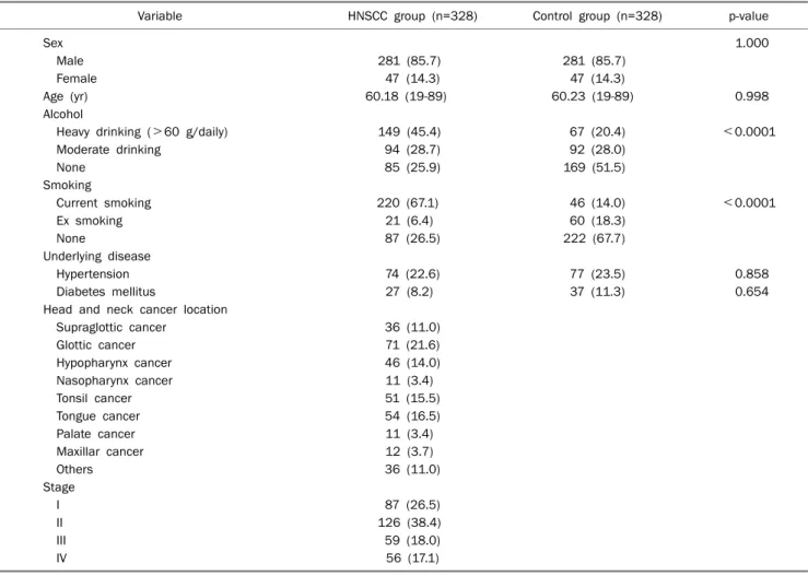
Baseline characteristics for study subjects are in Table 1.
By design there were 281 men and 47 women in both groups.
Table 1. Clinicopathological Data for Patients with HNSCC and the Control Group
Variable HNSCC group (n=328) Control group (n=328) p-value
Sex 1.000
Male 281 (85.7) 281 (85.7)
Female 47 (14.3) 47 (14.3)
Age (yr) 60.18 (19-89) 60.23 (19-89) 0.998
Alcohol
Heavy drinking (>60 g/daily) 149 (45.4) 67 (20.4) <0.0001
Moderate drinking 94 (28.7) 92 (28.0)
None 85 (25.9) 169 (51.5)
Smoking
Current smoking 220 (67.1) 46 (14.0) <0.0001
Ex smoking 21 (6.4) 60 (18.3)
None 87 (26.5) 222 (67.7)
Underlying disease
Hypertension 74 (22.6) 77 (23.5) 0.858
Diabetes mellitus 27 (8.2) 37 (11.3) 0.654
Head and neck cancer location
Supraglottic cancer 36 (11.0)
Glottic cancer 71 (21.6)
Hypopharynx cancer 46 (14.0)
Nasopharynx cancer 11 (3.4)
Tonsil cancer 51 (15.5)
Tongue cancer 54 (16.5)
Palate cancer 11 (3.4)
Maxillar cancer 12 (3.7)
Others 36 (11.0)
Stage
I 87 (26.5)
II 126 (38.4)
III 59 (18.0)
IV 56 (17.1)
Values are presented as n (%) or mean (range).
HNSCC, head and neck squamous cell carcinoma.
Table 2. Characteristics of Synchronous Gastrointestinal Neoplasm with HNSCC and the Control Group
Gastrointestinal neoplasm
HNSCC group
Control
group p-value Esophageal cancer 5/328 (1.5) 0/328 (0.0) 0.011 Gastric cancer 4/328 (1.2) 6/328 (1.8) 1.000 Gastric adenoma 3/328 (0.9) 2/328 (0.6) 0.400 Colon cancer 7/224 (3.1) 17/328 (5.2) 0.292 Colon adenoma 83/224 (37.1) 117/328 (35.7) 0.787 Values are presented as n (%).
HNSCC, head and neck squamous cell carcinoma.
The mean age was 60.18 years for men and 60.23 years for women. The HNSCC group had a greater proportion of heavy drinkers (>60 g/daily) (45.4% vs. 20.4%, p<0.0001) and current smokers (67.1% vs. 14.0%, p<0.001). In terms of un- derlying disease, hypertension and diabetes mellitus were not significantly different between the two groups. Based on the seventh TNM classification of malignant tumors of AJCC, 126 were stage II, the most common staging, with 38.4% of patients. Further, 87 were stage I (26.5%), 59 were stage III (18.0%), and 56 were stage IV (17.1%) (Table 1).
Of the patients with primary head and neck cancer, syn- chronous esophageal cancer was diagnosed in 5/328 (1.5%), gastric in 4/328 (1.2%), and colorectal cancer in 7/224 patients (3.1%). Of the 328 controls, there was no esophageal, 6 (1.8%) gastric, and 17 (5.2%) colorectal can- cers diagnosed. Only the prevalence of esophageal cancer with HNSCC was significantly higher than that of the control
group (1.5% vs. 0.0%, p=0.01) (Table 2).
The HNSCC patients with colorectal cancer were older than HNSCC patients without colorectal cancer (mean age of 71.29 years vs. 59.00 years, p<0.0001). There was no sig- nificant difference for sex between the HNSCC patients with colorectal cancer and those without colorectal cancer
Table 3. Univariate Analysis of Risk Factors in HNSCC Patients with or without Colon Cancer vs. with or without Colon Cancer or Adenoma
Variable With colon
cancer (n=7)
Without colon
cancer (n=217) p-value With colon cancer or adenoma (n=90)
Without colon cancer or adenoma (n=134) p-value
Age (yr) 71.29±5.16 59.00±11.93 <0.0001 62.01±10.03 57.62±12.85 0.003
Sex 0.37 0.001
Male 7 (100.0) 188 (86.6) 86 (95.6) 109 (81.3)
Female 0 (0.0) 29 (13.4) 4 (4.4) 25 (18.7)
Location of lesion 0.44 0.03
Supraglottic 2 (28.6) 19 (8.8) 11 (12.2) 10 (7.5)
Glottis 1 (14.3) 38 (17.5) 19 (21.1) 20 (14.9)
Hypopharynx 3 (42.9) 32 (14.7) 18 (20.0) 17 (12.7)
Nasopharynx 0 (0.0) 8 (3.7) 2 (2.2) 6 (4.5)
Tonsil 0 (0.0) 35 (16.1) 14 (15.6) 21 (15.7)
Tongue 1 (14.3) 39 (18.0) 7 (7.8) 33 (24.6)
Palate 0 (0.0) 9 (4.1) 2 (2.2) 7 (5.2)
Maxillar 0 (0.0) 12 (5.5) 4 (4.4) 8 (6.0)
Others 0 (0.0) 25 (11.5) 13 (14.4) 12 (9.0)
Alcohol 1.00 0.08
Heavy drinking (>60 g/daily) 3 (42.9) 100 (46.1) 48 (53.3) 55 (41.0)
Moderate drinking 2 (28.6) 66 (30.4) 22 (24.4) 46 (34.3)
None 2 (28.6) 51 (23.5) 20 (22.2) 33 (24.6)
Smoking 0.26 0.007
Current smoking 7 (100.0) 141 (65.0) 69 (76.7) 79 (59.0)
Ex smoking 0 (0.0) 16 (7.4) 6 (6.7) 10 (7.5)
None 0 (0.0) 60 (27.6) 15 (16.7) 45 (33.6)
Diabetes mellitus 0.53 0.33
Present 0 (0.0) 19 (8.8) 19 (21.1) 33 (24.6)
Absent 7 (100.0) 198 (91.2) 71 (78.9) 101 (75.4)
Hypertension 0.21 0.48
Present 3 (42.9) 49 (22.6) 7 (7.8) 12 (9.0)
Absent 4 (57.1) 168 (77.4) 83 (92.2) 122 (91.0)
Values are presented as mean±SD or n (%).
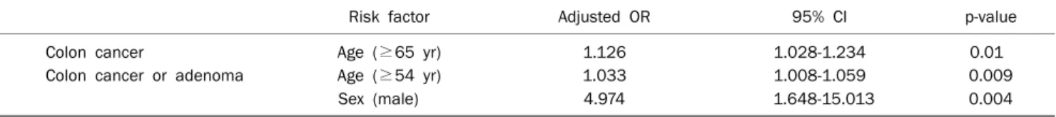
Table 4. Multivariate Logistic Regression Analysis of Risk Factors for Colon Cancer and Colon Cancer or Adenoma among the HNSCC Patients
Risk factor Adjusted OR 95% CI p-value
Colon cancer Age (≥65 yr) 1.126 1.028-1.234 0.01
Colon cancer or adenoma Age (≥54 yr) 1.033 1.008-1.059 0.009
Sex (male) 4.974 1.648-15.013 0.004
HNSCC, head and neck squamous cell carcinoma.
Including variables for adjustment: age, sex, location of lesion, smoking.
(p=0.37). There was no significant difference in alcohol con- sumption, smoking, hypertension, and diabetes mellitus be- tween groups (Table 3). The HNSCC patients with colorectal cancer or adenoma were older than those without colonic ne- oplasm (mean age of 62.01 years vs. 57.62 years, p=0.003).
The HNSCC subjects with colonic neoplasm had a greater proportion of men (95.6% vs. 81.3%, p=0.001). The location of HNSCC was different between the HNSCC patients with co- lonic neoplasm and those without colonic neoplasm (p=0.03). Current smokers were more prevalent in the HNSCC with colonic neoplasm than the non-colonic neoplasm group
(76.7% vs. 59.0%, p=0.007). There was no significant differ- ence in terms of alcohol consumption, diabetes mellitus, and hypertension between groups (Table 3).
In the multivariate logistic regression analysis, old age (≥
65 years) was an independent risk factor for concomitant col- orectal cancer in the HNSCC patients (OR, 1.126; 95% CI, 1.028-1.234; p=0.01). In addition, an age of 54 years or more (OR, 1.033; 95% CI, 1.008-1.059; p=0.009) and male gender (OR, 4.974; 95% CI, 1.648-15.013; p=0.004) were risk factors for concomitant colorectal cancer or adenomas in the HNSCC patients (Table 4).
Table 5. Positive and Negative Predictive Value of the Risk Factors for Colon Cancer and Colon Cancer or Adenoma among the HNSCC Patients
Risk factor n/n PPV (%) n/n NPV (%)
For colon cancer Age (≥65 yr) 7/78 9.0 146/146 100.0
For colon cancer or adenoma Age (≥54 yr) 68/145 46.9 57/79 72.2
Sex (male) 86/195 44.1 25/29 86.2
HNSCC, head and neck squamous cell carcinoma; PPV, positive predictive value; NPV, negative predictive value.
The risk factor of old age (≥65 years) had a positive pre- dictive value (PPV) of 9.0% and a negative predictive value (NPV) of 100.0% for colorectal cancer in the HNSCC patients.
An age of 54 years or more had a PPV of 46.9% and an NPV of 72.2% for predicting colorectal cancer or adenoma in the HNSCC patients. Male sex had a PPV of 44.1% and an NPV of 86.2% for predicting colorectal cancer or adenoma in the HNSCC patients (Table 5).
DISCUSSION
The independent risk factors for concomitant colorectal ne- oplasm (cancer or adenoma) in HNSCC patients were an age of 54 years or more (OR, 1.033; 95% CI, 1.008-1.059;
p=0.009) and the male sex (OR, 4.974; 95% CI, 1.648-15.013;
p=0.004). Although the overall prevalence of colorectal neo- plasm was not higher in the HNSCC group than the control group in this study, the same result as an earlier report,6 this association between HNSCC and colorectal neoplasm can be explained by the shared carcinogens of cigarette smoking and alcohol drinking.1,5 Advances in the prevention, treat- ment, and surveillance of HNSCC await a more complete un- derstanding of the mechanism of the multifocality of this tumor. According to the field carcinogenesis concept, multi- ple cell groups independently undergo neoplastic trans- formation under the stress of regional carcinogenic activity.16 Considering that NPV of age and sex was higher than PPV (72.2% vs. 46.9%; 86.2% vs. 44.1%, respectively), pre- operative colonoscopy for detecting colonic neoplasm may not be recommended in patients less than 54 years of age and women. However, screening colonoscopy is strongly rec- ommended for early detection of colorectal cancer in aver- age-risk persons aged 50 years and older in Korean guidelines.17 The colorectal cancer incidence decreased by 76-90% in a cohort that underwent colonoscopy and poly- pectomy compared to reference populations.18 Moreover, in- creased colonoscopy use was related to colorectal mortality
reduction.19 Thus, in combination with our results and color- ectal cancer screening guidelines, preoperative colonoscopy could be beneficial in all HNSCC men regardless of age or ≥50 years HNSCC women for detecting concomitant colorectal neoplasm.
Esophageal cancer was the most common malignant gastro- intestinal SPT with a significantly higher prevalence in the HNSCC group in this study (2.2% vs. none in the control group, p=0.01). Several studies provide evidence for this result.1,3,20-23 The previous prospective study reported that the rate of sec- ond esophageal primary tumors was 1.9% in the 268 HNSCC patients.23 One recent study reported that esophageal sur- veillance was recommended because early detection of esophageal cancer in HNSCC patients led to a good prognosis.24 The co-existence of either a colorectal malig- nancy or adenoma with esophageal cancer is also not un- common (1.5%).25 Therefore, EGD and colonoscopy as a screening tool for detecting SPT in HNSCC is crucial in diag- nosis and treatment.
Our study has several strengths. First, to the best of our knowledge, this is the first study assessing the risk factors for colorectal neoplasm as SPTs in HNSCC. Second, we analyzed detailed clinical parameters for each patient using our registry. This enabled our study to adjust for potential confounders. There are some limitations in this study. First, this study has possible selection bias in our exposed (HNSCC) and control group. In fact, the prevalence of colorectal cancer in our control group was too high (5.2%). Our control group who visited the gastroenterology center for various gastro- intestinal symptoms was not a general population. However, this bias might be trivial because we used our HNSCC registry in which the HNSCC patients were enrolled consecutively.
Second, the prevalence of colorectal cancer as an SPT is like- ly to be underestimated because we did not conduct a colono- scopy in all patients due to patient refusal.
In conclusion, preoperative colonoscopy can be recom- mended for detecting synchronous second primary color-
ectal lesions in HNSCC patients with male sex regardless of age, and EGD is necessary in all HNSCC patients for detecting esophageal cancer.
REFERENCES
1. Chuang SC, Scelo G, Tonita JM, et al. Risk of second primary can- cer among patients with head and neck cancers: a pooled analy- sis of 13 cancer registries. Int J Cancer 2008;123:2390-2396.
2. Guardiola E, Pivot X, Dassonville O, et al. Is routine triple endos- copy for head and neck carcinoma patients necessary in light of a negative chest computed tomography scan? Cancer 2004;
101:2028-2033.
3. Jones AS, Morar P, Phillips DE, Field JK, Husband D, Helliwell TR.
Second primary tumors in patients with head and neck squ- amous cell carcinoma. Cancer 1995;75:1343-1353.
4. Davidson J, Gilbert R, Irish J, et al. The role of panendoscopy in the management of mucosal head and neck malignancy-a pro- spective evaluation. Head Neck 2000;22:449-454; discussion 454-455.
5. McGarry GW, Mackenzie K, Finlay I. Colonic second primary can- cers in patients with index tumours of the head and neck. Br J Surg 1994;81:1481.
6. Sikora AG, Morris LG, Sturgis EM. Bidirectional association of anogenital and oral cavity/pharyngeal carcinomas in men. Arch Otolaryngol Head Neck Surg 2009;135:402-405.
7. Yamamoto E, Shibuya H, Yoshimura R, Miura M. Site specific de- pendency of second primary cancer in early stage head and neck squamous cell carcinoma. Cancer 2002;94:2007-2014.
8. Sturgis EM, Miller RH. Second primary malignancies in the head and neck cancer patient. Ann Otol Rhinol Laryngol 1995;104:
946-954.
9. Jung KW, Won YJ, Kong HJ, Oh CM, Lee DH, Lee JS. Cancer sta- tistics in Korea: incidence, mortality, survival, and prevalence in 2011. Cancer Res Treat 2014;46:109-123.
10. Pisani P, Bray F, Parkin DM. Estimates of the world-wide preva- lence of cancer for 25 sites in the adult population. Int J Cancer 2002;97:72-81.
11. Ramos M, Benavente S, Giralt J. Management of squamous cell carcinoma of the head and neck: updated European treatment recommendations. Expert Rev Anticancer Ther 2010;10:339- 344.
12. Shiozaki H, Tahara H, Kobayashi K, et al. Endoscopic screening of early esophageal cancer with the Lugol dye method in patients with head and neck cancers. Cancer 1990;66:2068-2071.
13. Makuuchi H, Tanaka H, Shimada H, et al. Esophageal cancer and multiple primary cancer. Gan To Kagaku Ryoho 1997;24:1-7.
14. Benninger MS, Shariff A, Blazoff K. Symptom-directed selective endoscopy: long-term efficacy. Arch Otolaryngol Head Neck Surg 2001;127:770-773.
15. Lo OS, Law S, Wei WI, et al. Esophageal cancers with synchro- nous or antecedent head and neck cancers: a more formidable challenge? Ann Surg Oncol 2008;15:1750-1756.
16. Slaughter DP, Southwick HW, Smejkal W. Field cancerization in oral stratified squamous epithelium; clinical implications of mul- ticentric origin. Cancer 1953;6:963-968.
17. Lee BI, Hong SP, Kim SE, et al. Korean guidelines for colorectal cancer screening and polyp detection. Clin Endosc 2012;45:
25-43.
18. Winawer SJ, Zauber AG, Ho MN, et al. Prevention of colorectal cancer by colonoscopic polypectomy. The National Polyp Study Workgroup. N Engl J Med 1993;329:1977-1981.
19. Rabeneck L, Paszat LF, Saskin R, Stukel TA. Association between colonoscopy rates and colorectal cancer mortality. Am J Gastroenterol 2010;105:1627-1632.
20. Chow TL, Lee DT, Choi CY, Chan TT, Lam SH. Prediction of simulta- neous esophageal lesions in head and neck squamous cell car- cinoma: a multivariate analysis. Arch Otolaryngol Head Neck Surg 2009;135:882-885.
21. Watanabe A, Hosokawa M, Taniguchi M, Tsujie H, Sasaki S. Head and neck cancer associated with esophageal cancer. Auris Nasus Larynx 2007;34:207-211.
22. León X, Ferlito A, Myer CM 3rd, et al. Second primary tumors in head and neck cancer patients. Acta Otolaryngol 2002;122:
765-778.
23. Grossman TW. The incidence and diagnosis of secondary esoph- ageal carcinoma in the head and neck cancer patient.
Laryngoscope 1989;99:1052-1056.
24. Lim H, Kim do H, Jung HY, et al. Clinical significance of early de- tection of esophageal cancer in patients with head and neck cancer. Gut Liver 2015;9:159-165.
25. Miyazaki T, Tanaka N, Sano A, et al. Clinical significance of total colonoscopy for screening of colon lesions in patients with esophageal cancer. Anticancer Res 2013;33:5113-5117.